Review
. 2017 Jun:44:178-185.
doi: 10.1016/j.conb.2017.04.011. Epub 2017 May 22.

Neural oscillations demonstrate that general anesthesia and sedative states are neurophysiologically distinct from sleep

Affiliations
Review

Neural oscillations demonstrate that general anesthesia and sedative states are neurophysiologically distinct from sleep

Oluwaseun Akeju et al. Curr Opin Neurobiol. 2017 Jun.

Abstract

General anesthesia is a man-made neurophysiological state comprised of unconsciousness, amnesia, analgesia, and immobility along with maintenance of physiological stability. Growing evidence suggests that anesthetic-induced neural oscillations are a primary mechanism of anesthetic action. Each anesthetic drug class produces distinct oscillatory dynamics that can be related to the circuit mechanisms of drug action. Sleep is a naturally occurring state of decreased arousal that is essential for normal health. Physiological measurements (electrooculogram, electromyogram) and neural oscillatory (electroencephalogram) dynamics are used to empirically characterize sleep into rapid eye movement sleep and the three stages of non-rapid eye movement sleep. In this review, we discuss the differences between anesthesia- and sleep-induced altered states from the perspective of neural oscillations.


Figures

Figure 1
Figure 1
Sleep stages have distinct EEG signatures that result from differences in the neural circuits that are involved in their generation and maintenance. The spectrogram, which is the decomposition of the EEG signal by frequency as a function of time, makes these differences clear. These signatures are also visible in the raw EEG signal (black traces represent first 10 seconds of data shown in spectrogram). A. EEG slowing and the loss of the awake state alpha oscillations are distinguishing features of N1 sleep. B. Slow-delta (0.1-4Hz) oscillations, K-complexes (black arrow on spectrogram and raw EEG), and spindle oscillations (12-16 Hz, red arrow on spectrogram and raw EEG) are distinguishing features of N2 sleep. C. The predominance of slow-delta oscillations is a distinguishing feature of N3 sleep. D. Activated “saw-tooth” EEG without the awake-state alpha oscillations are distinguishing features of REM sleep. dB; decibels; EEG, electroencephalogram; Hz, Hertz; N1, non-rapid eye movement stage 1 sleep; N2, non-rapid eye movement stage 3 sleep; N3, non-rapid eye movement stage 3 sleep; REM, rapid eye movement.
Figure 2
Figure 2
General Anesthesia. Each anesthetic drug has a different EEG signature that results from differences in the neural circuits that are involved in state generation and maintenance. The spectrogram, which is the decomposition of the EEG signal by frequency as a function of time, makes these differences clear. These signatures are also visible in the raw EEG signal (black traces represent first 10 seconds of data shown in spectrogram). A. Slow-delta (0.1-4 Hz) and alpha (8-12 Hz) oscillations are the predominant EEG signatures of propofol-general. This finding is consistent with the EEG signatures of other intravenous GABAA receptor anesthetics (i.e. benzodiazepines, etomidate) during general anesthesia. B. Slow-delta oscillations, theta (4-8 Hz), and alpha oscillations are the predominant EEG signatures of sevoflurane-general anesthesia. This finding is consistent with the EEG signatures of other modern day derivatives of ether during general anesthesia (desflurane, isoflurane). The close similarities between the EEG signatures of propofol and modern day derivatives ether anesthesia has been suggested to result from enhancement of GABAA receptor IPSCs. C. Isoelectricity is observed when high doses of anesthetics such as sevoflurane and propofol are administered. Significantly enhancement of IPSCs in cortical circuits is a mechanism to explain isoelectricity. D. Gamma oscillations (-30-45 Hz) that are interspersed with slow-delta (black arrow on spectrogram and raw EEG) oscillations are the predominant EEG signatures of general anesthesia maintained with the NMDA receptor antagonist ketamine. dB; decibels; EEG, electroencephalogram; GABAA, gamma amino butyric acid A; Hz, Hertz; IPSCs, inhibitory post synaptic currents; NMDA, N-methyl-D-aspartate.
Figure 3
Figure 3
Sedation States. Each anesthetic drug has a different EEG signature that results from differences in the neural circuits that are involved in state generation and maintenance. The spectrogram, which is the decomposition of the EEG signal by frequency as a function of time, makes these differences clear. These signatures are also visible in the raw EEG signal (black traces represent first 10 seconds of data shown in spectrogram). The spectrogram, which is the decomposition of the EEG signal by frequency as a function of time, makes these differences clear. A. Beta (-13-30 Hz) oscillations are the predominant EEG signature of sedation maintained by propofol and other medications that enhance GABAA receptor IPSCs (i.e. ether anesthetics, benzodiazepines, Zolpidem). B. Slow-delta and spindle (12-16 Hz; red arrow on spectrogram and raw EEG) oscillations are the predominant EEG signatures of dexmedetomidine-sedation. These dexmedetomidine-induced EEG signatures very closely approximate the EEG signatures of N2 sleep (Fig. 1B). dB; decibels; EEG, electroencephalogram; GABAA, gamma amino butyric acid A; Hz, Hertz; IPSCs, inhibitory post synaptic currents;
Figure 4
Figure 4
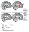
Anesthetics produce unconsciousness by acting on subcortical and cortical circuits. A. A mechanism to explain non-REM sleep is GABA- and galanin- mediated inhibition of brainstem arousal nuclei from sleep active cells in the preoptic area of the hypothalamus. Decreased firing rate of locus ceruleus cells and norepinephrine release from efferent projections to the preoptic area is a putative mechanism to explain activation of sleep active cells and non-REM sleep initiation. Decreased norepinephrine to the cortex, thalamus, and basal forebrain also contribute to decreased arousal. Other sites that are not depicted (i.e. cells in the parafacial zone) may play important roles in non-REM sleep. B. Propofol, ether anesthetics, and other medications that significantly enhance GABAA receptor IPSCs decrease arousal by enhancing the inhibitory activity of GABAergic interneurons in the cortex and thalamus. They also inhibit the major excitatory brainstem nuclei that project directly and indirectly to the cortex. C. Ketamine likely decreases the level of arousal by directly targeting the thalamus and also by blocking glutamatergic projections from the parabrachial nucleus to the thalamus. In the cortex, ketamine decreases arousal by blocking inhibitory interneurons, leading to markedly excited pyramidal neurons. This markedly excited state is associated with increased blood flow, cerebral glucose metabolism, and hallucinations. D. Dexmedetomidine impairs the release of norepinephrine from projection neurons from the LC to the POA, basal forebrain, and thalamus. Similar to sleep, this activates the endogenous sleep promoting cells in the POA. These sleep active cells inhibit of brainstem arousal nuclei. Dexmedetomidine may also impair the level of arousal by targeting autoreceptors on the LC and adrenergic receptors in other brain regions such as the thalamus. E. The principal anesthetic and sedative agents do not produce physiological sleep. GABAA, gamma amino butyric acid A; IPSCs, inhibitory post synaptic currents; LC, locus ceruleus; non-REM, non-rapid eye movement; POA, preoptic area

References

    1. Bigelow HJ. Insensibility during surgical operations produced by inhalation. Boston Med Surg J. 1846;XXXV:309–317. - PMC - PubMed
    1. Graham EA. Ether and humbug. J Am Med Assoc. 1947;133:97–100. - PubMed
    1. Brown EN, Lydic R, Schiff ND. General anesthesia, sleep, and coma. N Engl J Med. 2010;363:2638–2650. - PMC - PubMed
    1. Brown EN, Purdon PL, Van Dort CJ. General anesthesia and altered states of arousal: a systems neuroscience analysis. Annu Rev Neurosci. 2011;34:601–628. - PMC - PubMed
    1. Franks NP. General anaesthesia: from molecular targets to neuronal pathways of sleep and arousal. Nat Rev Neurosci. 2008;9:370–386. - PubMed

Substances