. 2017 Jul 11;8(28):46104-46120.
doi: 10.18632/oncotarget.17553.

An unusual intragenic promoter of PIWIL2 contributes to aberrant activation of oncogenic PL2L60

Affiliations

An unusual intragenic promoter of PIWIL2 contributes to aberrant activation of oncogenic PL2L60

Shan-Shan Liu et al. Oncotarget. .

Abstract

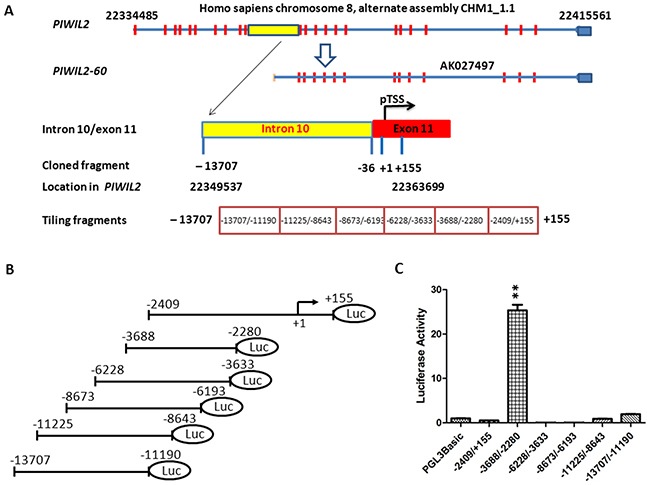
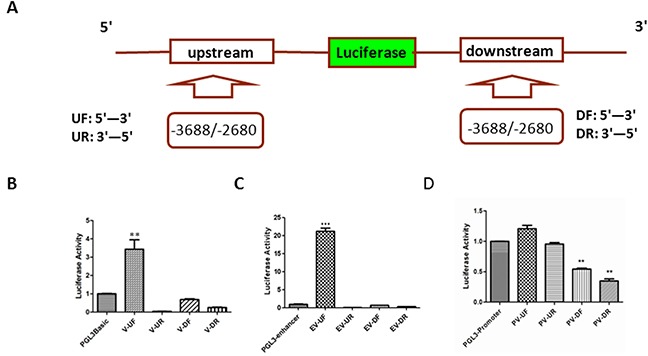
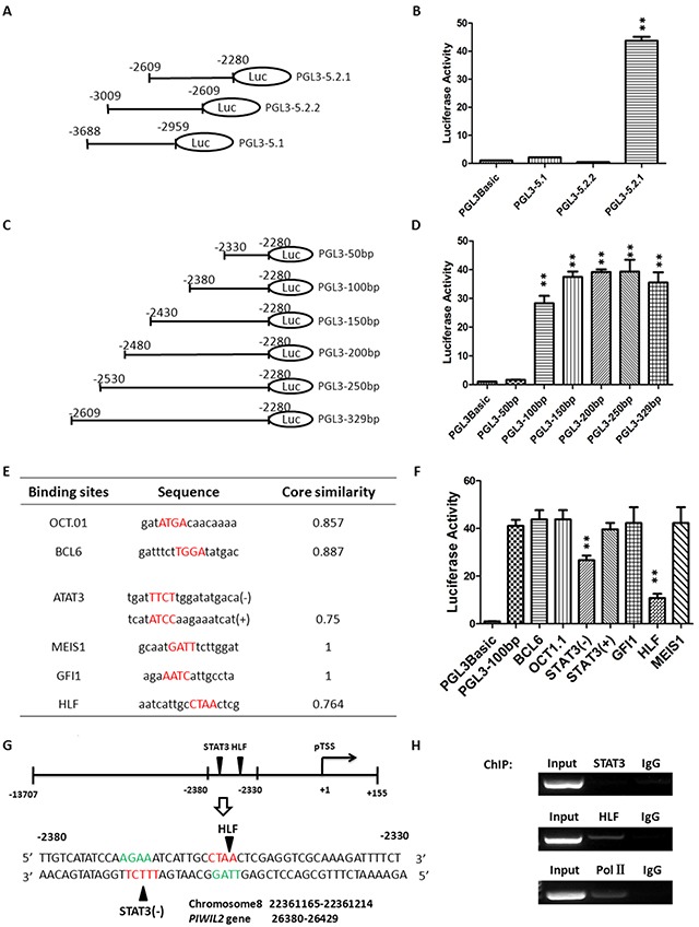
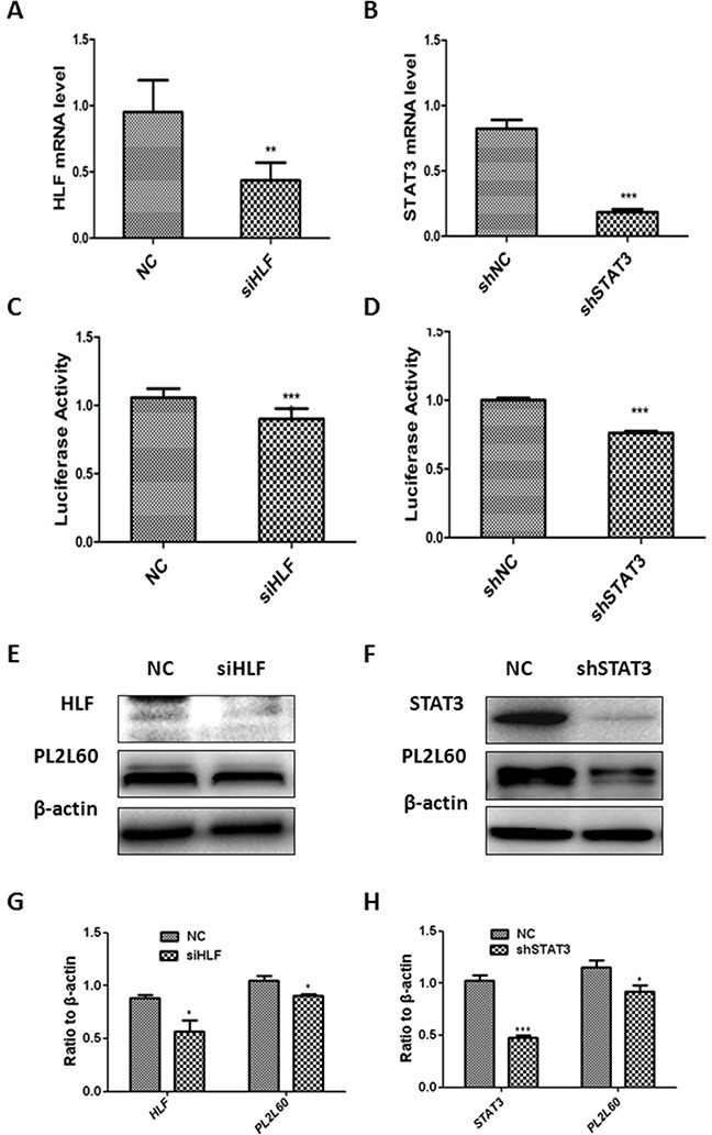
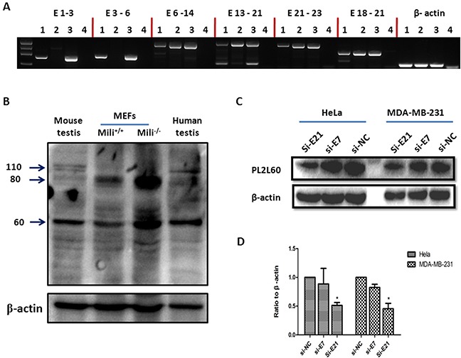
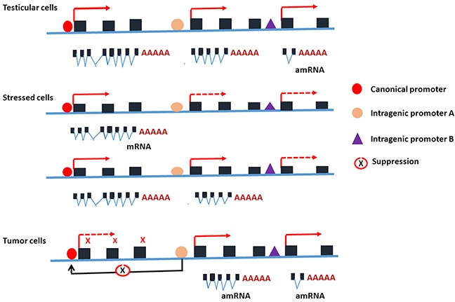
PIWIL2-like (PL2L) protein 60 (PL2L60), a product of aberrantly activated PIWIL2 gene, is widely expressed in various types of tumors and may promote tumorigenesis. However, the mechanisms underlying the activation of expression of PL2L60 remain unknown. In this study, an intragenic promoter responsible for the activation of PL2L60 within the human PIWIL2 gene has been identified, cloned and characterized. The promoter of PL2L60 is located in the intron 10 of the host gene PIWIL2. Bioinformatic and mutagenic analysis reveals that this intragenic promoter within the sequence of 50 nucleotides contains two closely arranged cis-acting elements specific for the hepatic leukemia factor (HLF) in the positive strand and signal transducer and activator of transcription 3 (STAT3) in the negative strand. Chromatin immunoprecipitation analysis demonstrates that both the HLF and polymerase II (Pol II), a hallmark of active promoters, directly bind to the sequence, although STAT3 does not. Knockdown of HLF and STAT3 alone or both by RNA interference significantly reduced both promoter activity and the PL2L60 protein expression, although there is no additive effect. The expression of PL2L60 proteins was enhanced when host gene Piwil2 was genetically disrupted in a murine cell model. Taken together, we have identified a PL2L60-specific intragenic promoter in the host gene of PIWIL2, which is interdependently activated by HLF and STAT3 through steric interaction. This activation is dependent on cellular milieu rather than the integrity of host gene PIWIL2, highlighting a novel, important mechanism for a cancer-causing gene to be activated during tumorigenesis.

Keywords: PIWIL2; PL2L60; STAT3; alienated gene activation; intragenic promoter.


Figures

Figure 1
Figure 1. Identification of PL2L60-specific intragenic promoter in the host gene of PIWIL2
(A) Schematic diagram of the predicted region of intragenic promoter specific for PL2L60 in human PIWIL2 genes: Full length PIWIL2 gene is located in chromosome 8 range from 22334485 to 22415561 (alternate assembly CHM1_1.1). Based on the PL2L60 mRNA (AK027497) [1], putative transcriptional start site (pTSS) (+1) was determined and a range of nucleotides (nt) from 22349537 to 22363699 was cloned (−13707/+155) and divided into six tiling fragments: -2409/+155, -3688/−2280, -6228/−3633, -8673/−6193, -11225/−8643 and -13707/−11190. (B) Structure of vectors harboring relevant fragments: Each tiling fragment was cloned into a pGL3-basic luciferase report vector. (C) Compare luciferase activity of the vectors containing tiling fragments: Each vector was transfected into HEK-293T cells and luciferase activity was measured 48 h later. Only does the vector harboring the fragment -3688/−2280 display strong luciferase activity. The data shown are the luciferase activity from three independent experiments (mean ± SE). Yellow square: part of intron 10; **, p < 0.01 and ***, p < 0.001, as compared to control and determined by Student T-test.
Figure 2
Figure 2. Distinguishing the intragenic promoter from enhancer
(A) Schematic diagram of construct for enhancer activity assay: A series of vectors were constructed with the fragment -3688/−2680, which was cloned into pGL3-basic or pGL3-enhancer report vector up (U) or down (D) stream of luciferase gene in forward (F: 5′-3′) or reverse (R: 3′-5′) direction. UF: the fragment -3688/−2680 from 5′ to 3′ was cloned into upstream of the luciferase gene; UR: the fragment -3688/−2680 from 3′ to 5′ was cloned into upstream of the luciferase gene; DF: the fragment -3688/−2680 from 5′ to 3′ was cloned into downstream of the luciferase gene; and DR: the fragment -3688/−2680 from 3′ to 5′ was cloned into downstream of the luciferase gene. (B) Luciferase activity of the fragment -3688/−2680 in the pGL3-basic report vectors (V): HEK-293T cells were transiently co-transfected with 1 μg V-UF, V-UR, V-DF or V-DR luciferase report vectors and 20 ng expression plasmids for Renilla luciferase (pRL-null). Transfection efficiency was normalized and the data shown are the luciferase activity from three experiments (mean ± SD). **, p < 0.01. (C) Luciferase activity of the fragment -3688/−2680 in the pGL3-enhancer report vectors (EV): HEK-293T cells were transiently co-transfected with 1 μg EV-UF, EV-UR, EV-DF or EV-DR luciferase report vectors and 20 ng expression plasmids for Renilla luciferase (pRL-null). Transfection efficiency and luciferase activity was determined as described in (B) and the data shown are the luciferase activity from three experiments (Mean ± SD). ***, p < 0.001. (D) Luciferase activity of the fragment -3688/−2680 in the pGL3-promoter report vectors (PV): HEK-293T cells were transiently co-transfected with 1 μg PV-UF, PV-UR, PV-DF or PV-DR luciferase report vectors and 20 ng expression plasmids for Renilla luciferase (pRL-null). Transfection efficiency and luciferase activity was determined as described in (B) and the data shown are the luciferase activity from three experiments (Mean ± SD). **, p < 0.01.
Figure 3
Figure 3. Identification of the potential binding sites for transcription factors in PL2L60-specific intragenic promoter
(A) Constructs containing truncates of the fragment -3688/−2680: The region from -3688 to -2680 with promoter activity was truncated into three fragments, which was cloned, respectively, into pGL3-basic report vectors: PGL3-5.2.1 (−2609/−2280), PGL3-5.2.2 (−3009/−2609) and PGL3-5.1 (−3688/−2959). (B) Luciferase activity of the vectors harboring truncates from the fragment -3688/−2680: HEK-293T cells were transiently co-transfected with 1 μg luciferase report plasmids (PGL3-5.2.1, PGL3-5.2.2 or PGL3-5.1) plus 20 ng expression plasmids for Renilla luciferase (pRL-null), which was used to normalize transfection efficiency. Only does the vector PGL3-5.2.1 (−2609/−2280) exhibit high luciferase activity. **, p < 0.01 as compared to control vectors (PGL3Basic). (C) Serial deletional analysis of the fragment -2609/−2280: The fragment was sequentially truncated off about 50 nt, six truncates were obtained and each of them was cloned into the pGL3-basic luciferase report vectors. (D) Identification of 50-nt-long truncates with luciferase activity: The vectors harboring a truncate of interest was transfected, respectively, into HEK-293T cells, and the luciferase activity was measured 48 h later. The fragment -2330/−2280 (PGL-3-50bp) completely lost luciferase activity, suggesting that promoter activity resided in the fragment between -2380 and -2330 (50 nt). **, p < 0.01, as compared to control vectors (PGL3Basic). (E) Bio-informatic prediction of binding sites potentially for transcription factors: Six potential binding sites for transcription factors in the fragment -2380/−2330 were predicted using Genomatix Software, including OCT1, BCL6, STAT3, MEIS1, GFI1 and HLF. (F) Identification of the specific binding sites responsible for promoter activity by site-direct point mutagenesis: Site-direct mutagenesis was performed on predicted binding sites for BCL6, OCT1, STAT3(−), STAT3(+), GFI1, HLF and MEIS1, and the mutated fragments (Supplementary Table 7) were cloned into pGL3-basic report vectors, respectively. Each vector harboring a mutated fragment was transfected into HEK-293T cells and luciferase activity was determined. The sites for STAT3 and HLF displayed significantly reduced activity after point mutation, indicating that they were the binding sites for STAT3 and HLF, respectively. **, p < 0.01, as compared to control vectors (PGL3Basic). (G) Positions of binding sites specific for transcription factors STAT3 and HLF in the chromosome and PIWIL2 gene: Schematic diagram showing the binding sites specific for STAT3 and HLF in the promoter region (−2380/−2330) as well as their position in chromosome 8 (NC_018919.2: 22361165-22361214) and PIWIL2 gene (26380-26429). (H) Validation of binding sites for STAT3 and HLF in PIWIL2-60-specific promoter: Chromatin Immunoprecipitation (ChIP) assay followed by DNA PCR were performed to detect the binding sites, respectively, for STAT3, HLF and Pol II in the fragment -2380/−2330. Samples of “input” and “IgG” were used, respectively, as positive and negative controls. Input: samples without treatment with antibody; IgG: samples treated with isotype IgG instead of specific antibody. All the data shown in B, D and F are mean ± SD from 3 independent experiments. Primers used for DNA PCR are shown in Supplementary Table 4.
Figure 4
Figure 4. Activation of the PL2L60-specific intragenic promoter by STAT3 and HLF
(A, C, E & G) Activation of PL2L60-specific intragenic promoter by HLF: HEK-293T cells were transfected with HLF siRNA or NC siRNA plus vectors PGL3-100bp. The cells were harvested 48 h after transfection and subjected to real-time PCR analyses of PL2L60 mRNAs (A), leuciferase activity assays for PL2L60-specific intragenic promoter activity (C) and Western blotting analyses of HLF and PL2L60 expressions (E & G). (B, D, F & H) Activation of PL2L60-specific promoter by STAT3: HEK-293T cells were infected with lentiviral STAT3 shRNAs or lentiviral NC shRNAs. Once stable shSTAT3 and shNC cell lines had been established, they were transfected with PGL-3 100bp plasmids for 48 hrs and subjected to analysis of STAT3 mRNAs (B), PL2L60-specific promoter activity (D) and protein expressions of STAT3 and PL2L60 (F & H) as described above. The data shown are mean ± SE from three independent experiments (A, B, C, D, G & H) and a representative from at least three independent experiments (E & F). siHLF: HLF siRNAs; NC: NC siRNAs; shSTAT3: stable cell lines expressing STAT3 shRNAs; shNC: stable cell lines expressing mock (NC) shRNAs. *, p < 0.05; **, p < 0.01; ***, p < 0.001, when compared to control groups.
Figure 5
Figure 5. Aberrant activation of PIWIL2 gene by intragenic promoters independently of the integrity of host gene
(A) Expression of short form mRMAs of Piwil2 in the primary mili−/− MEFs: RNAs were extracted from the testicular tissues and MEFs of wild-type (wt) or Piwil2-deficient (mili−/−) mice [5] and subjected to GEM RT-PCR analysis for the length of Piwil2 mRNAs [1]. As results, mili−/− MEFs expressed a short form mRNAs covering from exons 6 to exon 23, whereas the testicular tissues and MEFs from wt mice expressed full length of Piwil2 mRNAs, suggesting that intragenic promoter activation is independent of the integrity of host gene. Lane 1: wt MEF, Lane 2: mili−/− MEF; Lane 3: wt testicular tissues and Lane 4: H2O. The primer pairs specific for E1-3, E3-6, E6-14, E13-21 and E21-23 of Piwil2 were used for GEM RT-PCR analysis, and the primer pair specific for E18-21 was used as a positive control (Supplementary Table 5). (B) Piwil2 and PL2L proteins expression in Piwil2-deficient MEFs: Primary mili−/− MEFs, wt MEFs (both were at 30 generations of in vitro culture), and testicular tissues from a wt mouse and a patient were analyzed for the expressions of Piwil2 and PL2L proteins by Western blot, using a mouse mAb to a peptide common for Piwil2 and PL2L of both mouse and human (clone Kao3 [1]). PL2L60 and PL2L80 but not Piwil2 were detected in mili−/− MEFs. As comparisons, PL2L60, PL2L80 and Piwil2 were detected in wt MEFs, and testicular tissues of human and mice. However, PL2L60 and PL2L80 were also detected in mili−/− MEFs in a much higher level than in wt MEFs and testicular tissues. The detection of a high level of PL2L80 reflected potentially a PIWIL2-80-specific intragenic promoter that was activated more strongly in mili−/− MEFs than in wt MEFs. (C & D) PL2L60 protein expression in E21-specific siRNA-treated tumor cell lines: Human cancer cell lines of breast (MDA-MB-231) and cervix (HeLa) were treated with siE21, siE7 and siNC for 72 h and analyzed for PL2L60 expression by Western blot. Top panel: a representative result of Western blot, using polyclonal rabbit antibody to PL2L peptides with the same specificity as mAb Kao3 [1]; bottom panel: a summary of three independent experiments (mean ± SD). siE7: siRNA targeting exon 7 of PIWIL2 transcripts; siE21: siRNAs targeting exon 21 of PIWIL2 transcripts; and siNC: scramble siRNAs used as a negative control. *, p < 0.05, compared to siE7 or siNC.
Figure 6
Figure 6. Schematic depiction for the potential mechanisms underlying the activation of intragenic promoter in various conditions and its relationship to the canonical promoter of the host gene PIWIL2
In the testicular cells, both the canonical promoter and intragenic promoters of PIWIL2 are activated during gametogenesis and the proteins of PIWILL2, PL2L80 and PL2L60 are expressed [1, 27]. In the somatic cells, the canonical promoter and intragenic promoters of PIWIL2 are silent, but activated when the cells become stressed after exposure to environmental agents including DNA-damage-inducing agents or carcinogens [13]. The canonical promoter of PIWIL2 may be activated transiently in responding to stresses, serving as a tumor barrier gene [2, 13], while the activation of its intragenic promoters may lead to cell transformation and tumor initiation [9, 17, 28]. In the tumor cells, the canonical promoter of PIWIL2 is usually silent, while intragenic promoters are activated to promote tumorigenesis [1, 13], probably through inhibition of canonical promoter of PIWIL2. However, PIWIL2 could be activated in the tumor cells under the stressed condition such as hypoxia. Solid line arrows: activated; dashed line arrows: silent; and black line arrows: inhibited.

Similar articles

Cited by

References

    1. Ye Y, Yin DT, Chen L, Zhou Q, Shen R, He G, Yan Q, Tong Z, Issekutz AC, Shapiro CL, Barsky SH, Lin H, Li JJ, Gao JX. Identification of Piwil2-like (PL2L) proteins that promote tumorigenesis. PLoS ONE. 2010;5:e13406. - PMC - PubMed
    1. Gao JX, Liu N, Wu HL. PIWIL2 (piwi-like RNA-mediated gene silencing 2) Atlas Genet Cytogenet Oncol Haematol. 2014;18:919–927.
    1. Lin H, Spradling AC. A novel group of pumilio mutations affects the asymmetric division of germline stem cells in the Drosophila ovary. Development. 1997;124:2463–2476. - PubMed
    1. Kuramochi-Miyagawa S, Kimura T, Yomogida K, Kuroiwa A, Tadokoro Y, Fujita Y, Sato M, Matsuda Y, Nakano T. Two mouse piwi-related genes: miwi and mili. Mech Dev. 2001;108:121–133. - PubMed
    1. Kuramochi-Miyagawa S, Kimura T, Ijiri TW, Isobe T, Asada N, Fujita Y, Ikawa M, Iwai N, Okabe M, Deng W, Lin H, Matsuda Y, Nakano T. Mili, a mammalian member of piwi family gene, is essential for spermatogenesis. Development. 2004;131:839–849. - PubMed

MeSH terms