. 2017 May 17;13(5):e1006375.
doi: 10.1371/journal.ppat.1006375. eCollection 2017 May.

UGGT1 enhances enterovirus 71 pathogenicity by promoting viral RNA synthesis and viral replication

Affiliations

UGGT1 enhances enterovirus 71 pathogenicity by promoting viral RNA synthesis and viral replication

Peng-Nien Huang et al. PLoS Pathog. .

Abstract

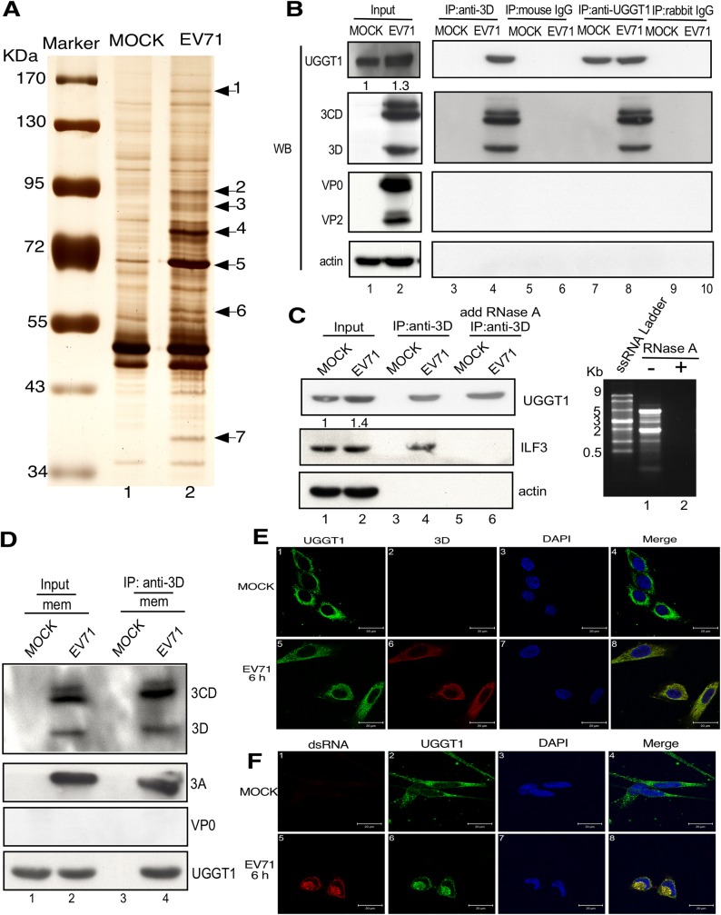
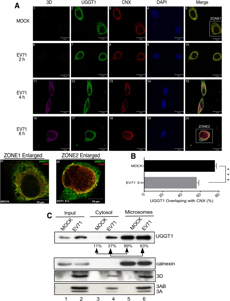
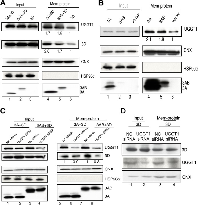
Positive-strand RNA virus infections can induce the stress-related unfolded protein response (UPR) in host cells. This study found that enterovirus A71 (EVA71) utilizes host UDP-glucose glycoprotein glucosyltransferase 1 (UGGT1), a key endoplasmic reticulum protein (ER) involved in UPR, to enhance viral replication and virulence. EVA71 forms replication complexes (RCs) on cellular membranes that contain a mix of host and viral proteins to facilitate viral replication, but the components and processes involved in the assembly and function of RCs are not fully understood. Using EVA71 as a model, this study found that host UGGT1 and viral 3D polymerase co-precipitate along with other factors on membranous replication complexes to enhance viral replication. Increased UGGT1 levels elevated viral growth rates, while viral pathogenicity was observed to be lower in heterozygous knockout mice (Uggt1 +/- mice). These findings provide important insight on the role of UPR and host UGGT1 in regulating RNA virus replication and pathogenicity.


Conflict of interest statement

The authors have declared that no competing interests exist.

Figures

Fig 1
Fig 1. Co-precipitation of UGGT1 and the EVA71 3D viral polymerase.
(A) At 6 hours post-infection, lysates from EVA71-infected or mock-infected cells were immunoprecipitated with anti-3D monoclonal antibody, and the precipitates were separated using SDS-PAGE, after which silver staining was applied for visualization. The seven labeled bands were excised, digested with trypsin, and analyzed by MALDI-TOF MS. (B) EVA71-infected and mock-infected cells were harvested and subjected to co-IP assays with anti-3D antibody (lanes 3 and 4) or mouse IgG (lanes 5 and 6); or anti-UGGT1 antibody (lanes 7 and 8) or rabbit IgG (lanes 9 and 10). The precipitates were analyzed using Western blotting with anti-UGGT1, anti-3D, anti-VP2, and anti-actin antibodies. (C) Cells were harvested at 6 h post-transfection, and lysates were treated with RNase A prior to being used in co-IP assays with an anti-3D antibody. Actin served as a loading control. Degradation of RNA was confirmed by RNA gel analysis. The precipitates were analyzed using Western blotting with anti-UGGT1, anti-ILF3, and anti-actin antibodies. (D) Membrane protein fractions were purified from EVA71-infected and mock-infected cells, and immunoprecipitation results with anti-3D antibody were analyzed by Western blotting with anti-3D, anti-3A, anti-VP2, and anti-UGGT1 antibodies. Expression of UGGT1, 3D, 3CD, 3AB, and 3A in the input lysate are shown. (E) EVA71-infected and mock-infected cells were fixed and stained with anti-UGGT1 and anti-3D antibodies at 6 h post-infection. An anti-UGGT1 antibody was used in panels 1 and 5, which were examined using a FITC filter. An anti-3D antibody was used in panels 2 and 6, which were examined using a rhodamine filter. Panels 3 and 7 display Hoechst 33258 staining results, and were examined using a 4’,6-diamidino-2-phenylindole (DAPI) filter. Panels 4 and 8 display merged rhodamine, FITC, and DAPI images. (F) EVA71-infected or mock-infected cells were fixed and stained with antibodies against UGGT1 and double strand RNA. Results with the anti-double strand RNA antibody are shown in panels 1 and 5, which were examined using a rhodamine filter. Anti-UGGT1 antibody was used for panels 2 and 6, which were examined using an FITC filter. Panels 3 and 7 display Hoechst 33258 staining results, which were examined using a DAPI filter. Panels 4 and 8 display merged rhodamine, FITC, and DAPI images.
Fig 2
Fig 2. In vivo interaction between 3D polymerase and UGGT1.
Uninfected RD cells were co-transfected with pFLAG-UGGT1 or pHA-3D DNA, and total cell lysates were harvested at 48 h after transfection for co-IP assays. (A) IP was performed with anti-HA, and the precipitates were analyzed by Western blotting (WB) with anti-FLAG. (B) IP was performed with anti-FLAG, and the precipitates were analyzed by WB with anti-HA. (C) Uninfected RD cells were transfected with vector or pHA-3D, and cell lysate proteins that were immunoprecipitated with anti-HA were subjected to WB with anti-UGGT1. The expression of FLAG-UGGT1 and HA-3D in the input lysates is indicated.
Fig 3
Fig 3. UGGT1 facilitates EVA71 replication and propagation.
(A) Confocal microscopy results of UGGT1 and 3D expression in NC or UGGT1 siRNA-treated RD cells that were infected with EVA71 and subjected to immunostaining at 6 h post-infection. Panels 1, 5, 9, and 13 were stained with anti-UGGT1 and examined using a FITC filter; panels 2, 6, 10, and 14 were stained with anti-3D and examined with a rhodamine filter; and panels 3, 7, 11, and 15 were subjected to Hoechst 33258 staining and examined with a DAPI filter. (B) and (C) RD cells were transfected with NC or UGGT1 siRNA for 48 h, and then challenged with EVA71 at an MOI of 10 or 0.1. A plaque assay was performed to measure viral propagation rates at various timepoints post-infection. The left panels show the knockdown of uggt1 following siRNA treatment. (D) RD cells were transfected with 1, 2, or 4 μg of pFLAG-UGGT1 or pFLAG-vector for 48 h, and then challenged with EVA71 at an MOI of 10. A plaque assay was performed to measure viral yields at 6 h post-infection. (E) UGGT1 was overexpressed by respectively transfecting 1, 2, or 4 μg of plasmid pFLAG-UGGT1 to RD cells, and the panels show the corresponding increase in UGGT1 levels following overexpression, with actin serving as a loading control. ***P < 0.001, **P < 0.01, and *P < 0.05, as calculated by two-tailed unpaired Student’s t-test.
Fig 4
Fig 4. Viral yield, neurological symptoms, and lethality in UGGT1 heterozygous knockout mice infected with EVA71.
(A) Phenotypes of UGGT1+/+ wild-type (WT) mice and UGGT1+/− heterozygous knockout mice. (B) UGGT1 expression levels in brain tissue homogenates of UGGT1+/+ WT mice and UGGT1+/− heterozygous knockout mice, as detected by western blotting. (C) and (D) 10-day-old WT or Uggt1 heterozygous knockout mice were injected with 105 PFU/mouse of EVA71 strain MP4, and on Day 3 after infection, EVA71 virus was extracted from brain and muscle tissues and quantitated. (E) CNS-like hind limb paralysis and (F) Survival rates in 10-day-old WT and Uggt1 heterozygous knockout mice injected with 105 PFU/mouse of the EVA71 MP4 strain were evaluated, and one-way ANOVA on ranks (Kruskal-Wallis H test) was used to determine statistical significance. The number (n) of mice in each group is shown.
Fig 5
Fig 5. UGGT1 enhances viral RNA replication.
(A) NC or UGGT1 siRNA-treated RD cells were transfected with EVA71-Luc replicon RNA, and cells were assayed for firefly luciferase signals (FLuc) at 6 h post-transfection. The right panel indicates the knockdown efficiency of Uggt1. (B) Monocistronic mRNA containing EVA71 IRES and FLuc was transfected to cells pretreated with NC or UGGT1 siRNA. At 6 h post-transfection, cell lysates were assayed for FLuc activity. Western blotting data indicates siRNA knockdown efficiency. Experiments were performed in triplicate to obtain the bar graph. (C) NC or UGGT1 siRNA-treated RD cells were infected with EVA71 at an MOI of 10. Intracellular viral RNA was isolated at 4, 6, 8, 10, 12, and 14 h post-infection, and quantitated using real-time RT-PCR. The amount of viral RNA at 14 h post-infection in NC siRNA-transfected cells was taken as 100%, and the relative amount of viral RNA isolated at each timepoint is presented as a percentage of this. The right panel indicates knockdown efficiency of Uggt1. (D) RD cells were transfected with NC or UGGT1 siRNA for 48 h and then reseeded. After 24 h, cells were infected with EVA71 at an MOI of 10, and RNA was extracted at 2, 4, 6, 8, and 10 h post-infection. RNA was loaded onto a nitrocellulose sheet in the slot blot manifold. The right panel demonstrates Uggt1 knockdown efficiency. ***P < 0.001 and *P < 0.05, as calculated by two-tailed unpaired Student’s t-test.
Fig 6
Fig 6. UGGT1 deploys from the ER lumen to the cytosol during EVA71 infection.
(A) Mock-infected or EVA71-infected cells were fixed and stained with anti-3D, anti-UGGT1, and anti-CNX antibodies at 2, 4, and 6 h post-infection. Use of an anti-3D antibody is shown in panels 1, 6, 11, and 16, which were examined using a cy5 filter. Use of an anti-UGGT1 antibody is shown in panels 2, 7, 12, and 17, which were examined using an FITC filter. Use of an anti-CNX antibody is shown in panels 3, 8, 13, and 18, which were examined using a rhodamine filter. Panels 4, 9, 14, and 19 indicate Hoechst 33258 staining, and were examined with a DAPI filter. Panel 21 displays enlargement zone 1 from panel 5. Panel 22 displays enlargement zone 2 from panel 20. (B) Percentage of UGGT1 and CNX signal overlap as calculated with ImageJ JACoP plugins from images shown in (A). The data shown represent the average and standard deviation of ten randomly selected images. ***P < 0.001, as calculated by two-tailed unpaired Student’s t-test. (C) RD cells were infected with EVA71, harvested at 6 h post-infection, separated into cytosol and microsome fractions, and subjected to Western blot analysis using anti-UGGT1 antibody. The same blot was also probed with anti-calnexin, anti-3D, and anti-3A antibodies. The results are representative of at least three independent and reproducible experiments.
Fig 7
Fig 7. UGGT1 enhances 3D viral polymerase levels in a viral protein 3A-associated membrane fraction.
(A) RD cells were co-transfected with pFLAG-3A and pFLAG-3D; pFLAG-3AB and pFLAG-3D; or pFLAG-3D only, and harvested at 48 h post-transfection. Membrane fractions were isolated and subjected to Western blot analysis. (B) Cells were transfected with pFLAG-3A, pFLAG-3AB, and pFLAG-vector and harvested at 48 h post-transfection. Membrane fractions were separated and subjected to Western blot analysis. (C) Cells were transfected with NC or UGGT1 siRNA for 48 h, and then co-transfected with pFLAG-3A and pFLAG-3D, or pFLAG-3AB and pFLAG-3D. Cells were harvested at 48 h post-transfection, and the membrane fractions were extracted and subjected to Western blot analysis. Anti-UGGT1, anti-3D and anti-3A antibodies were used. The same blot was probed with anti-CNX and anti-HSP90 antibodies. (D) Cells were transfected with NC or UGGT1 siRNA for 48 h, and then co-transfected with pFLAG-3D. Cells were harvested at 48 h post-transfection, and the membrane fractions were extracted and subjected to Western blot analysis. Anti-3D, anti-UGGT1 and anti-CNX antibodies were used. Results are representative of at least three independent experiments.

Similar articles

Cited by

References

    1. Salonen A, Ahola T, Kaariainen L (2005) Viral RNA replication in association with cellular membranes. Curr Top Microbiol Immunol 285: 139–173. - PMC - PubMed
    1. Belov GA, van Kuppeveld FJ (2012) (+)RNA viruses rewire cellular pathways to build replication organelles. Curr Opin Virol 2: 740–747. doi: 10.1016/j.coviro.2012.09.006 - DOI - PMC - PubMed
    1. Koonin EV, Dolja VV (1993) Evolution and taxonomy of positive-strand RNA viruses: implications of comparative analysis of amino acid sequences. Crit Rev Biochem Mol Biol 28: 375–430. doi: 10.3109/10409239309078440 - DOI - PubMed
    1. Koonin EV, Wolf YI, Nagasaki K, Dolja VV (2008) The Big Bang of picorna-like virus evolution antedates the radiation of eukaryotic supergroups. Nat Rev Microbiol 6: 925–939. doi: 10.1038/nrmicro2030 - DOI - PubMed
    1. McMinn PC (2002) An overview of the evolution of enterovirus 71 and its clinical and public health significance. FEMS Microbiol Rev 26: 91–107. - PubMed

MeSH terms