. 2017 May 18;12(5):e0177161.
doi: 10.1371/journal.pone.0177161. eCollection 2017.

Structures of the inactive and active states of RIP2 kinase inform on the mechanism of activation

Affiliations

Structures of the inactive and active states of RIP2 kinase inform on the mechanism of activation

Erika Pellegrini et al. PLoS One. .

Abstract

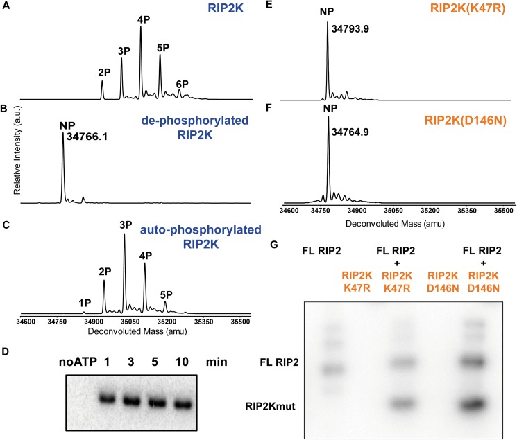
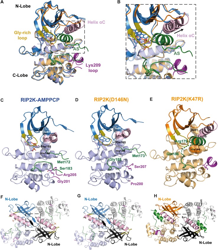
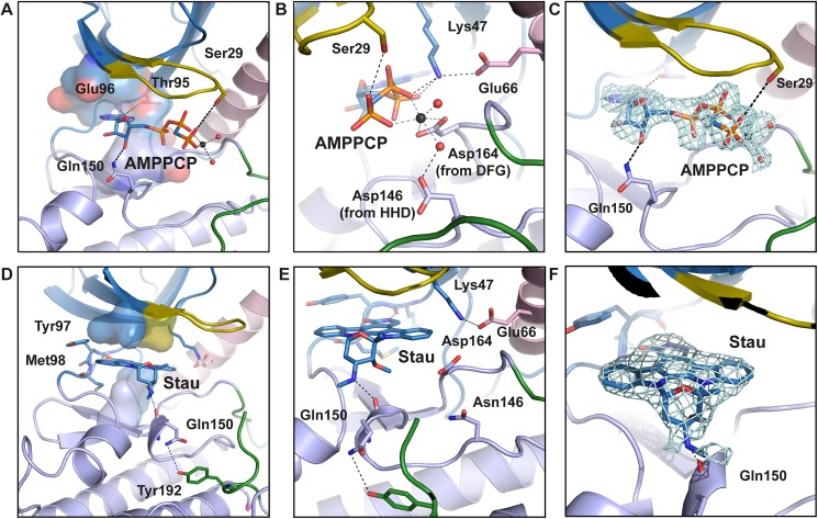
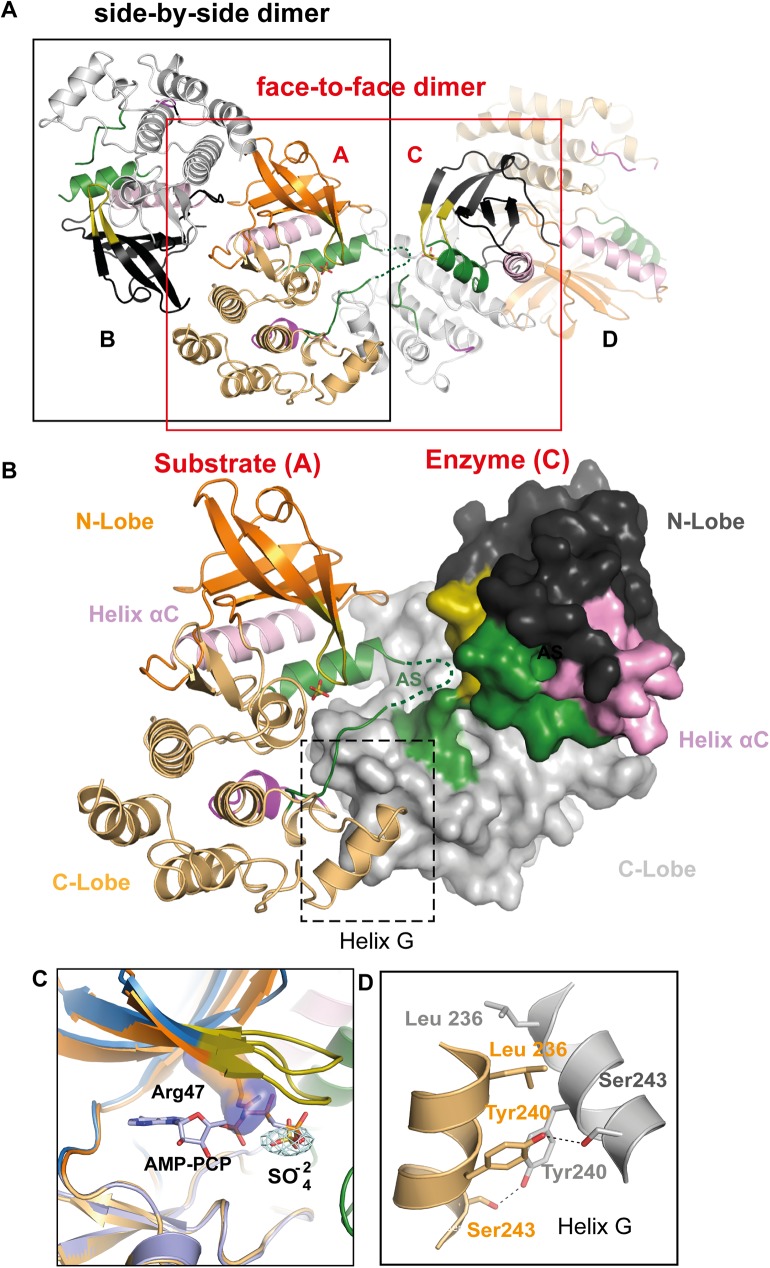
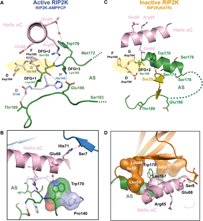
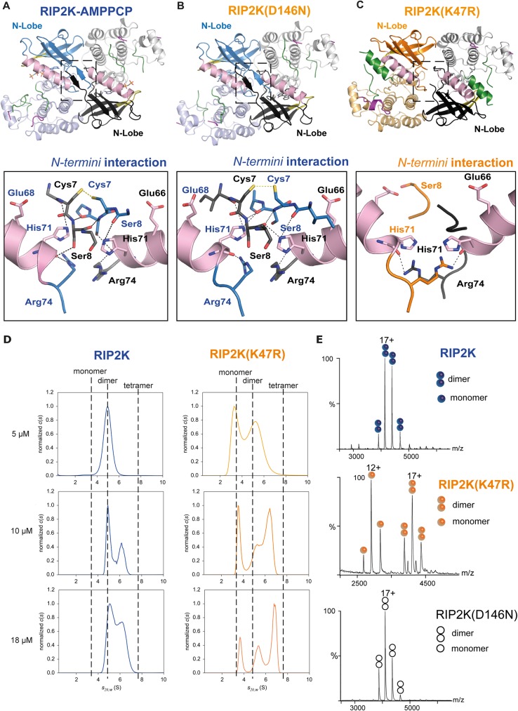
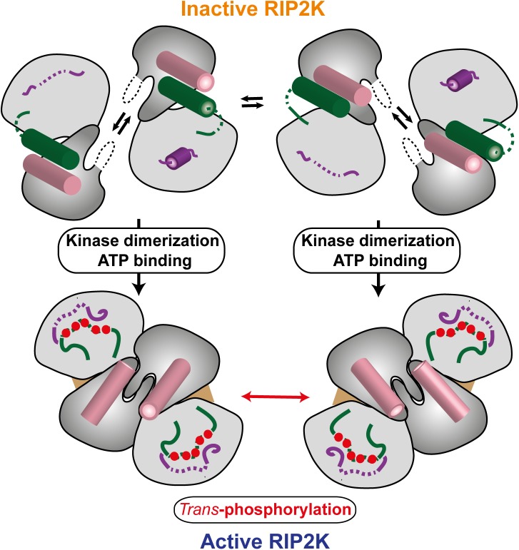
Innate immune receptors NOD1 and NOD2 are activated by bacterial peptidoglycans leading to recruitment of adaptor kinase RIP2, which, upon phosphorylation and ubiquitination, becomes a scaffold for downstream effectors. The kinase domain (RIP2K) is a pharmaceutical target for inflammatory diseases caused by aberrant NOD2-RIP2 signalling. Although structures of active RIP2K in complex with inhibitors have been reported, the mechanism of RIP2K activation remains to be elucidated. Here we analyse RIP2K activation by combining crystal structures of the active and inactive states with mass spectrometric characterization of their phosphorylation profiles. The active state has Helix αC inwardly displaced and the phosphorylated Activation Segment (AS) disordered, whilst in the inactive state Helix αC is outwardly displaced and packed against the helical, non-phosphorylated AS. Biophysical measurements show that the active state is a stable dimer whilst the inactive kinase is in a monomer-dimer equilibrium, consistent with the observed structural differences at the dimer interface. We conclude that RIP2 kinase auto-phosphorylation is intimately coupled to dimerization, similar to the case of BRAF. Our results will help drug design efforts targeting RIP2 as a potential treatment for NOD2-RIP2 related inflammatory diseases.


Conflict of interest statement

Competing Interests: Since leaving the project, co-author SS is employed by Unilever, a commercial company. Unilever has never had any role in the project. There are no patents, products in development or marketed products to declare. This does not alter our adherence to all the PLOS ONE policies on sharing data and materials.

Figures

Fig 1
Fig 1. Phosphorylation profile of WT RIP2K and RIP2K mutants using ESI-TOF MS and activity assays.
(A-C) MS spectra of RIP2K acquired (A) immediately after its purification from insect cells, (B) after lambda phosphatase treatment and (C) after auto-phosphorylation. Protein sequences used for mass calculations are reported in S1 Table; (D) In vitro kinase activity assay of the de-phosphorylated RIP2K using 10 μM ATP. The collected data indicated that RIP2K was completely auto-phosphorylated within 10 minutes. (E-F) MS spectra of (E) RIP2KK47R and (F) RIP2KD146N acquired after their purification from insect cells. The RIP2K mutants were not phosphorylated (NP). (G) In vitro end-point kinetic assays of RIP2K mutants at 10 μM ATP, in the presence of recombinant full length (FL) RIP2. RIP2K mutants were trans-phosphorylated by FL RIP2, which was also active on itself.
Fig 2
Fig 2. Overview of WT RIP2K, RIP2KD146N and RIP2KK47R structures.
(A) Overlay of the ribbon diagrams of RIP2K-AMPPCP, RIP2KD146N-STAU and RIP2KK47R. (B) The inset highlights the structural differences at αHelix C and at the AS. (C-E) Ribbon diagrams of (C) RIP2K-AMPPCP, (D) RIP2KD146N-STAU and (E) RIP2KK47R (E). N- and C-lobes are represented in dark and light colour respectively. The R-spine residues (Ile81, Leu70 from Helix αC, Phe165 from the DGF and His144 from the HHD motif) are shown with surface representation, AS is in green, Lys209 loop in magenta, Gly-rich loop in olive-orange, Helix αC in pink. Lys209 is represented as stick. (F-H) Ribbon diagrams of the side-by-side dimer of (F) RIP2K-AMPPCP, (G) RIP2KD146N-STAU and (H) RIP2KK47R. The interactions at the N-termini are highlighted. Labelling is kept consistent among the different structures.
Fig 3
Fig 3. View of the active site of RIP2K-AMPPCP and RIP2KD146N-STAU.
(A-B) ATP is placed at the interface between the two lobes, within the adenine ring inserted in a pocket formed by residues Ala45, Val46, Leu24, Leu79, Thr95, Tyr97, Leu153 and Ala163 (shown as surface). The adenine ring is hydrogen bonded with the hinge backbone, while N6 atom is coordinated by the hydroxyl group of Thr95 (the gatekeeper residue). The ribose is hydrogen bonded to Gln150, while the ATP-γP is anchored to Ser29, from the Gly-rich loop (residues 25–32). Glu66 in Helix αC forms a salt bridge with Lys47 from strand β3, which then coordinates the ATP-βP and ATP-αP by hydrogen bonds. ATP-βP and ATP-γP are further stabilized by a Mg2+ ion, coordinating by Asp164 from the DFG motif and by Asp146 via a water molecule. (C) Fo-Fc map of ATP and magnesium within water molecules is shown as counter level at 1.5 σ. Domain colouring is the same as in Fig 2. (D-F) In the kinase active site, the staurosporine molecule is sandwiched between hydrophobic residues from both N-Lobe (Leu24, Val32 in the Gly-rich loop) and C-Lobe (Leu79, Leu153). The inhibitor molecule is further H-bond to the Hinge backbone and Gln150 backbone. The Glu66-Lys47 salt bridge is shown. (E) Fo-Fc map of staurosporine is shown as contour level of 1 σ. H-bonds are represented in black dash-line. Helix αC is coloured in pink. Metal bonds are in dark grey dash-line. Magnesium is represented as black sphere, water molecules as red spheres.
Fig 4
Fig 4. RIP2KK47R asymmetric enzyme-substrate embrace.
(A) Ribbon diagram of the Rip2K47R side-by-side dimer and of the face-to-face dimer in cristallo. (B) Ribbon diagram of the RIP2KK47R asymmetric, putative enzyme-substrate embrace. The interaction interface is highlighted. (C) Overlay of RIP2KK47R with RIP2K-AMPPCP at the ATP site. The conformation of Arg47 is incompatible with nucleotide binding. Fo-Fc map is shown for the sulphate molecule, at 1.5σ. (D) Interaction at the Helix αG. H-bonds are represented by a black dashed line. Labelling is kept consistent among the different structures.
Fig 5
Fig 5. Helix αC, AS and kinase N-termini connections in active and inactive RIP2K.
(A-D) Detailed representation of the H-bond network between the Helix αC, AS, magnesium binding loop and N-terminus in (A-B) in RIP2K active state and (C-D) in RIP2K inactive state. For clarity, only one conformation of Ser168 side chain is shown (see S4 Fig). The insets show the interactions of Trp170 and with the N-terminus. Hydrophobic residues are shown as surface. H-bonds are represented as black dashed lines with those related to the DFG-HHD motif are in magenta. The residues belonging to the Magnesium binding loop are highlighted with a pale-yellow ellipse. Hydrophobic interactions are highlighted as surface representation. Missing AS residues are shown with a dashed line.
Fig 6
Fig 6. Dimerization of RIP2K and RIP2KK47R.
(A-C) The insets show the interactions at the N-termini for (A)RIP2K-AMPPCP, (B) RIP2KD146N-STAU, (C) RIP2KK47R); (C) Sedimentation-velocity AUC analysis of RIP2K and RIP2KK47R at three different protein concentrations. Normalized sedimentation-coefficient distributions are plotted. Non-interacting species analysis of the homogeneous RIP2K at 5 μM showed a unique symmetrical boundary in c(s), providing independent estimates of s20w = 4.9 S and M = 66 ± 6 kDa. The latter is close to the theoretical value of 69.6 kDa for the dimer and corresponds to f/fmin = 1.2, indicative of a globular compact shape. Using the same shape factor, we derived s20w values of 3.1 S for the monomer, close to the slow boundary observed in RIP2KK47R samples, and 7.8 S for the tetramer, smaller than the fast boundary observed in RIP2KK47R samples, and in RIP2K at 10 and 18 μM, which suggests that the latter is a reaction boundary and corresponds to a mixture of oligomers in equilibrium. (D) Native MS spectra of RIP2K, RIP2KK47R and RIP2KD146N-STAU at 5μM. 17+ and 12+ are the main charge states of the dimers and monomers, respectively. The signal intensity represented on the Y-axis is expressed in "arbitrary units".
Fig 7
Fig 7. Phosphorylation profile of RIP2K dimer interface mutants using ESI-TOF MS and activity assay.
(A-C) MS spectra of RIP2KR74A(A),RIP2KR74D (B) and RIP2KR74H (C) acquired immediately after tag cleavage. The dimer interface RIP2K mutants were not phosphorylated (NP). Protein sequences used for mass calculations are reported in S1 Table; (D) In vitro end-point kinetic assays of dimer interface RIP2K mutants (RIP2Kmut) and RIP2K at 10 μM ATP, in presence or absence of a recombinant form of full length (FL) RIP2. RIP2K has not previously de-phosphorylated (faint band).
Fig 8
Fig 8. Model of RIP2K activation (see text).

References

    1. Chen G, Shaw MH, Kim YG, Nunez G. NOD-like receptors: role in innate immunity and inflammatory disease. Annual review of pathology. 2009;4:365–98. doi: 10.1146/annurev.pathol.4.110807.092239 - DOI - PubMed
    1. Park JH, Kim YG, McDonald C, Kanneganti TD, Hasegawa M, Body-Malapel M, et al. RICK/RIP2 mediates innate immune responses induced through Nod1 and Nod2 but not TLRs. Journal of immunology. 2007;178(4):2380–6. - PubMed
    1. Grimes CL, Ariyananda Lde Z, Melnyk JE, O'Shea EK. The innate immune protein Nod2 binds directly to MDP, a bacterial cell wall fragment. Journal of the American Chemical Society. 2012;134(33):13535–7. PubMed Central PMCID: PMC3424850. doi: 10.1021/ja303883c - DOI - PMC - PubMed
    1. Tigno-Aranjuez JT, Asara JM, Abbott DW. Inhibition of RIP2's tyrosine kinase activity limits NOD2-driven cytokine responses. Genes & development. 2010;24(23):2666–77. PubMed Central PMCID: PMC2994040. - PMC - PubMed
    1. Girardin SE, Boneca IG, Viala J, Chamaillard M, Labigne A, Thomas G, et al. Nod2 is a general sensor of peptidoglycan through muramyl dipeptide (MDP) detection. The Journal of biological chemistry. 2003;278(11):8869–72. doi: 10.1074/jbc.C200651200 - DOI - PubMed

MeSH terms

Substances