. 2018 May;41(3):281-291.
doi: 10.1080/10790268.2017.1329075. Epub 2017 May 25.

Optimized methods for rapidly dissecting spinal cords and harvesting spinal motor neurons with high survival and purity from rats at different embryonic stages

Affiliations

Optimized methods for rapidly dissecting spinal cords and harvesting spinal motor neurons with high survival and purity from rats at different embryonic stages

Shudong Chen et al. J Spinal Cord Med. 2018 May.

Abstract

Study design: Experimental study, protocol optimization.

Objectives: To investigate and compare the isolation of spinal motor neurons from embryonic rats at different embryonic stages, and develop optimized methods for rapidly dissecting spinal cords and harvesting spinal motor neurons with high survival and purity.

Setting: Guangdong Provincial Academy of Chinese Medical Sciences, Guangzhou, China.

Methods: Embryonic rats at different embryonic stages (12-18 days) were used to isolate spinal motor neurons. Their shape and corresponding dissection procedures, time needed and skills were compared. After dissecting and dissociating spinal cords, cells were randomly divided into immunopanning group and control group, in which antibodies to p75NTR were used or not. After plating cells, different recipe were added at different stages in serum-free culture media. Morphological features of cells were observed during development. Immunoflurorescence assay was performed to indentify motor neurons and the proportion of motor neurons in both control and immunopanning group were evaluated and compared.

Results: We summarized the operation essentials for rapid isolation of spinal cords, as well as compared anatomical features and dissection procedures of embryos at different embryonic stages, which help us to better evaluate the developmental profile and isolate cells by adopting corresponding skills. Through the fast isolation procedure and optimized culture media, cells grow in good viability. Moreover, compared with control group, the purity of spinal motor neurons in the immunopanning group was significantly increased, reaching a proportion of over 95%.

Keywords: Embryos; High purity; Immunopanning; Spinal cords; Spinal motor neurons; Survival of motor neuron.


Figures

Figure 1
Figure 1
Operating procedures and the skills during dissection of embryonic spinal cords (A) Dissect the lower abdomen and take the uteri out (B) Separate embryo from foetal sac (C) Decapitate the embryo from occiput, the yellow line shows the approximate level for decapitation (D) Place the embryo backside up so that its spine can be clearly exposed, dotted line shows the focus (E) Remove the skin overlying the spinal cord from cranial to caudal (F) Remove the fascia upon the spinal canal (G) gently lift out the spinal cord (H) spinal cords taken from all embryos (I) for each spinal cord, carefully strip the vessels and meninges (indicated by arrows) from cranial to caudal (J) the dissected spinal cord tissues.
Figure 2
Figure 2
Shape of embryos at different stages, and the spinal cords taken from embryos (A) In 12 d, the embryo is very tiny. When an embryo is 14–16 d, it would be twice or triple the size of that in 12 d. But when an embryo is 18 d, it grows more than five times bigger than before. Also, the blood supply gets better as embryos grow. (B) Spinal cords in 12 d embryos can't be dissected because their spinal cord tissues have not developed and look just like watery sacs. Spinal cord tissues taken from all embryos were placed in ice-cold L15. Those taken from E14 are too soft and easily hurt (indicated by yellow arrows). While in E18, the fused lamina become solid, so the process of opening the lamina may also break the spinal cord (indicated by yellow arrows).
Figure 3
Figure 3
Key differences of the spinal cord dissection procedures of embryos at different stages (A) In the 14 d embryo, skin and fascia overlying the spinal cord are very thin and soft (indicated by black arrow), which can be together removed easily. But the tissue adhesion (indicated by white arrow) makes it a little hard to lift out the spinal cord, so the most important thing to do is resolve adhesion to avoid damaging spinal cord. (B) In the 16 d embryo, there is only an overlying layer of toughness fascia (indicated by black arrow), we can prick the fascia from the edge (indicated by white arrow), then the spinal cord can be easily lifted out (indicated by yellow arrow). (C) In the 18 d embryo, the tissue back of neck has grown too thick (indicated by black arrow), and the lamina (vertebral plate) has fused (indicated by white arrow), so it needs greater efforts to remove fascia and open lamina before lifting out spinal cord (indicated by yellow arrows).
Figure 4
Figure 4
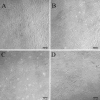
Phase contrast micrographs of purified cells obtained from 16 d embryonic rats. (A) 2 h after plating, almost all the cells had been adherent to the bottom and even several already grew tiny neurites; (B) Until 24 h, most cells had grown neurites and exhibited a bright and ovoid cell body, with a ring-shaped halo, but all cells were solitary; (C) 6 d in culture, cells grew with big body and high refraction, and formed obvious neurite networks; (D) 10 days in culture, cells grew more mature and showed vastly interconnected neurite networks. Scale bars represent 100 μm.
Figure 5
Figure 5
Confocal images of cultured cells stained with CHAT (red) and SMI-32 (green) antibody for identification of motor neurons. DAPI stains the nuclei, and the CHAT and SMI-32 staining clearly defines the cell soma, dendrites and large axons. Scale bars represent 50 μm (top row) and 20 μm (bottom row) respectively.
Figure 6
Figure 6
Cells stained with the SMI-32 (green) and GFAP (red) antibody in immunopanning and control group (from 16 d embryonic rats). (A1, A2) SMI-32 immunopositive cells. (B1, B2) GFAP immunopositive cells. (C1, C2) DAPI stains the nuclei. (D1, D2) Merged photos depicting co-localization of the two antigens and the nuclei. (E) The percentage of SMI-32 and GFAP immunopositive cells in both immunopanning and control group were calculated. Scale bars represent 50 μm.

Similar articles

Cited by

References

    1. Wagenaar DA, Pine J, Potter SM.. An extremely rich repertoire of bursting patterns during the development of cortical cultures. BMC Neurosci 2006;7:11. doi: 10.1186/1471-2202-7-11 - DOI - PMC - PubMed
    1. Voigt T, Baier H, Dolabela de Lima A.. Synchronization of neuronal activity promotes survival of individual rat neocortical neurons in early development. Eur J Neurosci 1997;9(5):990–9. doi: 10.1111/j.1460-9568.1997.tb01449.x - DOI - PubMed
    1. Sun JJ, Kilb W, Luhmann HJ.. Self-organization of repetitive spike patterns in developing neuronal networks in vitro. Eur J Neurosci 2010;32(8):1289–99. doi: 10.1111/j.1460-9568.2010.07383.x - DOI - PubMed
    1. Kamioka H, Maeda E, Jimbo Y, Robinson HP, Kawana A.. Spontaneous periodic synchronized bursting during formation of mature patterns of connections in cortical cultures. Neurosci Lett 1996;206(2–3):109–12. doi: 10.1016/S0304-3940(96)12448-4 - DOI - PubMed
    1. Baltz T, de Lima AD, Voigt T.. Contribution of GABAergic interneurons to the development of spontaneous activity patterns in cultured neocortical networks. Front Cell Neurosci 2010;4:15. - PMC - PubMed

Publication types

LinkOut - more resources