Review
. 2017 May 25;12(1):42.
doi: 10.1186/s13024-017-0183-y.

Endoplasmic reticulum stress and inflammation in the central nervous system

Affiliations
Review

Endoplasmic reticulum stress and inflammation in the central nervous system

Neil T Sprenkle et al. Mol Neurodegener. .

Abstract

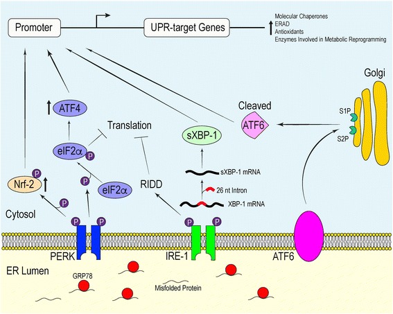
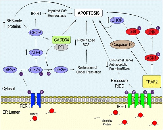
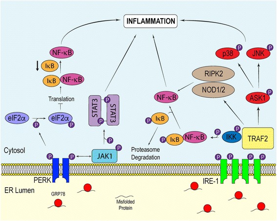
Persistent endoplasmic reticulum (ER) stress is thought to drive the pathology of many chronic disorders due to its potential to elicit aberrant inflammatory signaling and facilitate cell death. In neurodegenerative diseases, the accumulation of misfolded proteins and concomitant induction of ER stress in neurons contributes to neuronal dysfunction. In addition, ER stress responses induced in the surrounding neuroglia may promote disease progression by coordinating damaging inflammatory responses, which help fuel a neurotoxic milieu. Nevertheless, there still remains a gap in knowledge regarding the cell-specific mechanisms by which ER stress mediates neuroinflammation. In this review, we will discuss recently uncovered inflammatory pathways linked to the ER stress response. Moreover, we will summarize the present literature delineating how ER stress is generated in Alzheimer's disease, Parkinson's disease, Amyotrophic Lateral Sclerosis, and Multiple Sclerosis, and highlight how ER stress and neuroinflammation intersect mechanistically within the central nervous system. The mechanisms by which stress-induced inflammation contributes to the pathogenesis and progression of neurodegenerative diseases remain poorly understood. Further examination of this interplay could present unappreciated insights into the development of neurodegenerative diseases, and reveal new therapeutic targets.

Keywords: Endoplasmic reticulum stress; Neurodegeneration; Neuroinflammation; Unfolded protein response.


Figures

Fig. 1
Fig. 1
The Adaptive Signals of the Mammalian UPR. The activation of PERK, IRE-1α and ATF6 in response to protein misfolding stress primarily requires the dissociation of the molecular chaperone GRP78 from each of the ER stress sensors. This initiates signaling cascades which orchestrate the transcriptional and translational landscape of the cell in an effort to maintain homeostasis
Fig. 2
Fig. 2
Apoptotic Signals Associated with Chronic UPR Activation. Persistent ER stress triggers the apoptotic component of the UPR. PERK and IRE-1α drive UPR-induced apoptosis by initiating pathways which facilitate enhanced ROS production, Ca2+ dysregulation and caspase activation
Fig. 3
Fig. 3
Inflammatory Pathways Induced by the UPR. The UPR stimulates various inflammatory pathways to alert surrounding cells of potential danger. The transient interaction between impaired proteostasis and inflammation is considered a beneficial feature of the UPR. Nevertheless, sustained UPR-induced inflammation is considered a pathological factor in many chronic disorders, such as neurodegenerative diseases. Inflammatory pathways associated with the UPR include the NF-κB, JAK1/STAT3, NOD1/2-RIPK2, JNK and p38 signaling pathways

References

    1. Heneka MT, Kummer MP, Latz E. Innate immune activation in neurodegenerative disease. Nat Rev Immunol. 2014;14(7):463–477. doi: 10.1038/nri3705. - DOI - PubMed
    1. Glass CK, Saijo K, Winner B, Marchetto MC, Gage FH. Mechanisms underlying inflammation in neurodegeneration. Cell. 2010;140(6):918–934. doi: 10.1016/j.cell.2010.02.016. - DOI - PMC - PubMed
    1. Sofroniew MV. Astrocyte barriers to neurotoxic inflammation. Nat Rev Neurosci. 2015;16(5):249–263. doi: 10.1038/nrn3898. - DOI - PMC - PubMed
    1. Prinz M, Priller J. Microglia and brain macrophages in the molecular age: from origin to neuropsychiatric disease. Nat Rev Neurosci. 2014;15(5):300–312. doi: 10.1038/nrn3722. - DOI - PubMed
    1. Block ML, Hong JS. Microglia and inflammation-mediated neurodegeneration: multiple triggers with a common mechanism. Prog Neurobiol. 2005;76(2):77–98. doi: 10.1016/j.pneurobio.2005.06.004. - DOI - PubMed

MeSH terms