Review
. 2017 Nov:179:142-157.
doi: 10.1016/j.pharmthera.2017.05.011. Epub 2017 May 23.

Targeting nuclear receptors for the treatment of fatty liver disease

Affiliations
Review

Targeting nuclear receptors for the treatment of fatty liver disease

Naoki Tanaka et al. Pharmacol Ther. 2017 Nov.

Abstract

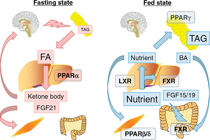
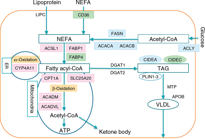
Ligand-activated nuclear receptors, including peroxisome proliferator-activated receptor alpha (PPARα), pregnane X receptor, and constitutive androstane receptor, were first identified as key regulators of the responses against chemical toxicants. However, numerous studies using mouse disease models and human samples have revealed critical roles for these receptors and others, such as PPARβ/δ, PPARγ, farnesoid X receptor (FXR), and liver X receptor (LXR), in maintaining nutrient/energy homeostasis in part through modulation of the gut-liver-adipose axis. Recently, disorders associated with disrupted nutrient/energy homeostasis, e.g., obesity, metabolic syndrome, and non-alcoholic fatty liver disease (NAFLD), are increasing worldwide. Notably, in NAFLD, a progressive subtype exists, designated as non-alcoholic steatohepatitis (NASH) that is characterized by typical histological features resembling alcoholic steatohepatitis (ASH), and NASH/ASH are recognized as major causes of hepatitis virus-unrelated liver cirrhosis and hepatocellular carcinoma. Since hepatic steatosis is basically caused by an imbalance between fat/energy influx and utilization, abnormal signaling of these nuclear receptors contribute to the pathogenesis of fatty liver disease. Standard therapeutic interventions have not been fully established for fatty liver disease, but some new agents that activate or inhibit nuclear receptor signaling have shown promise as possible therapeutic targets. In this review, we summarize recent findings on the roles of nuclear receptors in fatty liver disease and discuss future perspectives to develop promising pharmacological strategies targeting nuclear receptors for NAFLD/NASH.

Keywords: Energy vector; Hepatocellular carcinoma; Liver fibrosis; Peroxisome proliferator-activated receptor; Steatohepatitis; Tissue-specific agonist/antagonist.


Conflict of interest statement

Conflict of interest statement

The authors declare that there are no conflicts of interest.

Figures

Fig. 1.
Fig. 1.
Clinical course of NAFLD/NASH. A. The patient had obesity, diabetes, hypertension, dyslipidemia, and persistent elevation of serum alanine aminotransferase (ALT) levels. Initial laparoscopic examination revealed yellowish enlarged liver with smooth surface and soft consistency (1st Exam, upper panel), while liver biopsy section showed steatosis without significant fibrosis (1st Exam, lower panel). Serum ALT levels did not improve and the laparoscopic examination carried out at 5 years after the initial biopsy revealed whitish liver with rough surface (5 years later, upper panel). Dense fibrotic bands were found in biopsied specimen, indicative of pre-cirrhotic phase (5 years later, lower panel). The sections in the lower panel are stained by the Azan-Mallory method, and collagen fiber are indicated as blue. B. Careful pathological examination of the first biopsied specimen detected hepatocyte ballooning (arrow) in addition to macrovesicular steatosis (*), leading to the diagnosis of steatohepatitis. This section is stained by the hematoxylin and eosin method.
Fig. 2.
Fig. 2.
Nuclear receptors as energy vectors. In a fasting state, triacylglycerol (TAG) stored in white adipose tissue is subjected to lipolysis and released into the circulation as fatty acids (FAs). FAs are taken into many organs as an energy source. In the liver, FAs activate PPARα and enhance FA catabolism, resulting in the production of ATP, ketone bodies, and hepatokine fibroblast growth factor (FGF) 21. Ketone body is consumed as an energy source in the brain and FGF21 serves as a stress messenger to prepare other organs for energy deprivation. In the fed state, energy flux is reversed and FXR, LXR, PPARβ/δ and PPARγ are mainly involved in nutrient absorption from the gut and distribution from gut/liver to peripheral tissues, such as adipose tissue and muscle. After meals, bile acids (BAs) activate intestinal FXR, promoting nutrient absorption and maintaining a barrier to the gut microbiome. Absorbed dietary lipids are transported into the circulation as chylomicron and its remnant. Hepatic FXR promotes post-prandial TAG-rich lipoprotein clearance. Excess cholesterol is removed from the body by reverse cholesterol transport under the control of the FXR-stimulated enterokine FGF19 (FGF15 in rodents) and/or activation of hepatic LXR by oxysterols. FGF15/19 attenuates post-prandial hyperglycemia through enhancing hepatic glycogenesis. Consequently, excess nutrients are either consumed in muscle or stored in WAT due to PPARβ/δ and PPARγ, respectively. The concept “dysfunction of energy vectors on the gut-liver-adipose axis” may explain the pathogenesis of NAFLD/NASH.
Fig. 3.
Fig. 3.
FA/TAG metabolism in hepatocytes. Non-esterified fatty acids (NEFAs) are taken from blood into hepatocytes or synthesized from glucose. NEFAs are converted to fatty acyl-CoA esters, then are subjected to β-oxidation (Only mitochondrial β-oxidation pathway is shown for simplification), or generation of triacylglycerol (TAG) and very-low-density lipoprotein (VLDL). TGA is stored in the hepatocytes in the form of lipid droplets, which are coated by perilipin 1–3 and cell death inducing DFFA like effector (CIDE) a-c to prevent excessive lipolysis. The genes regulated by PPARα, PPARγ, and SREBP1c are indicated in pink, green, and blue, respectively. PPARα activation drives FA elimination, but the signaling of PPARγ and SREBP1c stimulates lipogenesis and TAG storage. ACACA, acetyl-CoA carboxylase α; ACACB, acetyl-CoA carboxylase β; ACADM, medium-chain acyl-CoA dehydrogenase; ACADVL, very-long-chain acyl-CoA dehydrogenase; ACLY, ATP citrate lyase; ACSL1, long-chain acyl-CoA synthetase; APOB, apolipoprotein B; CIDEA, cell death inducing DFFA like effector a; CIDEC, cell death inducing DFFA like effector c; CPT1A, carnitine palmitoyl-CoA transferase 1α; CYP, cytochrome P450; DGAT, diacylglycerol acyltransferase; FABP, fatty acid-binding protein; FASN, fatty acid synthase; LIPC, hepatic lipase; MTP, microsomal triacylglycerol transfer protein; NEFA, non-esterified fatty acid; PLIN, perilipin; SLC25A20, carnitineacylcarnitine translocase; TAG, triacylglycerol; VLDL, very-low-density lipoprotein.
Fig. 4.
Fig. 4.
Effects of FGF21. A hepatokine FGF21 is secreted into blood in response to various stress, such as fasting and endoplasmic reticulum stress, and activation of PPARα. It binds to a plasma membrane receptor complex, mainly FGF receptor 1 and β-Klotho, and enhances expression of glucose transporter 1 in extra-hepatic tissues, leading to improvement of systemic insulin sensitivity and enhancement of lipid turnover. FGF21 may prolong lifespan through down-regulating growth hormone (GH)-insulin-like growth factor 1 (IGF-1) axis or other unknown mechanisms. FGF21 may inhibit osteoblastogenesis and promote osteoclastogenesis, causing increased fracture risk, while it remains controversial.

References

    1. de Aguiar Vallim TQ, Tarling EJ, & Edwards PA (2013). Pleiotropic roles of bile acids in metabolism. Cell Metabolism 17(5), 657–669. - PMC - PubMed
    1. Ahmadian M, Suh JM, Hah N, Liddle C, Atkins AR, Downes M, & Evans RM (2013). PPARγ signaling and metabolism: The good, the bad and the future. Nature Medicine 19(5), 557–566. - PMC - PubMed
    1. Ahn SB, Jang K, Jun DW, Lee BH, & Shin KJ (2014). Expression of liver X receptor correlates with intrahepatic inflammation and fibrosis in patients with nonalcoholic fatty liver disease. Digestive Diseases and Sciences 59(12), 2975–2982. - PubMed
    1. Altmann R, Hausmann M, Spöttl T, Gruber M, Bull AW, Menzel K, … Rogler G (2007). 13-Oxo-ODE is an endogenous ligand for PPARgamma in human colonic epithelial cells. Biochemical Pharmacology 74(4), 612–622. - PubMed
    1. Anderson SP, Dunn C, Laughter A, Yoon L, Swanson C, Stulnig TM, … Corton JC (2004). Overlapping transcriptional programs regulated by the nuclear receptors peroxisome proliferator-activated receptor alpha, retinoid X receptor, and liver X receptor in mouse liver. Molecular Pharmacology 66(6), 1440–1452. - PubMed

Substances