

Jugular Anomalies in Multiple Sclerosis Are Associated with Increased Collateral Venous Flow
- PMID: 28546249
- PMCID: PMC5557656
- DOI: 10.3174/ajnr.A5219
Jugular Anomalies in Multiple Sclerosis Are Associated with Increased Collateral Venous Flow
Abstract
Background and purpose: To date, research on extracranial venous collaterals has been focused on structure, with relatively little attention paid to hemodynamics. We addressed this limitation by quantitatively comparing collateral flow in patients with multiple sclerosis and healthy controls by using phase-contrast MR imaging. We hypothesize that patients with MS with structurally anomalous internal jugular veins will have elevated collateral venous flow compared with healthy controls.
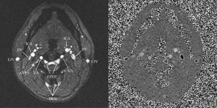
Materials and methods: The sample consisted of 276 patients with MS and 106 healthy controls. We used MRV to classify internal jugular veins as stenotic and nonstenotic based on an absolute cross-sectional area threshold in 276 patients with MS and 60 healthy controls; 46 healthy controls lacked this imaging. Individual and total vessel flows were quantified by using phase-contrast MR imaging on all patients. Veins were classified by extracranial drainage type: internal jugular veins (I), paraspinal (II), and superficial (III). Differences among healthy controls, patients with MS, nonstenotic patients, and stenotic subgroups in total venous flow by vessel type were evaluated in a general linear model for statistical analysis.
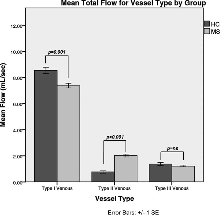
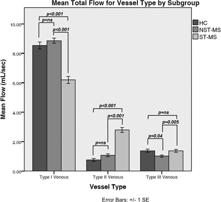
Results: In the MS group, 153 patients (55%) evidenced stenosis, whereas 12 (20%) healthy controls were classified as stenotic (P < .001). Compared with healthy controls, the MS group showed lower type I flow and increased type II flow. Stenosis was associated with reduced flow in the type I vessels [F(1272) = 68; P < .001]. The stenotic MS group had increased flow in the type II vessels compared with the nonstenotic MS group [F(1272) = 67; P < .001].
Conclusions: Compared with healthy controls, patients with MS exhibit reduced venous flow in the main extracerebral drainage vein (internal jugular vein). In contrast, flow in the paraspinal venous collaterals is elevated in patients with MS and exacerbated by venous stenosis. Collateral drainage may be a compensatory response to internal jugular vein flow reduction.
© 2017 by American Journal of Neuroradiology.
Figures
Similar articles
-
Extracranial Venous abnormalities: A true pathological finding in patients with multiple sclerosis or an anatomical variant?Eur Radiol. 2017 Jan;27(1):239-246. doi: 10.1007/s00330-016-4314-6. Epub 2016 Mar 24. Eur Radiol. 2017. PMID: 27011374
-
Patients with multiple sclerosis with structural venous abnormalities on MR imaging exhibit an abnormal flow distribution of the internal jugular veins.J Vasc Interv Radiol. 2012 Jan;23(1):60-8.e1-3. doi: 10.1016/j.jvir.2011.09.027. J Vasc Interv Radiol. 2012. PMID: 22221473
-
A comparative study of magnetic resonance venography techniques for the evaluation of the internal jugular veins in multiple sclerosis patients.Magn Reson Imaging. 2013 Dec;31(10):1668-76. doi: 10.1016/j.mri.2013.05.012. Epub 2013 Jul 12. Magn Reson Imaging. 2013. PMID: 23850076 Free PMC article.
-
Quantitative flow measurements in the internal jugular veins of multiple sclerosis patients using magnetic resonance imaging.Rev Recent Clin Trials. 2012 May;7(2):117-26. doi: 10.2174/157488712800100206. Rev Recent Clin Trials. 2012. PMID: 22356242 Review.
-
Screening for chronic cerebrospinal venous insufficiency (CCSVI) using ultrasound--recommendations for a protocol.Int Angiol. 2011 Dec;30(6):571-97. Int Angiol. 2011. PMID: 22233619
Cited by
-
Lower Arterial Cross-Sectional Area of Carotid and Vertebral Arteries and Higher Frequency of Secondary Neck Vessels Are Associated with Multiple Sclerosis.AJNR Am J Neuroradiol. 2018 Jan;39(1):123-130. doi: 10.3174/ajnr.A5469. Epub 2017 Dec 7. AJNR Am J Neuroradiol. 2018. PMID: 29217748 Free PMC article.
-
Internal jugular vein stenosis associated with elongated styloid process: five case reports and literature review.BMC Neurol. 2019 Jun 4;19(1):112. doi: 10.1186/s12883-019-1344-0. BMC Neurol. 2019. PMID: 31164090 Free PMC article. Review.
-
Clinical Classification and Collateral Circulation in Chronic Cerebrospinal Venous Insufficiency.Front Neurol. 2020 Sep 23;11:913. doi: 10.3389/fneur.2020.00913. eCollection 2020. Front Neurol. 2020. PMID: 33071925 Free PMC article.
-
The jugular-subclavian junction and venous drainage of the brain.Mediastinum. 2023 Dec 4;8:6. doi: 10.21037/med-23-15. eCollection 2024. Mediastinum. 2023. PMID: 38322186 Free PMC article. Review.
-
Internal jugular vein stenosis induced by tortuous internal carotid artery compression: two case reports and literature review.J Int Med Res. 2019 Aug;47(8):3926-3933. doi: 10.1177/0300060519860678. Epub 2019 Jul 15. J Int Med Res. 2019. PMID: 31304848 Free PMC article. Review.
References
-
- Haacke EM, Sethi SK, Jiang J, et al. . The role of magnetic resonance imaging in assessing venous vascular abnormalities in the head and neck: a demonstration of cerebrospinal venous insufficiency in a subset of multiple sclerosis patients. Veins Lymphatics 2015;4:5012–20 10.4081/vl.2015.5012 - DOI
MeSH terms
Grants and funding
LinkOut - more resources
Full Text Sources
Other Literature Sources
Medical