. 2017 Jun 28;37(26):6359-6371.
doi: 10.1523/JNEUROSCI.3029-16.2017. Epub 2017 May 25.

Dentate Gyrus Contributes to Retrieval as well as Encoding: Evidence from Context Fear Conditioning, Recall, and Extinction

Affiliations

Dentate Gyrus Contributes to Retrieval as well as Encoding: Evidence from Context Fear Conditioning, Recall, and Extinction

Brian E Bernier et al. J Neurosci. .

Abstract

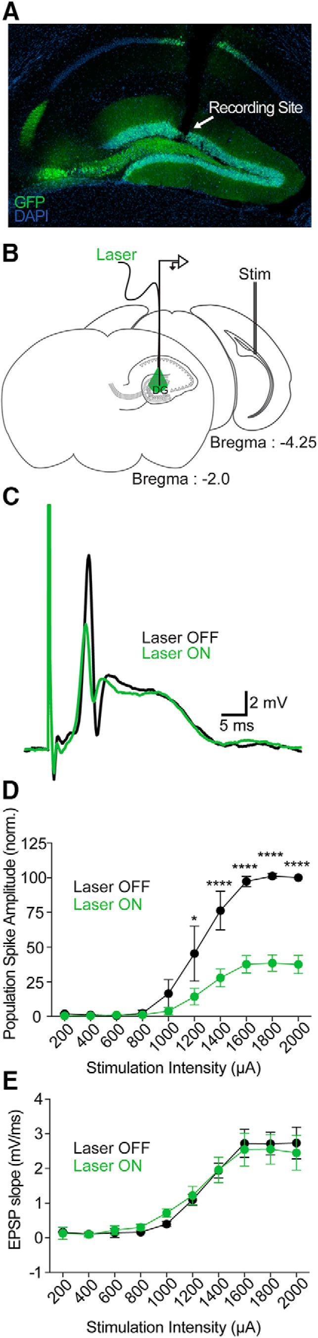
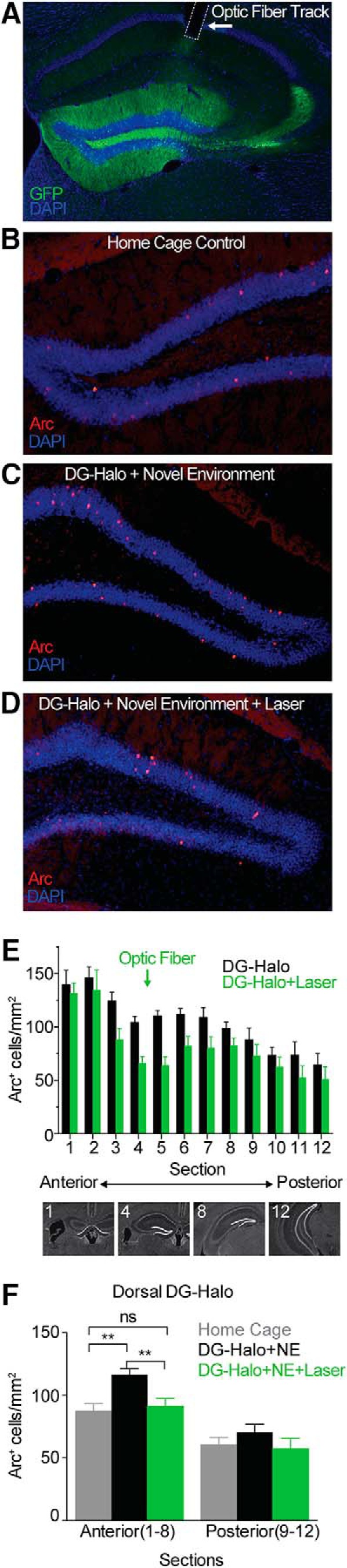
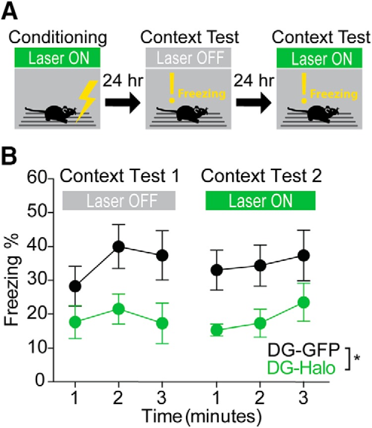
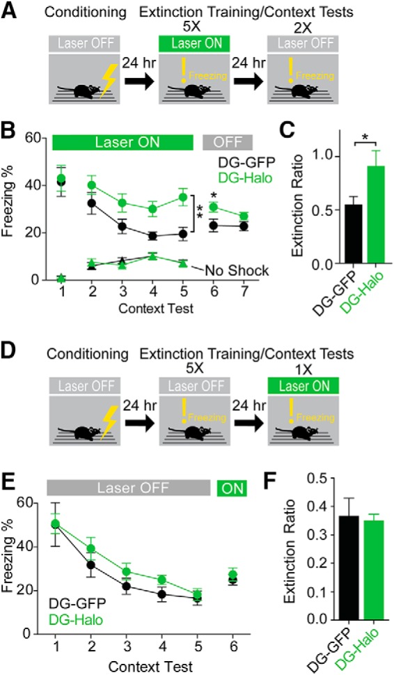
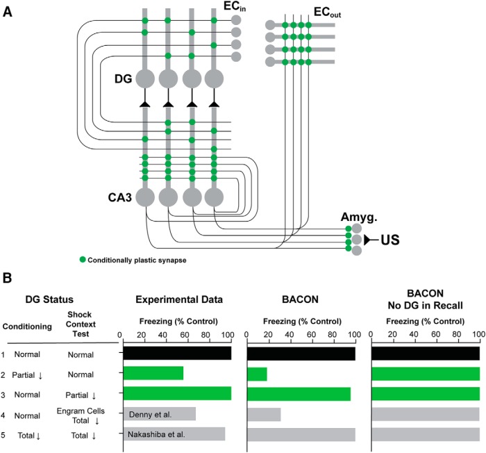
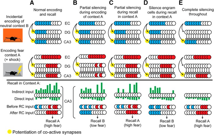
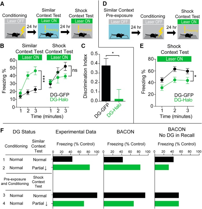
Dentate gyrus (DG) is widely thought to provide a teaching signal that enables hippocampal encoding of memories, but its role during retrieval is poorly understood. Some data and models suggest that DG plays no role in retrieval; others encourage the opposite conclusion. To resolve this controversy, we evaluated the effects of optogenetic inhibition of dorsal DG during context fear conditioning, recall, generalization, and extinction in male mice. We found that (1) inhibition during training impaired context fear acquisition; (2) inhibition during recall did not impair fear expression in the training context, unless mice had to distinguish between similar feared and neutral contexts; (3) inhibition increased generalization of fear to an unfamiliar context that was similar to a feared one and impaired fear expression in the conditioned context when it was similar to a neutral one; and (4) inhibition impaired fear extinction. These effects, as well as several seemingly contradictory published findings, could be reproduced by BACON (Bayesian Context Fear Algorithm), a physiologically realistic hippocampal model positing that acquisition and retrieval both involve coordinated activity in DG and CA3. Our findings thus suggest that DG contributes to retrieval and extinction, as well as to the initial establishment of context fear.SIGNIFICANCE STATEMENT Despite abundant evidence that the hippocampal dentate gyrus (DG) plays a critical role in memory, it remains unclear whether the role of DG relates to memory acquisition or retrieval. Using contextual fear conditioning and optogenetic inhibition, we show that DG contributes to both of these processes. Using computational simulations, we identify specific mechanisms through which the suppression of DG affects memory performance. Finally, we show that DG contributes to fear extinction learning, a process in which learned fear is attenuated through exposures to a fearful context in the absence of threat. Our data resolve a long-standing question about the role of DG in memory and provide insight into how disorders affecting DG, including aging, stress, and depression, influence cognitive processes.

Keywords: context; dentate gyrus; extinction; fear conditioning; hippocampus; memory.


Conflict of interest statement

The authors declare no competing financial interests.

Figures

Figure 1.
Figure 1.
In vivo optogenetic inhibition of perforant path-evoked population responses in DG. A, AAV microinjection produced robust eNpHR3.0–sfGFP expression in the dorsal DG. An electrolytic lesion was made to mark recording location. The electrode and optic fiber track can be seen above it. B, In vivo electrophysiological recording configuration. C, Sample traces of population response with and without laser inhibition. D, Input–output curve for population spike amplitude showing significant inhibition during laser illumination. E, Input–output curve for EPSP slope showing no effect of laser illumination (n = 3). *p < 0.05; ****p < 0.001. Data in D and E are represented as the mean ± SEM.
Figure 2.
Figure 2.
Optogenetic inhibition of dorsal DG activity in vivo. A, Representative example of eNpHR3.0–sfGFP expression in the dorsal DG. The optic fiber implant track is marked with dashed lines and arrow. B, Arc expression in home-cage control. C, D, Laser illumination blocks induction of Arc by novel environment exposure in DG-Halo mice. E, Arc+ cell density by section in DG-Halo mice with and without laser illumination. Representative sections are shown below the graph. F, Laser illumination prevented the novelty-evoked Arc induction in dorsal DG. DG-Halo mice with laser illumination, n = 10; DG-Halo mice without laser illumination, n = 11; home-cage controls, n = 8. **p < 0.01. Data in E and F are represented as the mean ± SEM.
Figure 3.
Figure 3.
Optogenetic inhibition of the dorsal DG impairs acquisition of CFC. A, Experimental design. B, Time course of freezing during successive context tests with and without laser illumination of the DG. DG inhibition during conditioning led to reduced freezing regardless of the status of DG during context test. DG-GFP mice, n = 8; DG-Halo mice, n = 7. Data are represented as the mean ± SEM.
Figure 4.
Figure 4.
Optogenetic inhibition of the dorsal DG impairs context fear extinction but does not impair the expression of fear or extinction. A, Experimental design. B, Freezing during the seven context exposures in the absence of shock. In unshocked mice, freezing remained low across all sessions and there was no effect of optogenetic inhibition (No shock DG-Halo mice, n = 7; No shock DG-GFP mice, n = 7). In shocked mice, freezing did not differ between DG-Halo mice (n = 15) and DG-GFP control mice (n = 12) in session 1, indicating that DG neural activity was not required for context fear expression. However, during sessions 2–5, freezing of DG-Halo mice exceeded that of DG-GFP controls, suggesting that DG suppression impaired extinction. C, Extinction ratio comparing freezing in session 1 to sessions 4 and 5 (shocked mice only). DG-GFP mice exhibited a significantly greater reduction in freezing over the course of extinction. D, Design of the extinction recall experiment. E, Time course of extinction showing total freezing per context test/extinction session. DG inhibition during session 6 did not affect recall of extinction learning. DG-GFP mice, n = 7; DG-Halo mice, n = 11. F, Extinction ratio comparing freezing in session 1 to session 6. Both groups of mice show equivalent levels of extinction. *p < 0.05; **p < 0.01. Data in B and C and E and F are represented as the mean ± SEM.
Figure 5.
Figure 5.
The BACON computational model of hippocampal function reproduces the behavioral effects of a variety of DG manipulations. A, The BACON cortex–hippocampus–amygdala circuit includes 1000 entorhinal cortex neurons (ECin) projecting to 10,000 DG granule cells. The 3000 CA3 neurons are innervated by ECin, DG, and recurrent collaterals from other CA3 pyramidal cells. CA1 is omitted from the model. ECout and amygdala are innervated directly by CA3 pyramidal neurons. The CA3–amygdala projection allows fear to become conditioned directly to hippocampal context representations. Context fear memory acquisition depends on plasticity at ECin–DG, ECin–CA3, CA3–CA3, and CA3–amygdala synapses, which are highlighted in green. The DG–CA3 projection operates during both memory acquisition and retrieval. A complete description is available in Krasne et al. (2015). B, Experimental data (left) from the current study (rows 1–3) and two previously published studies (Nakashiba et al., 2012; Denny et al., 2014; rows 4–5) compared with predictions from BACON simulations. The middle column shows the results of simulations in which BACON used DG during recall. The right column represents simulations in which BACON was prevented from using DG during recall. The labels at left denote the degree of DG suppression during conditioning and test sessions. Bars represent freezing during the test session as a percentage of freezing in control conditions. BACON recapitulates the seemingly contradictory behavioral results only when it is allowed to use DG during recall.
Figure 6.
Figure 6.
The effects of DG silencing on acquisition and recall of context fear: proposed mechanisms. A, To simulate incidental encoding of places other than the conditioning context, the subject is exposed to (and encodes) a neutral context (context B) before being fear conditioned in a different context (context A). Then the subject is tested for fear recall in context A. EC, DG, and CA3 are portrayed as linear arrays of cells, with cells involved in the processing of each context grouped. Active cells are filled in red or blue. The EC cells activated by contexts A and B overlap. During encoding, DG recodes the EC representations to form relatively sparse, nonoverlapping codes for each context, and each DG cell drives a corresponding CA3 neuron. Each DG and CA3 cell is innervated via Hebbian synapses by a random subset of EC neurons. EC–DG, EC–CA3, and CA3–CA3 [recurrent collateral (RC)] synapses of coactive cells become potentiated, providing a basis for later recognition of the encoded contexts. During tests in which only some fraction of the attributes of a context may have been sampled, CA3 cells are excited via the potentiated synapses of the indirect (EC–DG–CA3) and direct (EC–CA3) paths, but the indirect path is dominant. The k most excited CA3 cells fire, and the recurrent collateral system then completes the representation, which determines input to the amygdala and hence fear responses. B, When a random subset of DG neurons is suppressed during encoding of context A, a subnormal number of DG cells is incorporated into the context A representation. As a result, the context B representation dominates during recall. C, When a random subset of DG neurons is suppressed during recall, representation cells of both context A and B receive less indirect path input, but the remaining direct and indirect inputs are sufficient to cause the representation of context A to be completed. D, When all DG cells that were active during the encoding of context A (engram cells) are suppressed during recall, the active cells are necessarily those representing context B. The context B representation is completed, and fear is low. E, When DG activity is totally suppressed during both encoding and recall, direct input to CA3 is adequate to cause representation of context A to emerge and evoke fear.
Figure 7.
Figure 7.
Optogenetic inhibition of the dorsal DG affects fear expression when similar contexts must be distinguished. A, Design of an experiment testing fear generalization in a context similar to the conditioning context. B, Time course of freezing during the generalization test and shock context test with laser inhibition of the DG. DG inhibition during fear expression caused elevated freezing in the neutral generalization context relative to controls but did not affect freezing in the shock context. C, Discrimination index ([Shock–Neutral]/ [Neutral + Shock]) demonstrating that DG-GFP mice discriminated between contexts but DG-Halo mice did not. DG-GFP mice, n = 6; DG-Halo mice, n = 5. D, Design of an experiment in which mice were pre-exposed to a neutral context before conditioning in a similar context. E, Time course of freezing during shock context test with DG inhibition. DG inhibition reduced fear expression. DG-GFP mice, n = 16; DG-Halo mice, n = 16. *p < 0.05; ***p < 0.005. Data are represented as the mean ± SEM. F, Comparison of behavioral results and the corresponding BACON simulations. Behavioral results are shown in the left column. BACON simulations are shown in the middle and left columns. The simulations were performed with BACON using DG during recall (middle) or with BACON prevented from using DG during recall (right). The generalization experiment depicted in A is shown in rows 1–2. The pre-exposure experiment depicted in D is shown in rows 3–4. Bars represent freezing during the test session as a percentage of freezing in control animals. The simulations recapitulate the observed effects of DG suppression only when BACON uses DG during recall.

Similar articles

Cited by

References

    1. Aurnhammer C, Haase M, Muether N, Hausl M, Rauschhuber C, Huber I, Nitschko H, Busch U, Sing A, Ehrhardt A, Baiker A (2012) Universal real-time PCR for the detection and quantification of adeno-associated virus serotype 2-derived inverted terminal repeat sequences. Hum Gene Ther Methods 23:18–28. 10.1089/hgtb.2011.034 - DOI - PubMed
    1. Bernier BE, Lacagnina AF, Drew MR (2014) Potent attenuation of context fear by extinction training contiguous with acquisition. Learn Mem 22:31–38. 10.1101/lm.036673.114 - DOI - PMC - PubMed
    1. Bouton ME. (2002) Context, ambiguity, and unlearning: sources of relapse after behavioral extinction. Biol Psychiatry 52:976–986. 10.1016/S0006-3223(02)01546-9 - DOI - PubMed
    1. Bouton ME. (2004) Context and behavioral processes in extinction. Learn Mem 11:485–494. 10.1101/lm.78804 - DOI - PubMed
    1. Bouton ME, Bolles RC (1979) Role of conditioned contextual stimuli in reinstatement of extinguished fear. J Exp Psychol Anim Behav Process 5:368–378. 10.1037/0097-7403.5.4.368 - DOI - PubMed

Publication types