Review
. 2017 May 26;120(11):1825-1841.
doi: 10.1161/CIRCRESAHA.117.309633.

AMP-Activated Protein Kinase: An Ubiquitous Signaling Pathway With Key Roles in the Cardiovascular System

Affiliations
Review

AMP-Activated Protein Kinase: An Ubiquitous Signaling Pathway With Key Roles in the Cardiovascular System

Ian P Salt et al. Circ Res. .

Abstract

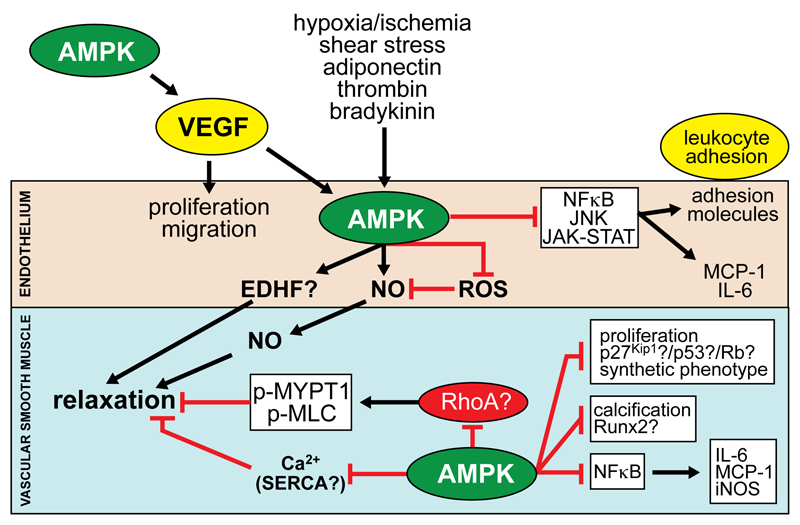
The AMP-activated protein kinase (AMPK) is a key regulator of cellular and whole-body energy homeostasis, which acts to restore energy homoeostasis whenever cellular energy charge is depleted. Over the last 2 decades, it has become apparent that AMPK regulates several other cellular functions and has specific roles in cardiovascular tissues, acting to regulate cardiac metabolism and contractile function, as well as promoting anticontractile, anti-inflammatory, and antiatherogenic actions in blood vessels. In this review, we discuss the role of AMPK in the cardiovascular system, including the molecular basis of mutations in AMPK that alter cardiac physiology and the proposed mechanisms by which AMPK regulates vascular function under physiological and pathophysiological conditions.

Keywords: AMP-activated protein kinase; heart; heart defects (congenital); metabolism; vasculature.


Figures

Figure 1
Figure 1. Domain layouts of AMPK subunits and their isoforms.
The linear layout of domains is shown, approximately to scale and with similar color coding as in Fig. 2. Note that both β subunits are N-myristoylated, and that the γ2 and γ3 subunit isoforms have unrelated N-terminal extensions of unknown function, although both are also reported to exist as shorter, N-terminally truncated versions due to alternate start sites and/or splicing.
Figure 2
Figure 2. Structure of human AMPK (α1β2γ1 complex).
The model was created in spacefilling mode using PyMol v1.7.4.2 with the co-ordinates in PDB file 4RER, and with color coding similar to Fig. 1. The heterotrimer was crystallized in the presence of β-cyclodextrin, which occupies the glycogen-binding site, staurosporine, which occupies the active site, and AMP, which occupies sites 1, 3 and 4 on the γ subunit (sites 1 and 4 are round the back in this view). Although Thr172 was phosphorylated, it is not visible in this view but lies in the cleft between the α subunit C lobe and the β-CTD, just over the right-hand “shoulder” of the C-lobe.
Figure 3
Figure 3. Two views of the structure of the four CBS repeats of the γ1 subunit.
The model used the co-ordinates in PDB file 2V8Q, and was rendered in PyMol v1.7.4.2 with the γ1 subunit in cartoon view. The two views are rotated 180° around the x axis (dashed line) with respect to each other, with the orientation of the top view being similar to that in Fig. 2. Note the pseudosymmetrical layout of the four CBS repeats, which are colored differently (and differently to Figs. 1 and 2). Residues equivalent to those mutated in γ2 are highlighted using the “dots” version of space-filling representation, and are numbered using the human γ2 numbering. The three molecules of bound AMP are labelled and are shown in standard space-filling view, with C atoms green, O red and N blue.
Figure 4
Figure 4. Regulation of endothelial NO and superoxide synthesis by AMPK.
AMPK activation stimulates NO synthesis via multiple mechanisms: (i) phosphorylation of eNOS at Ser633 and Ser1177; (ii) increasing Hsp90 association with eNOS; (iii) increasing BH4 concentrations via GTP cyclohydrolase I (GTPCH1); (iv) reducing superoxide synthesis via inhibition of Nox and increasing antioxidant protein (superoxide dismutase (SOD), thioredoxin and catalase) levels. NO itself is also reported to activate AMPK.
Figure 5
Figure 5. Actions of AMPK in vascular cells.
Physiological activators of AMPK in ECs include hypoxia/ischemia, shear stress, adiponectin, thrombin, bradykinin and VEGF (synthesis of which is stimulated by AMPK in other tissues). AMPK is reported to stimulate VSMC relaxation through: (i) increased NO synthesis and possibly endothelium-dependent hypopolarising factor (EDHF); (ii) inhibition of MYPT1/MLC phosphorylation and Ca2+ levels in VSMCs, reported to be mediated by reduced RhoA activity and increased sarco/endoplasmic Ca2+ ATPase (SERCA) activity respectively. AMPK activation in ECs stimulates proliferation and migration, whereas in VSMCs, proliferation and migration are inhibited, associated with p53 phosphorylation, Rb dephosphorylation and p27(Kip1) stabilisation. AMPK also inhibits VSMC calcification by reducing Runx2 and pro-inflammatory signalling pathways leading to leukocyte adhesion and cytokine/chemokine synthesis.

References

    1. Hardie DG, Schaffer BE, Brunet A. AMPK: an energy-sensing pathway with multiple inputs and outputs. Trends Cell Biol. 2016;26:190–201. - PMC - PubMed
    1. Hardie DG, Ashford ML. AMPK: regulating energy balance at the cellular and whole body levels. Physiology. 2014;29:99–107. - PMC - PubMed
    1. Hardie DG, Ross FA, Hawley SA. AMPK: a nutrient and energy sensor that maintains energy homeostasis. Nature Rev Mol Cell Biol. 2012;13:251–262. - PMC - PubMed
    1. Daskalopoulos EP, Dufeys C, Beauloye C, Bertrand L, Horman S. AMPK in cardiovascular diseases. EXS. 2016;107:179–201. - PubMed
    1. Daskalopoulos EP, Dufeys C, Bertrand L, Beauloye C, Horman S. AMPK in cardiac fibrosis and repair: Actions beyond metabolic regulation. J Mol Cell Cardiol. 2016;91:188–200. - PubMed

MeSH terms

Substances