. 2017 Jul;206(3):1445-1458.
doi: 10.1534/genetics.116.193102. Epub 2017 May 25.

Behavioral Deficits Following Withdrawal from Chronic Ethanol Are Influenced by SLO Channel Function in Caenorhabditis elegans

Affiliations

Behavioral Deficits Following Withdrawal from Chronic Ethanol Are Influenced by SLO Channel Function in Caenorhabditis elegans

Luisa L Scott et al. Genetics. 2017 Jul.

Abstract

Symptoms of withdrawal from chronic alcohol use are a driving force for relapse in alcohol dependence. Thus, uncovering molecular targets to lessen their severity is key to breaking the cycle of dependence. Using the nematode Caenorhabditis elegans, we tested whether one highly conserved ethanol target, the large-conductance, calcium-activated potassium channel (known as the BK channel or Slo1), modulates ethanol withdrawal. Consistent with a previous report, we found that C. elegans displays withdrawal-related behavioral impairments after cessation of chronic ethanol exposure. We found that the degree of impairment is exacerbated in worms lacking the worm BK channel, SLO-1, and is reduced by selective rescue of this channel in the nervous system. Enhanced SLO-1 function, via gain-of-function mutation or overexpression, also dramatically reduced behavioral impairment during withdrawal. Consistent with these results, we found that chronic ethanol exposure decreased SLO-1 expression in a subset of neurons. In addition, we found that the function of a distinct, conserved Slo family channel, SLO-2, showed an inverse relationship to withdrawal behavior, and this influence depended on SLO-1 function. Together, our findings show that modulation of either Slo family ion channel bidirectionally regulates withdrawal behaviors in worm, supporting further exploration of the Slo family as targets for normalizing behaviors during alcohol withdrawal.

Keywords: alcohol; behavior; ethanol; potassium channel; slo-1; withdrawal.


Figures

Figure 1
Figure 1
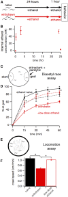
Two behavioral deficits during alcohol withdrawal recovered by low-dose ethanol. Worms withdrawn from chronic ethanol exposure display behavioral deficits. (A) Schematic showing the exposure paradigm used for the two treatment groups, naïve (black) and withdrawn (red), starting with age-matched L4-stage larvae. Worms assayed for behaviors are young adults 25 hr later. (B) Gas chromatography determined internal ethanol concentration after 0, 20 min, 3, and 24 hr of ethanol treatment, and after 1 hr of withdrawal. (C) Schematic of the diacetyl-race assay. Diacetyl was used as a volatile attractant and sodium azide was used as a paralytic trapping worms that reached the goal. (D) The mean fraction of WT worms that reached the attractant ± SEM plotted every 15 min for 1 hr. At all timepoints, withdrawn worms (solid red line) performed less well than naïve worms (solid black line, ****P < 0.001). Withdrawn worms treated with a low dose of ethanol during the race (dashed red line) performed significantly better than withdrawn worms (*P < 0.05). Naïve worms treated with a low dose of ethanol during the race (dashed black line) performed similarly to naïve worms. (E) Schematic of locomotion assay. Worms were allowed to move freely on a blank agar surface within a copper ring. (F) Histogram of mean speed ± SEM. Locomotion was also impaired during withdrawal. Withdrawn worms moved slower than naïve worms (naïve vs. withdrawn, 1.10 ± 0.026 vs. 0.68 ± 0.028 cm/min; ****P < 0.001). Again, this withdrawal-induced impairment was improved when worms were placed on low-dose ethanol during the assay (withdrawn vs. + low-dose ethanol, 0.68 ± 0.028 vs. 1.0 ± 0.025 cm/min; ****P < 0.001).
Figure 2
Figure 2
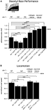
Reduced neuronal SLO-1 channel function exacerbated behavioral impairments during alcohol withdrawal. (A) Schematic above indicates how the time course of performance was quantified by the AUC for the percent of worms at the goal vs. time for the diacetyl race. Treatment groups: withdrawn (black area), naïve (gray + black areas). Histogram below shows the mean AUC for withdrawn worms normalized to the mean AUC for naïve worms (dashed horizontal line) ± SEM. The slo-1 genotype for each strain is indicated above each bar for reference. Two slo-1 strains with null alleles (js379 and js118) showed more withdrawal-related impairment for the diacetyl-race assay than WT strain N2. A heterozygous slo-1(+/js379) strain performed similarly to WT. Rescue strains with slo-1(+) driven by the endogenous promoter (pslo-1; JPS344=#1, JPS345=#2) or a pan-neuronal promoter (punc-119) all showed substantially improved withdrawn performance on the diacetyl-race assay compared to the background slo-1 null strain containing slo-1(js379). Two of these rescue strains (pslo-1:slo-1(+) #2, punc-119:slo-1(+)) also showed substantially less withdrawal-related impairment than WT. A dgk-1(sy428) null strain showed substantially less withdrawal-related impairment than WT or either slo-1 null strains (P < 0.001). (B) Locomotion during withdrawal also worsened with reduced BK channel function. Histogram shows mean speed during withdrawal for different strains normalized to mean speed for naïve worms (dashed horizontal line) ±SEM. Two slo-1 null strains were more impaired upon withdrawal for locomotion than WT. Rescue strains with slo-1(+) driven by the endogenous promoter or a pan-neuronal promoter showed substantially improved performance compared to the background null strain containing slo-1(js379). For A and B, *P < 0.05, **P < 0.01, ***P < 0.005, ****P < 0.001.
Figure 3
Figure 3
Enhanced SLO-1 channel function ameliorated behavioral impairment during alcohol withdrawal. (A) Schematic above indicates how performance was quantified by the AUC for the percent of worms at the goal vs. time for the diacetyl race. Treatment groups: withdrawn (black area), naïve (gray + black areas). Histogram below shows the mean AUC for withdrawn worms normalized to the mean AUC for naïve worms (dashed horizontal line) ± SEM. The slo-1 genotype for each strain is indicated above each bar for reference. The slo-1(ky399) gain-of-function mutant and two strains with slo-1(+) overexpressed in a WT background were significantly less impaired upon withdrawal for the diacetyl-race assay than WT strain N2. (B) Enhancing SLO-1 channel function also improved locomotion during withdrawal. Histogram shows mean normalized crawl speed ± SEM. For A and B, *P < 0.05, ***P < 0.005, ****P < 0.001.
Figure 4
Figure 4
A different large-conductance potassium channel, SLO-2, influences withdrawal impairments via a SLO-1 channel-dependent mechanism. Knockout of slo-2 improved behavior during alcohol withdrawal. (A) Histogram shows the mean AUC values of different strains for diacetyl-race performance; withdrawn performance normalized to naïve performance (dashed lines) ± SEM. Two slo-2 strains with null alleles (nf100 and nf101) were significantly less impaired upon withdrawal for the diacetyl race than WT (**P < 0.001). A strain with genomic slo-2(+) driven by the endogenous promoter (pslo-2) on background slo-2 null strain containing slo-2(nf100) showed substantially impaired withdrawn performance on the diacetyl-race <assay compared to the background strain (##P < 0.001) and WT (**P < 0.001). (B) Epistasis between slo-1 and slo-2 for alcohol withdrawal. A strain carrying the null alleles slo-1(js379) and slo-2(nf100) was more impaired in the diacetyl race during withdrawal than WT, similar to the parent slo-1(js379) null strain. Independent rescue strains (#1 and #2) with slo-1(+) introduced on the slo-1(js379);slo-2(nf100) double null mutant background were less impaired than the parent strain during withdrawal. For B, *P < 0.025, **P < 0.001.
Figure 5
Figure 5
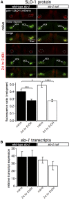
Chronic ethanol treatment suppresses neuronal SLO-1 channel expression. (A and B) Confocal microscopy stacks were summed to produce the photomicrographs showing translational slo-1 reporter tagged with mCherry in a slo-1(js379) null background. The red:green fluorescence decreased by half in GFP-labeled VC4 and VC5 neurons after 24-hr exposure to ethanol (A, ***P < 0.0001), but not in GFP-labeled AWA olfactory neurons (B). (C) Confocal photomicrographs showing a GFP transcriptional reporter of slo-1 in the green channel and mCherry-labeled VC4 and VC5 motorneurons in a WT background. Ratiometric analysis showed no change in whole body green:red ratios in the VC4 and VC5 neurons following chronic ethanol treatment. Bar, 10 μm in A–C.
Figure 6
Figure 6
Loss-of-function mutation in slo-2 enhances neuronal SLO-1 channel expression. (A) Confocal microscopy stacks were summed to produce the photomicrographs showing translational slo-1 reporter tagged with mCherry in a slo-1(js379) null (left, solid bar) or a slo-1(js379);slo-2(nf100) double null mutant (right, open bar) background. In both strains, the red:green fluorescence decreased in GFP-labeled VC4 and VC5 neurons after 24-hr exposure to ethanol (***P < 0.005, ****P < 0.001). In naïve worms, the amount of VC4 and VC5 neuron red:green fluorescence was greater in the slo-1;slo-2 double null mutant than the slo-1 null background (*P < 0.05), while the fluorescence ratio was the same in the strain after a 24-hr ethanol treatment. (B) Relative total slo-1 transcript expression in whole worm. qPCR measured slo-1 transcript expression relative to the control gene cdc-42 in WT (solid bar) and a slo-2(nf100) null strain (open bar). Chronic ethanol treatment did not alter slo-1 transcript expression in either strain. A loss-of-function mutation in slo-2 did not alter slo-1 transcript expression in either naïve or chronic ethanol-treated worms.

Similar articles

Cited by

References

    1. Alaimo J. T., Davis S. J., Song S. S., Burnette C. R., Grotewiel M., et al. , 2012. Ethanol metabolism and osmolarity modify behavioral responses to ethanol in C. elegans. Alcohol. Clin. Exp. Res. 36: 1840–1850. - PMC - PubMed
    1. Alqadah A., Hsieh Y. W., Schumacher J. A., Wang X., Merrill S. A., et al. , 2016. SLO BK potassium channels couple gap junctions to inhibition of calcium signaling in olfactory neuron diversification. PLoS Genet. 12(1): e1005654. - PMC - PubMed
    1. Bargmann, C. I., E. Hartwieg, and H. R. Horvitz, 1993 Odorant-selective genes and neurons mediate olfaction in C. elegans. Cell 74: 515–527. - PubMed
    1. Bhattacharjee A., Kaczmarek L. K., 2005. For K+ channels, Na+ is the new Ca2+. Trends Neurosci. 28(8): 422–428. - PubMed
    1. Becker H. C., Mulholland P. J., 2014. Neurochemical mechanisms of alcohol withdrawal. Handb. Clin. Neurol. 125: 133–156. - PMC - PubMed

Publication types

MeSH terms

Substances

LinkOut - more resources