. 2017 May 25;7(1):2417.
doi: 10.1038/s41598-017-02679-z.

Melatonin-mediated mitophagy protects against early brain injury after subarachnoid hemorrhage through inhibition of NLRP3 inflammasome activation

Affiliations

Melatonin-mediated mitophagy protects against early brain injury after subarachnoid hemorrhage through inhibition of NLRP3 inflammasome activation

Shenglong Cao et al. Sci Rep. .

Erratum in

Abstract

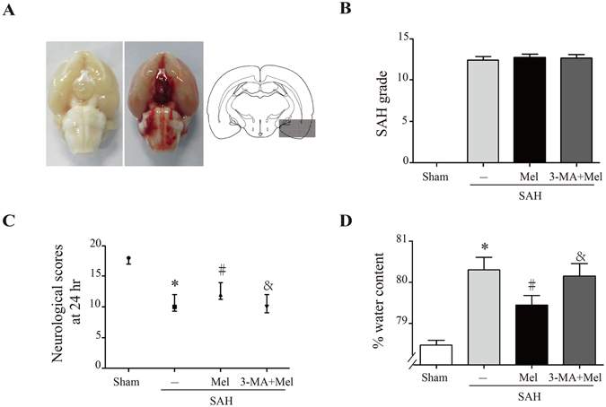
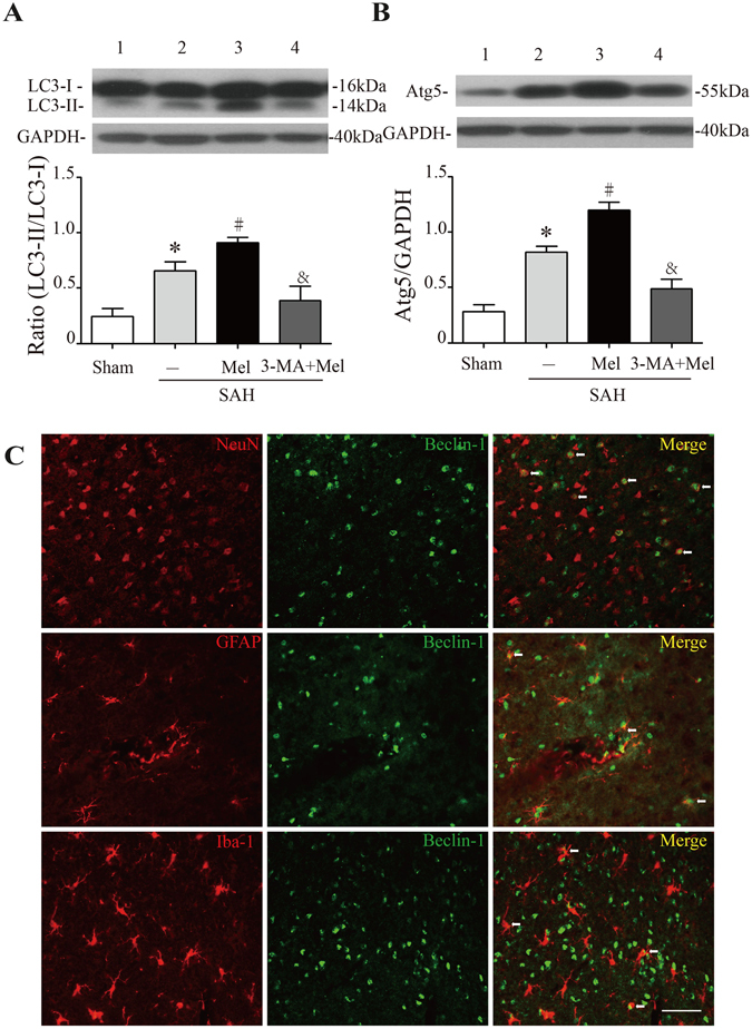
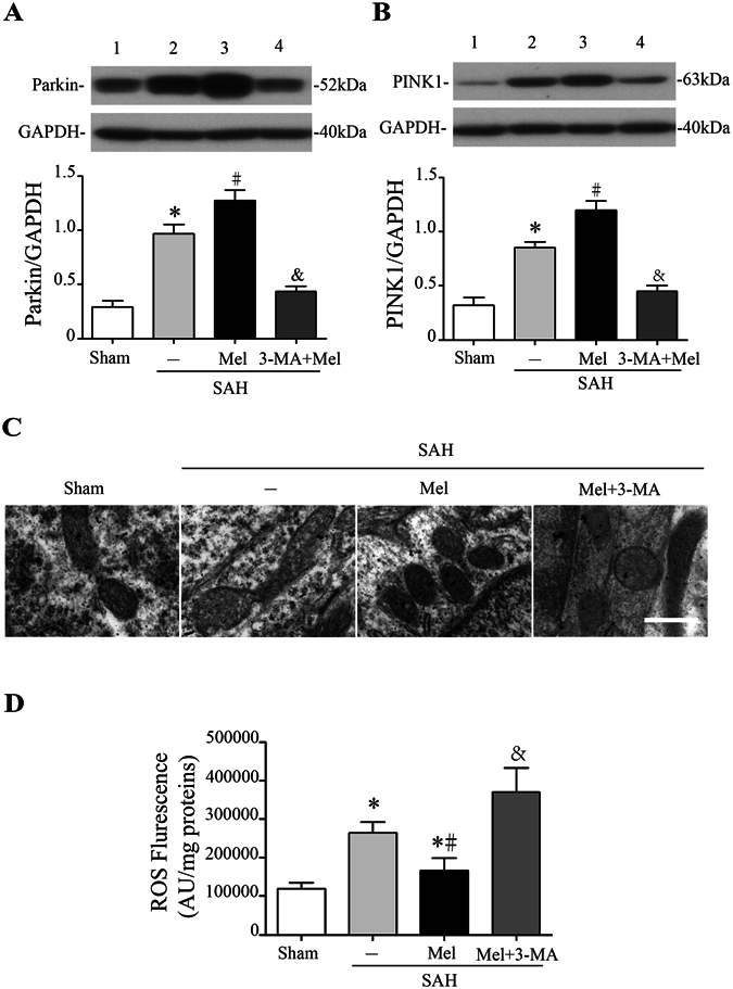
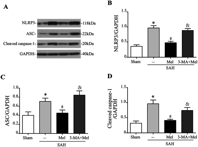
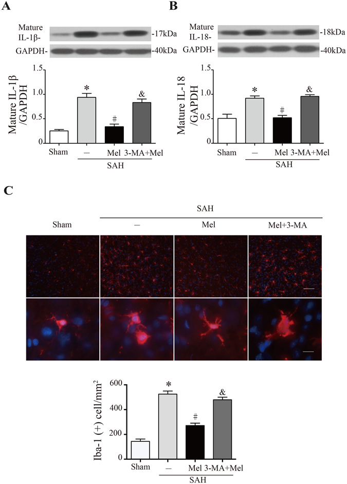
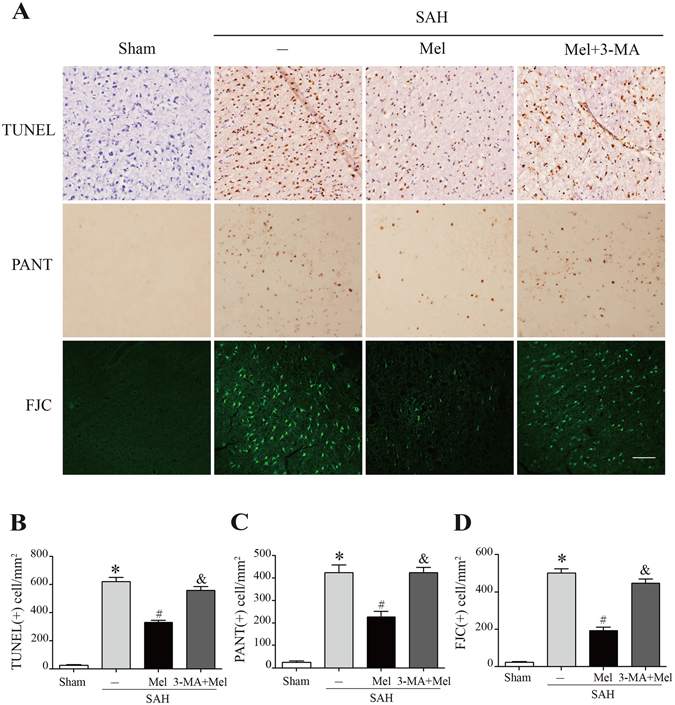
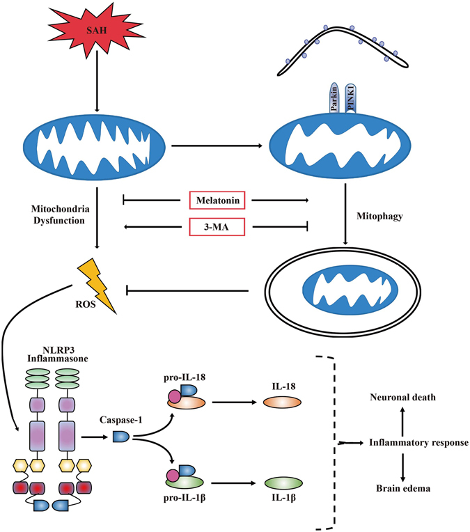
The NLRP3 inflammasome is activated in the early period following subarachnoid hemorrhage(SAH), resulting in inflammatory responses. Recent studies have shown that activation of NLRP3 inflammasome is suppressed by autophagy, but the potential mechanism is unclear. In this study, we examined whether mitophagy was involved in the beneficial effect of melatonin and its relationship with NLRP3 inflammasome activation after SAH. In total, 130 adult-male SD rats were randomly divided into four groups: sham group, SAH + vehicle group, SAH + melatonin group, and SAH + 3-methyladenine (3-MA) + melatonin group. Brain samples were used for brain water content analysis, ROS assay, Western blot, immunohistochemistry and transmission electron microscopy. The results showed that melatonin treatment markedly increased the expression of both autophagy markers(LC3-II/LC3-I and Atg 5), and mitophagy markers(Parkin and PINK-1) following SAH induction. Additionally, melatonin treatment attenuated pathological changes in mitochondria and reduced ROS generation, which are closely related to NLRP3 inflammasome activation. Consequently, melatonin-mediated upregulation of proteins associated with mitophagy inhibited NLRP3 inflammasome activation and significantly reduced pro-inflammatory cytokine levels after SAH. Conversely, 3-MA, an autophagy inhibitor, reversed these beneficial effects of melatonin on mitophagy and the NLRP3 inflammasome. These results suggest that mitophagy-associated NLRP3 inflammasome inhibition by melatonin is neuroprotective against early brain injury post-SAH in rats.


Conflict of interest statement

The authors declare that they have no competing interests.

Figures

Figure 1
Figure 1
Effect of melatonin treatment on brain injury 24 h after SAH induction. (A) Representative brains from the sham and SAH groups, and the optimal region of brain section for immunochemistry. (B) The quantification of SAH severity, n = 24 per group. (C) The quantification of neurological scores. Values are presented as medians (interquartile range); n = 24 per group. (D) Brain water content measured by the wet-dry method; n = 6 per group. *P < 0.05 versus sham group, #P < 0.05 versus SAH + vehicle group, and &P < 0.05 versus SAH + Mel group.
Figure 2
Figure 2
Effect of melatonin treatment on autophagy. (A) The Western blot relative band density of LC3; n = 6 per group. (B) The Western blot relative band density of Atg5; n = 6 per group. The cropped bands had been run under the same experimental conditions. (C) Representative micrographs showing double immunofluorescence labeling of Beclin-1 with NeuN (neuronal marker), GFAP (astrocyte marker) and Iba-1 (microglia marker) in the ipsilateral basal cortex at 24 h after SAH. White arrows indicated double-labeled cells. Scale bar = 50 μm. *P < 0.05 versus sham group, #P < 0.05 versus SAH + vehicle group, and &P < 0.05 versus SAH + Mel group.
Figure 3
Figure 3
Effect of melatonin treatment on mitophagy and ROS generation. (A) The Western blot relative band density of Parkin; n = 6 per group. (B) The Western blot relative band density of PINK1; n = 6 per group. The cropped bands had been run under the same experimental conditions. (C) Ultrastructural changes in mitochondria in the ipsilateral basal cortex at 24 h after SAH induction. (D) The quantification of ROS levels in the ipsilateral basal cortex at 24 h after SAH; n = 6 per group. *P < 0.05 versus sham group, #P < 0.05 versus SAH + vehicle group, and &P < 0.05 versus SAH + Mel group.
Figure 4
Figure 4
Melatonin treatment inhibits NLRP3 inflammasome activation. (A) Representative Western blot bands of NLRP3, ASC, and cleaved caspase-1. (BD) The relative band densities of NLRP3, ASC, and cleaved caspase-1; n = 6 per group. The cropped bands had been run under the same experimental conditions. *P < 0.05 versus sham group, #P < 0.05 versus SAH + vehicle group, and &P < 0.05 versus SAH + Mel group.
Figure 5
Figure 5
Melatonin treatment inhibits microglia activation and pro-inflammatory cytokine secretion. (A) The Western blot relative band density of IL-1β; n = 6 per group. (B) The Western blot relative band density of IL-18; n = 6 per group. The cropped bands had been run under the same experimental conditions. (C) Upper: representative micrographs showing microglial activation in the lesion area at 24 h after SAH, with immunofluorescence for Iba-1. Upper scale bar = 50 μm, lower scale bar = 10 μm. Lower: quantification of Iba-1-positive cells. *P < 0.05 versus sham group, #P < 0.05 versus SAH + vehicle group, and &P < 0.05 versus SAH + Mel group.
Figure 6
Figure 6
Effect of melatonin treatment on neuronal cell death. (A) Representative micrographs showing TUNEL, PANT and Fluoro-Jade C staining in the ipsilateral basal cortex at 24 h after SAH. Scale bar = 50 μm. (BD) The quantification of TUNEL-positive cells, PANT-positive cells, and Fluoro-Jade C-positive cells. *P < 0.05 versus sham group, #P < 0.05 versus SAH + vehicle group, and &P < 0.05 versus SAH + Mel group.
Figure 7
Figure 7
A potential process illustrating the effect of melatonin on mitophagy and the NLRP3 inflammasome following SAH. Briefly, SAH-induced damaged mitochondria released ROS and then activated the NLRP3 inflammmasome and secondary inflammatory responses. Treatment with melatonin upregulated mitophagy and reduced ROS generation, which resulted in positive feedback to inhibit the NLRP3-mediated inflammatory response. Mitophagy inhibitor 3-MA pretreatment reversed the beneficial effect of melatonin on the inhibition of the NLRP3-induced inflammatory response.

Similar articles

Cited by

References

    1. Connolly, E. S. Jr. et al. Guidelines for the management of aneurysmal subarachnoid hemorrhage: a guideline for healthcare professionals from the American Heart Association/american Stroke Association. Stroke43, 1711–1737, doi:10.1161/STR.0b013e3182587839 (2012). 10.1161/STR.0b013e3182587839 - DOI - PubMed
    1. Sercombe, R., Dinh, Y. R. & Gomis, P. Cerebrovascular inflammation following subarachnoid hemorrhage. Jpn J Pharmacol88, 227–249 (2002). 10.1254/jjp.88.227 - DOI - PubMed
    1. Dumont, A. S. et al. Cerebral vasospasm after subarachnoid hemorrhage: putative role of inflammation. Neurosurgery53, 123–133, discussion 133–125 (2003). - PubMed
    1. Ostrowski, R. P., Colohan, A. R. & Zhang, J. H. Molecular mechanisms of early brain injury after subarachnoid hemorrhage. Neurol Res28, 399–414, doi:10.1179/016164106X115008 (2006). 10.1179/016164106X115008 - DOI - PubMed
    1. Ma, Q. et al. NLRP3 inflammasome contributes to inflammation after intracerebral hemorrhage. Ann Neurol75, 209–219, doi:10.1002/ana.24070 (2014). 10.1002/ana.24070 - DOI - PMC - PubMed

Publication types

MeSH terms

LinkOut - more resources