. 2017 May 25;7(1):2429.
doi: 10.1038/s41598-017-02677-1.

Probing the Roles of Calcium-Binding Sites during the Folding of Human Peptidylarginine Deiminase 4

Affiliations

Probing the Roles of Calcium-Binding Sites during the Folding of Human Peptidylarginine Deiminase 4

Yi-Liang Liu et al. Sci Rep. .

Abstract

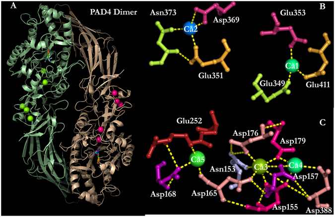
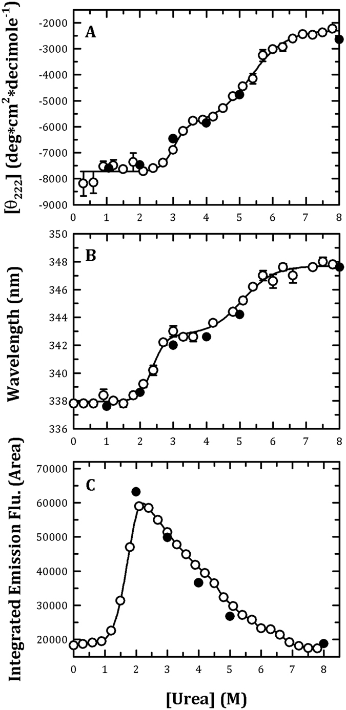
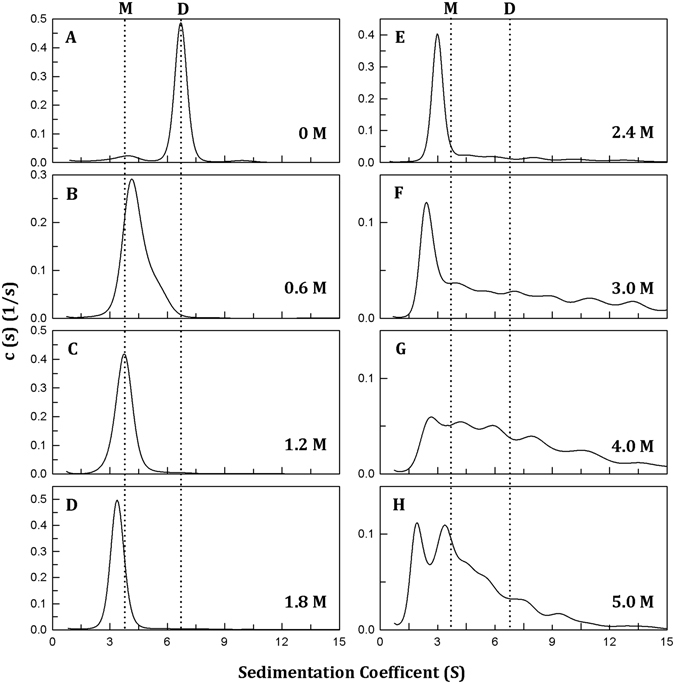
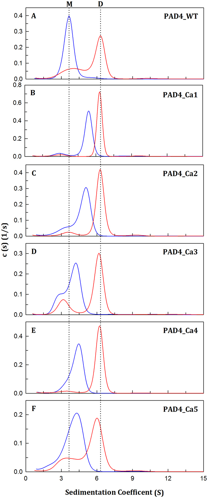
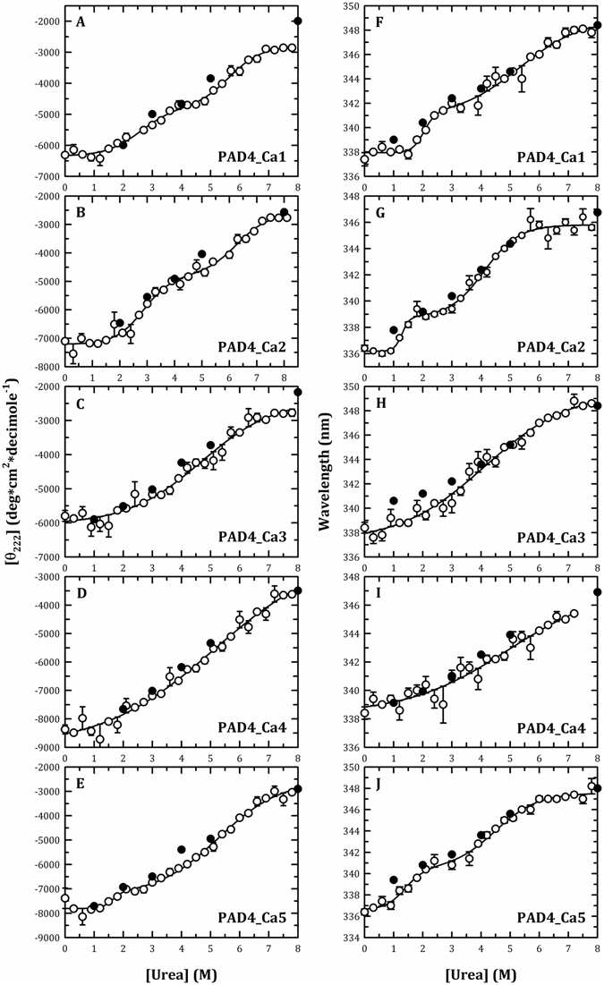
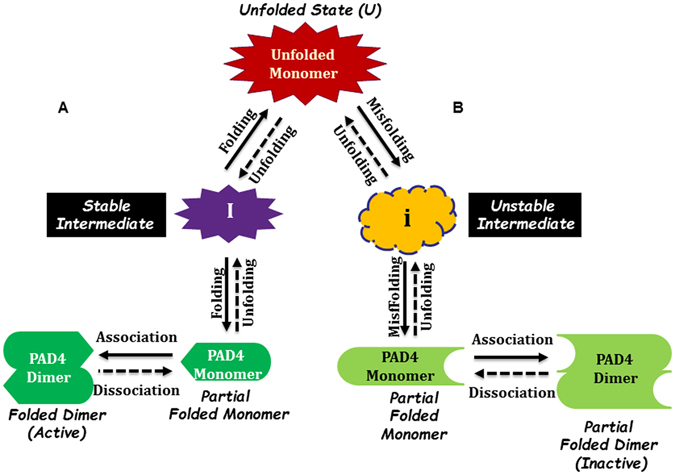
Our recent studies of peptidylarginine deiminase 4 (PAD4) demonstrate that its non-catalytic Ca2+-binding sites play a crucial role in the assembly of the correct geometry of the enzyme. Here, we examined the folding mechanism of PAD4 and the role of Ca2+ ions in the folding pathway. Multiple mutations were introduced into the calcium-binding sites, and these mutants were termed the Ca1_site, Ca2_site, Ca3_site, Ca4_site and Ca5_site mutants. Our data indicate that during the unfolding process, the PAD4 dimer first dissociates into monomers, and the monomers then undergo a three-state denaturation process via an intermediate state formation. In addition, Ca2+ ions assist in stabilizing the folding intermediate, particularly through binding to the Ca3_site and Ca4_site to ensure the correct and active conformation of PAD4. The binding of calcium ions to the Ca1_site and Ca2_site is directly involved in the catalytic action of the enzyme. Finally, this study proposes a model for the folding of PAD4. The nascent polypeptide chains of PAD4 are first folded into monomeric intermediate states, then continue to fold into monomers, and ultimately assemble into a functional and dimeric PAD4 enzyme, and cellular Ca2+ ions may be the critical factor governing the interchange.


Conflict of interest statement

The authors declare that they have no competing interests.

Figures

Figure 1
Figure 1
Five Ca2+-binding sites in the human PAD4 enzyme. Binding ligands for the five Ca2+-binding sites, which include the C-terminal calcium-binding sites, i.e., the Ca1_site and Ca2_site, and the N-terminal calcium-binding sites, i.e., the Ca3_site, Ca4_site and Ca5_site. This figure was generated using PyMOL.
Figure 2
Figure 2
Monitoring of the urea-induced unfolding and refolding of human PAD4 WT enzyme by CD spectrometry and the intrinsic protein fluorescence. The PAD4 WT enzyme in the presence of 10 mM Ca2+ was treated with various concentrations of urea in 50 mM Tris-HCl buffer (pH 7.4) at 25 °C for 16 h and then monitored through CD spectrometry (A), fluorescence (B) or ANS fluorescence (C). Open circles: the PAD4 enzyme was denatured with different concentrations of urea. Closed circles: the PAD4 enzyme was completely denatured with 8 M urea and then renatured by diluting the urea concentration to 5, 4, 3, 2 and 1 M, as indicated in the figures. The experimental data in (A,B) were fitted by either a two-state or three-state model. The fit results and residues are shown as a solid line with error bars.
Figure 3
Figure 3
Continuous sedimentation coefficient distributions of human PAD4 WT enzyme during urea denaturation. The PAD4 WT enzyme (0.3 mg/ml) in the presence of 10 mM Ca2+ was treated with various concentrations of urea in 50 mM Tris-HCl buffer (pH 7.4) at 25 °C for 16 h: (A) 0 M urea, (B) 0.6 M urea, (C) 1.2 M urea, (D) 1.8 M urea, (E) 2.4 M urea, (F) 3.0 M urea, (G) 4.0 M urea and (H) 5.0 M urea.
Figure 4
Figure 4
Dissociation-reassociation of human PAD4 WT and the calcium-binding-site mutant enzymes. The PAD4 WT and the calcium-binding-site mutant enzymes were treated with 1.2 M urea in 50 mM Tris-HCl buffer (pH 7.4) at 25 °C for 16 h, and the protein samples were then diluted 10-fold to reduce the urea concentration to 0.12 M. (A) PAD4_WT. (B) PAD4_Ca1. (C) PAD4_Ca2. (D) PAD4_Ca3. (E) PAD4_Ca4. (F) PAD4_Ca5. Blue line: the enzyme treated with 1.2 M urea. Red line: 10-fold dilution of the 1.2 M urea-treated enzyme (0.12 M urea).
Figure 5
Figure 5
Monitoring of the urea-induced unfolding and refolding of the human PAD4 calcium-binding-site mutant enzymes by CD spectrometry and the intrinsic protein fluorescence. The PAD4 calcium-binding-site mutant enzymes in the presence of 10 mM Ca2+ were treated with various concentrations of urea in 50 mM Tris-HCl buffer (pH 7.4) at 25 °C for 16 h and then monitored through CD spectrometry (Panels (A–E)) or fluorescence (Panels (F–J)). Panels (A and F) PAD4_Ca1. Panels (B and G) PAD4_Ca2. Panels (C and H) PAD4_Ca3. Panels (D and I) PAD4_Ca4. Panels (E and J) PAD4_Ca5. Open circles: the PAD4 enzyme was denatured with different concentrations of urea. Closed circles: the PAD4 enzyme was completely denatured with 8 M urea and then renatured by diluting the urea concentration as indicated in the figures. All the data were fitted by either a two-state or three-state model. The fit results and residues are shown as a solid line with error bars.
Figure 6
Figure 6
Proposed model of the PAD4 folding pathway. (A) Three-state folding pathway with the formation of a stable intermediate and assembly of dimers of the PAD4 WT, PAD4_Ca1, PAD4_Ca2 and PAD4_Ca5 mutant enzymes. (B) Two-state folding pathway with the formation of an unstable intermediate and assembly of dimers of PAD4_Ca3 and PAD4_Ca4 mutant enzymes.

Similar articles

Cited by

References

    1. Lu X, Galkin A, Herzberg O, Dunaway-Mariano D. Arginine deiminase uses an active-site cysteine in nucleophilic catalysis of L-arginine hydrolysis. J. Am. Chem. Soc. 2004;126:5374–5375. doi: 10.1021/ja049543p. - DOI - PubMed
    1. Knuckley B, Bhatia M, Thompson PR. Protein arginine deiminase 4: evidence for a reverse protonation mechanism. Biochemistry. 2007;46:6578–6587. doi: 10.1021/bi700095s. - DOI - PMC - PubMed
    1. Anzilotti C, Pratesi F, Tommasi C, Migliorini P. Peptidylarginine deiminase 4 and citrullination in health and disease. Autoimmun. Rev. 2010;9:158–160. doi: 10.1016/j.autrev.2009.06.002. - DOI - PubMed
    1. Gyorgy B, Toth E, Tarcsa E, Falus A, Buzas EI. Citrullination: a posttranslational modification in health and disease. Int. J. Biochem. Cell Biol. 2006;38:1662–1677. doi: 10.1016/j.biocel.2006.03.008. - DOI - PubMed
    1. Ishigami A, Maruyama N. Importance of research on peptidylarginine deiminase and citrullinated proteins in age-related disease. Geriatr. Gerontol. Int. 2010;10(Suppl 1):S53–58. doi: 10.1111/j.1447-0594.2010.00593.x. - DOI - PubMed

Publication types

MeSH terms