Review
. 2017 May 26;49(5):e338.
doi: 10.1038/emm.2017.20.

Roles of intestinal epithelial cells in the maintenance of gut homeostasis

Affiliations
Review

Roles of intestinal epithelial cells in the maintenance of gut homeostasis

Ryu Okumura et al. Exp Mol Med. .

Abstract

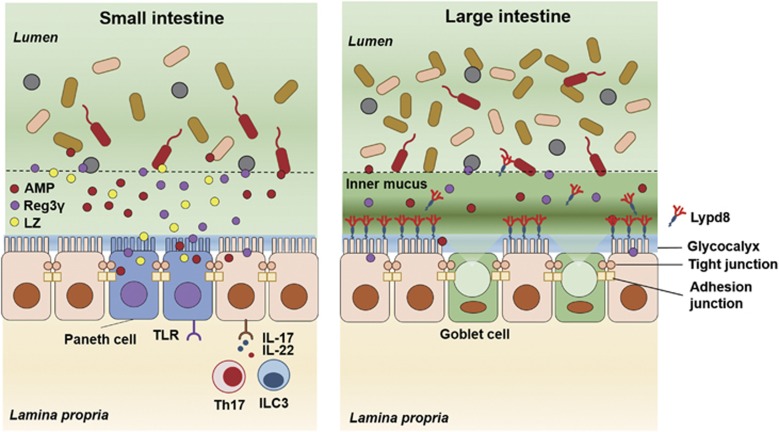
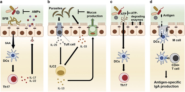
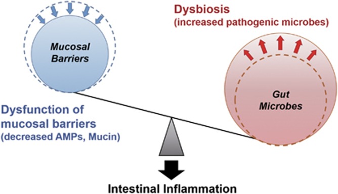
The intestine is a unique organ inhabited by a tremendous number of microorganisms. Intestinal epithelial cells greatly contribute to the maintenance of the symbiotic relationship between gut microbiota and the host by constructing mucosal barriers, secreting various immunological mediators and delivering bacterial antigens. Mucosal barriers, including physical barriers and chemical barriers, spatially segregate gut microbiota and the host immune system to avoid unnecessary immune responses to gut microbes, leading to the intestinal inflammation. In addition, various immunological mediators, including cytokines and chemokines, secreted from intestinal epithelial cells stimulated by gut microbiota modulate host immune responses, maintaining a well-balanced relationship between gut microbes and the host immune system. Therefore, impairment of the innate immune functions of intestinal epithelial cells is associated with intestinal inflammation.


Conflict of interest statement

The authors declare no conflict of interest.

Figures

Figure 1
Figure 1
Mucosal barriers in the gut. In the small intestine, chemical barriers, including AMPs produced by Paneth cells, have major roles in the segregation of intestinal bacteria and intestinal epithelial cells. In contrast, in the large intestine, which is inhabited by a tremendous number of bacteria, intestinal bacteria and intestinal epithelial cells are separated by the inner mucus layer containing polymerized MUC2. Lypd8, a GPI-anchored protein expressed in the epithelial cells, promotes the segregation of the two by binding to intestinal bacteria, especially flagellated bacteria. AMP, antimicrobial peptides; LZ, lysozyme; TLR, toll-like receptor; ILC3, type 3 innate lymphoid cell.
Figure 2
Figure 2
Intestinal epithelial cells mediate the crosstalk between gut microbes and the host immunity. Intestinal epithelial cells stimulated by gut environmental factors modulate gut immune cell responses by secreting humoral factors, such as serum amyloid A (SAA) and several kinds of cytokines (a, b). Intestinal epithelial cells also inactivate bacteria-derived stimulators to host immune cells, such as adenosine triphosphate (ATP), to regulate excessive host immune responses (c). In addition, M cells, which specialize in the uptake and delivery of antigens in the intestinal lumen, contribute to the induction of antigen-specific IgA responses by delivering bacterial or dietary antigens to dendritic cells (DCs) (d).
Figure 3
Figure 3
The imbalance between mucosal barriers and gut microbes promotes susceptibility to intestinal inflammation. Dysfunction of mucosal barriers because of genetic predisposition, such as the decreased production of AMPs and mucin, allows intestinal bacteria to gain access to gut immune cells, thereby contributing to the development of intestinal inflammation. Dysbiosis induced by environmental factors, such as a high-fat diet and various medicines, accelerates intestinal inflammation in situations in which the mucosal barrier is disrupted. AMPs, antimicrobial peptides.

Similar articles

Cited by

References

    1. Furusawa Y, Obata Y, Fukuda S, Endo TA, Nakato G, Takahashi D et al. Commensal microbe-derived butyrate induces the differentiation of colonic regulatory T cells. Nature 2013; 504: 446–450. - PubMed
    1. Atarashi K, Nishimura J, Shima T, Umesaki Y, Yamamoto M, Onoue M et al. ATP drives lamina propria T(H)17 cell differentiation. Nature 2008; 455: 808–812. - PubMed
    1. Kim YS, Ho SB. Intestinal goblet cells and mucins in health and disease: recent insights and progress. Curr Gastroenterol Rep 2010; 12: 319–330. - PMC - PubMed
    1. Moro K, Yamada T, Tanabe M, Takeuchi T, Ikawa T, Kawamoto H et al. Innate production of T(H)2 cytokines by adipose tissue-associated c-Kit(+)Sca-1(+) lymphoid cells. Nature 2010; 463: 540–544. - PubMed
    1. Price AE, Liang HE, Sullivan BM, Reinhardt RL, Eisley CJ, Erle DJ et al. Systemically dispersed innate IL-13-expressing cells in type 2 immunity. Proc Natl Acad Sci USA 2010; 107: 11489–11494. - PMC - PubMed