

Retinal Vascular Occlusion Secondary to Retrobulbar Injection: Case Report and Literature Review
- PMID: 28546695
- PMCID: PMC5433131
- DOI: 10.4103/meajo.MEAJO_37_16
Retinal Vascular Occlusion Secondary to Retrobulbar Injection: Case Report and Literature Review
Abstract
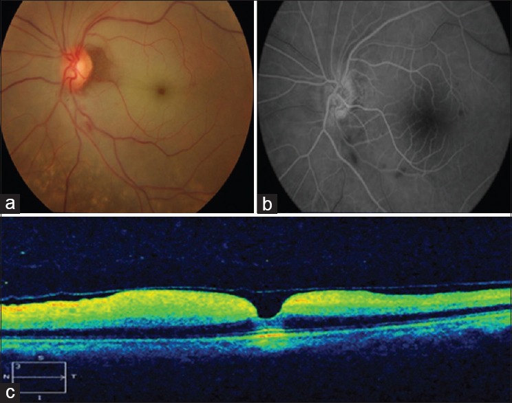
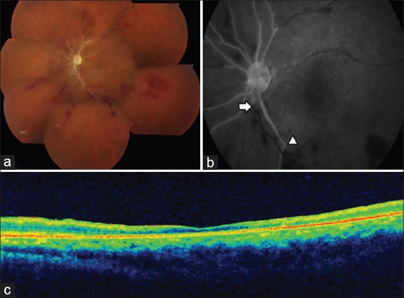
Retrobulbar injection has been widely practiced as a technique of ocular anesthesia for many decades. Nevertheless, the technique is not free from complications. Vascular occlusion secondary to retrobulbar injection is rare but can be vision threatening. We report a case series of two such patients who presented with poor vision following retrobulbar injection. Fundus showed pale retina with cherry red spot suggestive of central retinal artery occlusion in case 1 and pale disc with sclerosed vessels and multiple superficial hemorrhages suggestive of a combined occlusion of retinal artery and vein in case 2. Optical coherence tomography (OCT) showed thickened inner retinal layers with intact outer retinal layers in case 1 and thinning in case 2. We conclude that retrobulbar injections can rarely be associated with dreadful vision-threatening complications like in our patients. We also report the role of OCT in assessing the prognosis following vascular occlusion.
Keywords: Combined occlusion; retrobulbar injection; vascular occlusion.
Conflict of interest statement
There are no conflicts of interest.
Figures
References
-
- Park SJ, Choi NK, Seo KH, Park KH, Woo SJ. Nationwide incidence of clinically diagnosed central retinal artery occlusion in Korea, 2008 to 2011. Ophthalmology. 2014;121:1933–8. - PubMed
-
- Katsev DA, Drews RC, Rose BT. An anatomic study of retrobulbar needle path length. Ophthalmology. 1989;96:1221–4. - PubMed
-
- Morgan CM, Schatz H, Vine AK, Cantrill HL, Davidorf FH, Gitter KA, et al. Ocular complications associated with retrobulbar injections. Ophthalmology. 1988;95:660–5. - PubMed
-
- Torres RJ, Luchini A, Weis W, Frecceiro PR, Casella M. Combined central retinal vein and artery occlusion after retrobulbar anesthesia – report of two cases. Arq Bras Oftalmol. 2005;68:257–61. - PubMed
-
- Mameletzi E, Pournaras JA, Ambresin A, Nguyen C. Retinal embolisation with localised retinal detachment following retrobulbar anaesthesia. Klin Monbl Augenheilkd. 2008;225:476–8. - PubMed
Publication types
MeSH terms
LinkOut - more resources
Full Text Sources
