

Kinematic and Electromyographic Activity Changes during Back Squat with Submaximal and Maximal Loading
- PMID: 28546738
- PMCID: PMC5435978
- DOI: 10.1155/2017/9084725
Kinematic and Electromyographic Activity Changes during Back Squat with Submaximal and Maximal Loading
Abstract
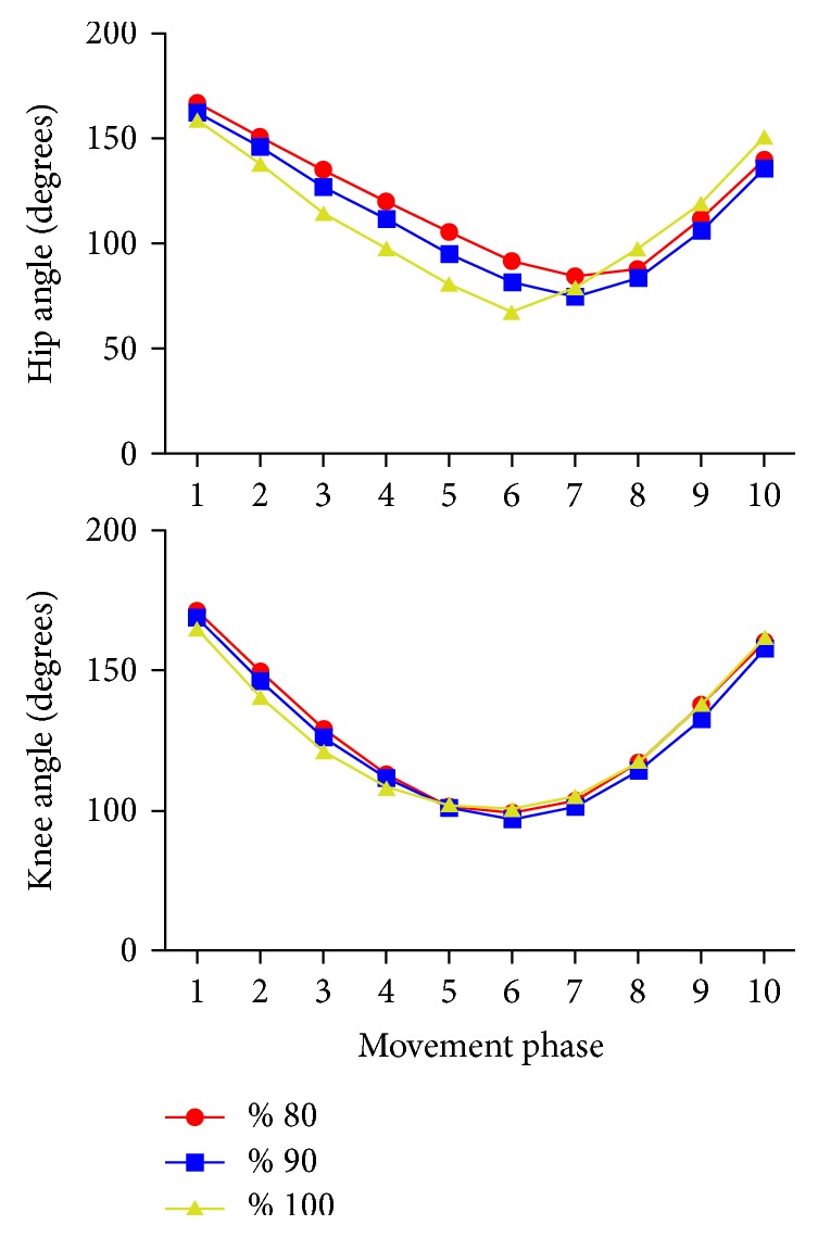
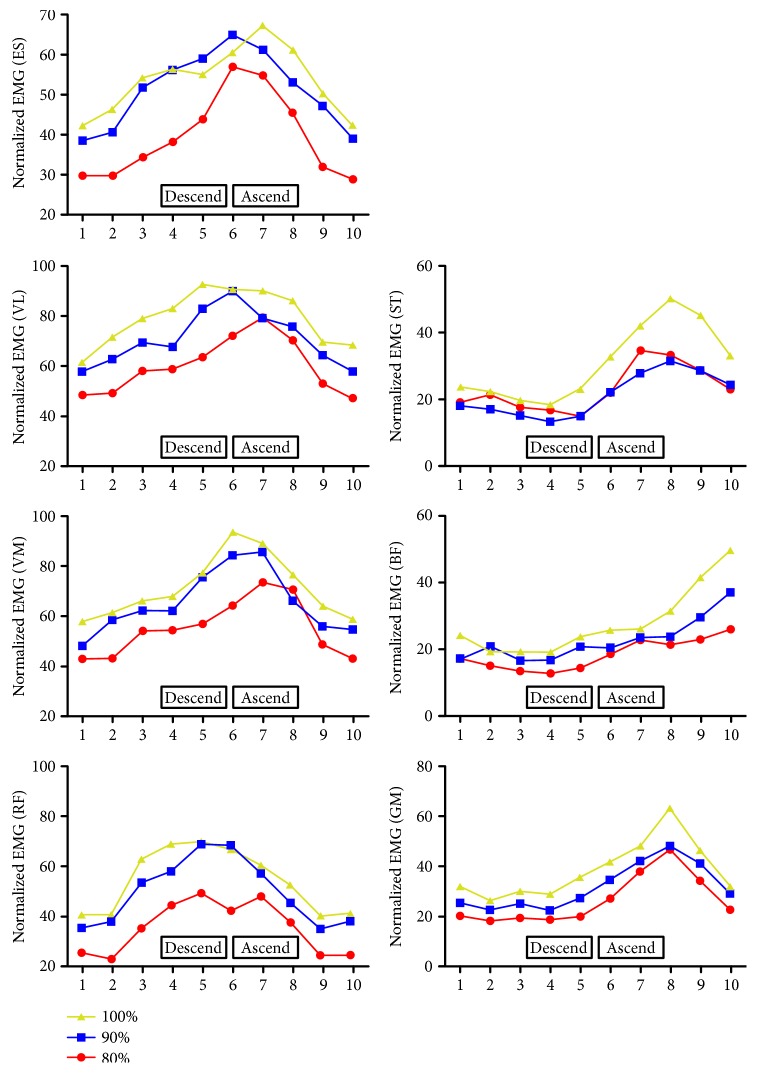
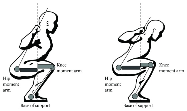
The aim of this study was to investigate the possible kinematic and muscular activity changes with maximal loading during squat maneuver. Fourteen healthy male individuals, who were experienced at performing squats, participated in this study. Each subject performed squats with 80%, 90%, and 100% of the previously established 1 repetition maximum (1RM). Electromyographic (EMG) activities were measured for the vastus lateralis, vastus medialis, rectus femoris, semitendinosus, biceps femoris, gluteus maximus, and erector spinae by using an 8-channel dual-mode portable EMG and physiological signal data acquisition system (Myomonitor IV, Delsys Inc., Boston, MA, USA). Kinematical data were analyzed by using saSuite 2D kinematical analysis program. Data were analyzed with repeated measures analysis of variance (p < 0.05). Overall muscle activities increased with increasing loads, but significant increases were seen only for vastus medialis and gluteus maximus during 90% and 100% of 1RM compared to 80% while there was no significant difference between 90% and 100% for any muscle. The movement pattern in the hip joint changed with an increase in forward lean during maximal loading. Results may suggest that maximal loading during squat may not be necessary for focusing on knee extensor improvement and may increase the lumbar injury risk.
Figures
Similar articles
-
Comparison of muscle activation and kinematics during free-weight back squats with different loads.PLoS One. 2019 May 16;14(5):e0217044. doi: 10.1371/journal.pone.0217044. eCollection 2019. PLoS One. 2019. PMID: 31095625 Free PMC article.
-
Electromyographic activity in the gluteus medius, gluteus maximus, biceps femoris, vastus lateralis, vastus medialis and rectus femoris during the Monopodal Squat, Forward Lunge and Lateral Step-Up exercises.PLoS One. 2020 Apr 1;15(4):e0230841. doi: 10.1371/journal.pone.0230841. eCollection 2020. PLoS One. 2020. PMID: 32236133 Free PMC article.
-
Kinematic and EMG activities during front and back squat variations in maximum loads.J Sports Sci. 2015;33(10):1058-66. doi: 10.1080/02640414.2014.984240. Epub 2015 Jan 29. J Sports Sci. 2015. PMID: 25630691
-
The effect of stance width on the electromyographical activity of eight superficial thigh muscles during back squat with different bar loads.J Strength Cond Res. 2009 Jan;23(1):246-50. doi: 10.1519/jsc.0b013e3181876811. J Strength Cond Res. 2009. PMID: 19130646
-
A Comparison of Gluteus Maximus, Biceps Femoris, and Vastus Lateralis Electromyographic Activity in the Back Squat and Barbell Hip Thrust Exercises.J Appl Biomech. 2015 Dec;31(6):452-8. doi: 10.1123/jab.2014-0301. Epub 2015 Jul 24. J Appl Biomech. 2015. PMID: 26214739
Cited by
-
Comparison of muscle activation and kinematics during free-weight back squats with different loads.PLoS One. 2019 May 16;14(5):e0217044. doi: 10.1371/journal.pone.0217044. eCollection 2019. PLoS One. 2019. PMID: 31095625 Free PMC article.
-
Electromyographic activity in the gluteus medius, gluteus maximus, biceps femoris, vastus lateralis, vastus medialis and rectus femoris during the Monopodal Squat, Forward Lunge and Lateral Step-Up exercises.PLoS One. 2020 Apr 1;15(4):e0230841. doi: 10.1371/journal.pone.0230841. eCollection 2020. PLoS One. 2020. PMID: 32236133 Free PMC article.
-
The effects of fatigue on linear and angular kinematics during bilateral squat.PLoS One. 2023 Nov 27;18(11):e0289089. doi: 10.1371/journal.pone.0289089. eCollection 2023. PLoS One. 2023. PMID: 38011209 Free PMC article.
-
Sex differences in intra-set kinematics and electromyography during different maximum repetition sets in the barbell back squat?PLoS One. 2024 Aug 7;19(8):e0308344. doi: 10.1371/journal.pone.0308344. eCollection 2024. PLoS One. 2024. PMID: 39110682 Free PMC article.
-
Relationship between agility and lower limb muscle strength, targeting university badminton players.J Phys Ther Sci. 2018 Feb;30(2):320-323. doi: 10.1589/jpts.30.320. Epub 2018 Feb 28. J Phys Ther Sci. 2018. PMID: 29545704 Free PMC article.
References
-
- Contreras B., Vigotsky A. D., Schoenfeld B. J., Beardsley C., Cronin J. A comparison of gluteus maximus, biceps femoris, and vastus lateralis electromyography amplitude in the parallel, full, and front squat variations in resistance-trained females. Journal of Applied Biomechanics. 2016;32(1):16–22. doi: 10.1123/jab.2015-0113. - DOI - PubMed
-
- Escamilla R. F. Knee biomechanics of the dynamic squat exercise. Medicine and Science in Sports and Exercise. 2001;33(1):127–141. - PubMed
LinkOut - more resources
Full Text Sources
Other Literature Sources
