

Homocysteine mediates transcriptional changes of the inflammatory pathway signature genes in human retinal pigment epithelial cells
- PMID: 28546923
- PMCID: PMC5437454
- DOI: 10.18240/ijo.2017.05.06
Homocysteine mediates transcriptional changes of the inflammatory pathway signature genes in human retinal pigment epithelial cells
Abstract
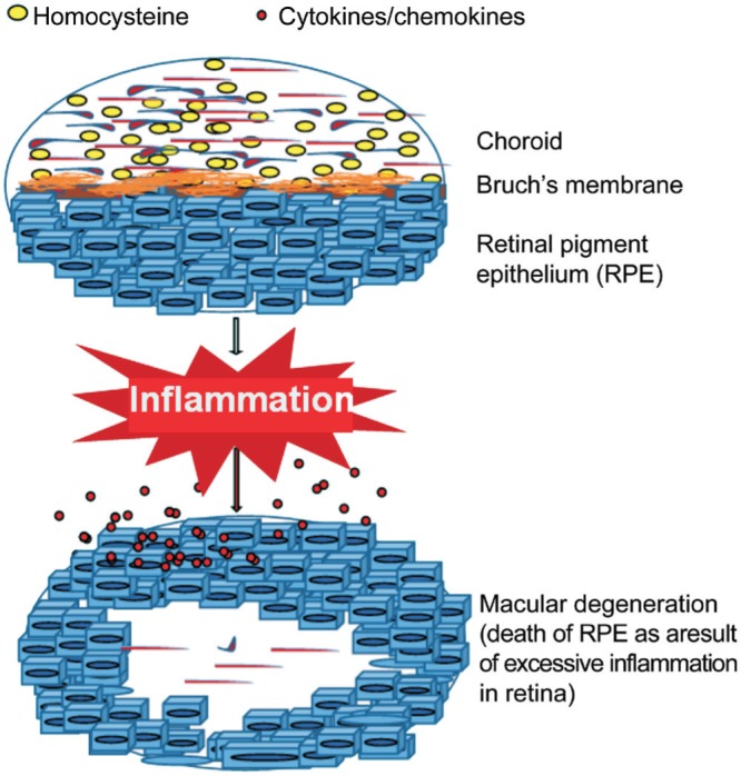
Aim: To test whether homocysteine (Hcy) can influence the transcriptional profile, we hypothesized that Hcy can lead to the induction of proinflammatory molecules in the retinal cells of aging people.
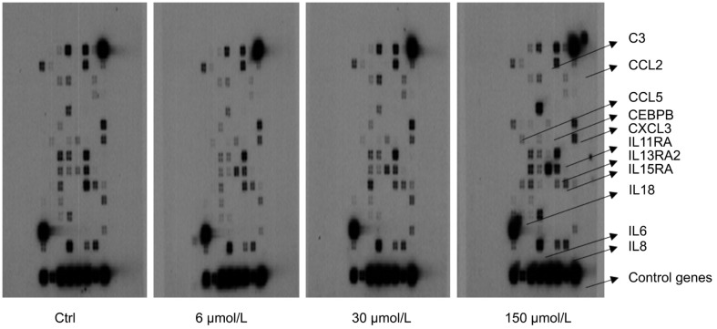
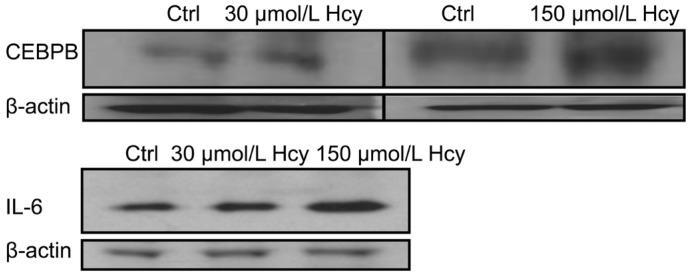
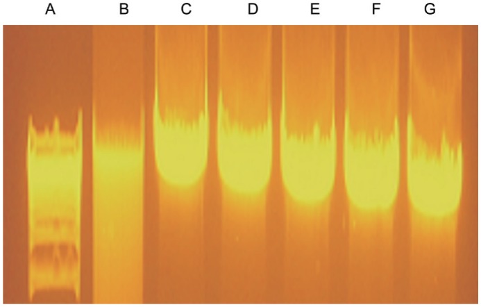
Methods: An unbiased in vitro inflammatory pathway focused study was designed employing retinal pigment epithelial (RPE) cell line, ARPE-19. Cells were cultured in the presence or absence of Hcy to capture target genes' expression profile. Three different concentrations of Hcy were added in the culture medium of confluent monolayers. cRNAs were made from the isolated total RNAs and the labeled cRNA probes were hybridized to microarrays specific for human disease pathway inflammatory cytokines, chemokines and their receptor gene micro-array panels as per manufacture's recommendations. Two Hcy up-regulated molecules: IL6 and CEBPB were further validated via Western blot analysis. Hcy's effect on ARPE-19 cellular morphology and genomic DNA integrity were also evaluated.
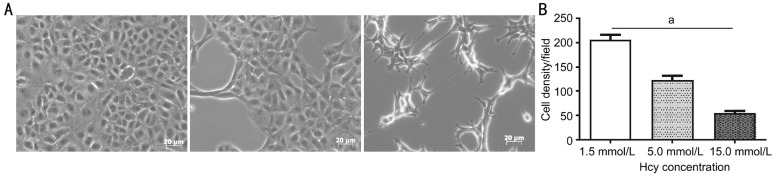
Results: Gene microarray analyses of RPE cells in response to Hcy treatment revealed alterations in the expressions of several inflammatory gene transcripts such as CCL5, CEBPB, IL13RA2, IL15RA, IL6, IL8 and CXCL3 that were up-regulated. The transcripts for C3, CCL2, IL11RA and IL18 genes exhibited down-regulation. The IL6 and CEBPB expressions were subsequently validated at the protein levels. Treatment of the retinal cells with increasing Hcy concentration influenced their density in culture however their morphology and DNA integrity remained unaffected.
Conclusion: These findings suggest that Hcy can potentially mediate the expression of chemokines, cytokines and interleukins receptors in the retinal cells without having any debilitating effects on their morphology and the genomic DNA integrity.
Keywords: chemokine; choroidal neovascularization; cytokine; gene expression; hyperhomocysteinemia; inflammation; macular degeneration; retinal remodeling.
Figures
References
-
- Viktorov IV, Aleksandrova OP, Alekseeva NY. Homocysteine toxicity in organotypic cultures of rat retina. Bull Exp Biol Med. 2006;141(4):471–474. - PubMed
-
- Flott-Rahmel B, Schurmann M, Schluff P, Fingerhut R, Musshoff U, Fowler B, Ullrich K. Homocysteic and homocysteine sulphinic acid exhibit excitotoxicity in organotypic cultures from rat brain. Eur J Pediatr. 1998;157:112–117. Suppl 2. - PubMed
-
- Bharathselvi M, Biswas J, Selvi R, Coral K, Narayanasamy A, Ramakrishnan S, Sulochana KN. Increased homocysteine, homocysteine-thiolactone, protein homocysteinylation and oxidative stress in the circulation of patients with Eales' disease. Ann Clin Biochem. 2013;50(4):330–338. - PubMed
Grants and funding
LinkOut - more resources
Full Text Sources
Other Literature Sources
Miscellaneous