

New quantitative radiographic parameters for vertical and horizontal instability in acromioclavicular joint dislocations
- PMID: 28547587
- PMCID: PMC5754414
- DOI: 10.1007/s00167-017-4579-6
New quantitative radiographic parameters for vertical and horizontal instability in acromioclavicular joint dislocations
Abstract
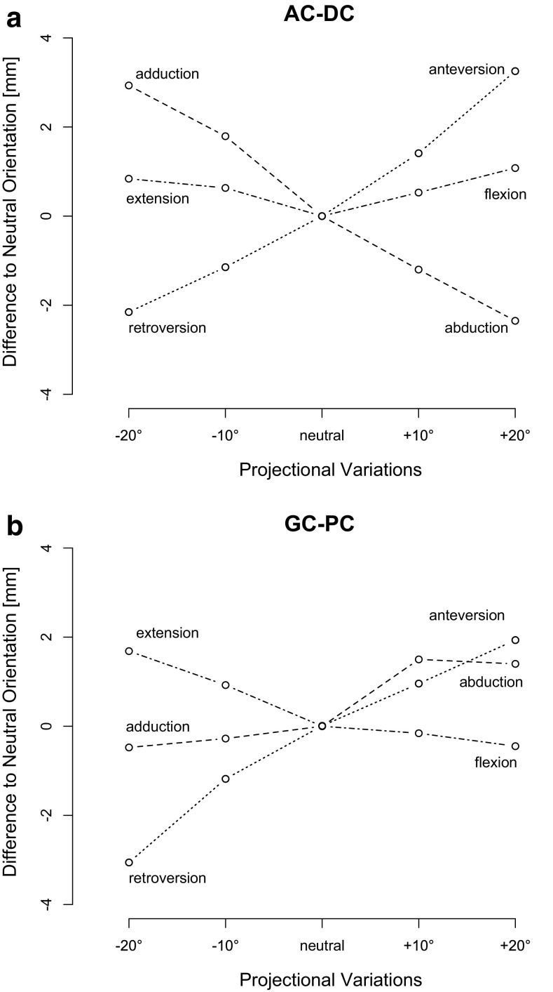
Purpose: The aim of this study was to identify the most accurate and reliable quantitative radiographic parameters for assessing vertical and horizontal instability in different Rockwood grades of acromioclavicular joint (ACJ) separations. Furthermore, the effect of projectional variation on these parameters was investigated in obtaining lateral Alexander view radiographs.
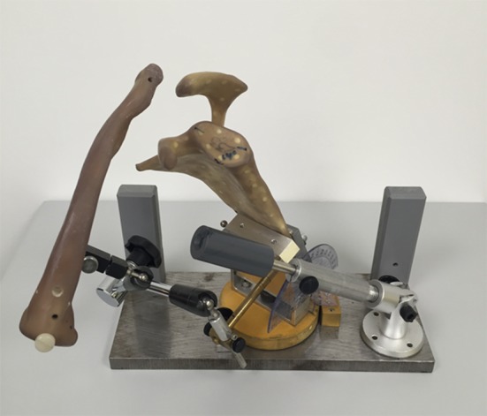
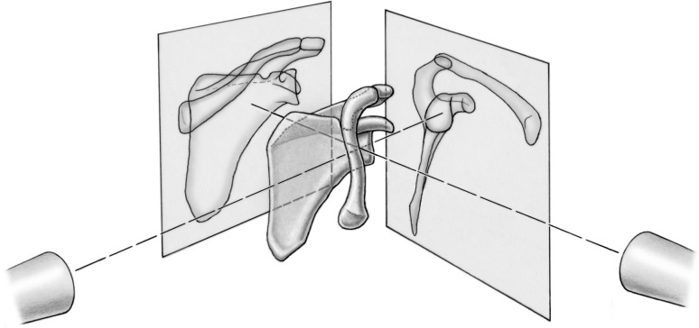
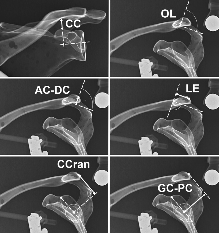
Methods: A Sawbone model of a scapula with clavicle was mounted on a holding device, and acromioclavicular dislocations as per the Rockwood classification system were simulated with the addition of horizontal posterior displacement. Projectional variations for each injury type were performed by tilting/rotating the Sawbone construct in the coronal, sagittal or axial plane. Radiographic imaging in the form of an anterior-posterior Zanca view and a lateral Alexander view were taken for each injury type and each projectional variation. Five newly defined radiographic parameters for assessing horizontal and vertical displacement as well as commonly used coracoclavicular distance view were measured. Reliability, validity and the effect of projectional variation were investigated for these radiographic measurements.
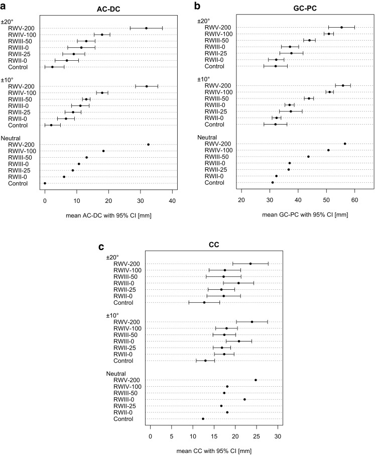
Results: All radiographic parameters showed excellent intra- and interobserver reliability. The validity was excellent for the acromial centre line to dorsal clavicle (AC-DC) in vertical displacement and for the glenoid centre line to posterior clavicle (GC-PC) in horizontal displacement, whilst the remaining measurements showed moderate validity. For AC-DC and GC-PC, convergent validity expressed strong correlation to the effective distance and discriminant validity demonstrated its ability to differentiate between various grades of ACJ dislocations. The effect of projectional variation increased with the degree of deviation and was maximal (3 mm) for AC-DC in 20° anteverted malpositioning and for GC-PC in 20° retroverted malpositioning.
Conclusions: AC-DC and the GC-PC are two novel quantitative radiographic parameters of vertical and horizontal instability in ACJ dislocations that demonstrate excellent reliability and validity with reasonable inertness to malpositioning. The use of AC-DC for assessing vertical displacement and GC-PC for assessing horizontal displacement in a single Alexander view is recommended to guide the appropriate management of ACJ dislocations. A better appreciation of the degree of horizontal instability, especially in lower Rockwood grades (II, III) of ACJ dislocations, may improve management of these controversial injuries.
Keywords: AC joint; AC joint separation; AC–DC; Acromioclavicular joint; Dislocation; GC–PC; Horizontal instability; Instability; Intra- and interobserver reliability; Radiographic parameters; Rockwood classification; Validity; Vertical instability.
Conflict of interest statement
Conflict of interest
All authors declare that they have no conflict of interest.
Funding
No grant was received for this study.
Ethical approval
This article does not contain any studies with human participants or animals performed by any of the authors.
Informed consent
For this type of study formal consent is not required.
Figures
Similar articles
-
Improved identification of unstable acromioclavicular joint injuries in a clinical population using the acromial center line to dorsal clavicle radiographic measurement.J Shoulder Elbow Surg. 2020 Aug;29(8):1599-1605. doi: 10.1016/j.jse.2019.12.014. Epub 2020 Mar 5. J Shoulder Elbow Surg. 2020. PMID: 32147334
-
Evaluation of the Circles Measurement and the ABC Classification of Acromioclavicular Joint Injuries.Am J Sports Med. 2021 May;49(6):1619-1625. doi: 10.1177/03635465211003300. Epub 2021 Apr 15. Am J Sports Med. 2021. PMID: 33856933
-
Is coracoclavicular stabilisation alone sufficient for the endoscopic treatment of severe acromioclavicular joint dislocation (Rockwood types III, IV, and V)?Orthop Traumatol Surg Res. 2015 Dec;101(8 Suppl):S297-303. doi: 10.1016/j.otsr.2015.09.003. Epub 2015 Oct 27. Orthop Traumatol Surg Res. 2015. PMID: 26514849
-
Management of acute acromioclavicular joint dislocations: current concepts.Arch Orthop Trauma Surg. 2013 Jul;133(7):985-95. doi: 10.1007/s00402-013-1748-z. Epub 2013 Apr 30. Arch Orthop Trauma Surg. 2013. PMID: 23632779 Review.
-
MR imaging appearances of acromioclavicular joint dislocation.Radiographics. 2008 Mar-Apr;28(2):463-79; quiz 619. doi: 10.1148/rg.282075714. Radiographics. 2008. PMID: 18349451 Review.
Cited by
-
[Current aspects and new techniques in dislocation of the shoulder joint].Orthopade. 2018 Feb;47(2):158-167. doi: 10.1007/s00132-017-3517-0. Orthopade. 2018. PMID: 29335760 Review. German.
-
Anatomic coracoclavicular ligament reconstruction with triple flip-buttons leads to good functional outcomes and low reduction loss: a case series.Clin Shoulder Elb. 2023 Jun;26(2):140-147. doi: 10.5397/cise.2022.01298. Epub 2023 May 3. Clin Shoulder Elb. 2023. PMID: 37150589 Free PMC article.
-
Better Radiographic Reduction and Lower Complication Rates With Combined Coracoclavicular and Acromioclavicular Ligament Reconstruction Than With Isolated Coracoclavicular Reconstruction.Arthrosc Sports Med Rehabil. 2021 Feb 24;3(2):e441-e448. doi: 10.1016/j.asmr.2020.10.009. eCollection 2021 Apr. Arthrosc Sports Med Rehabil. 2021. PMID: 34027453 Free PMC article.
-
Current concepts in acromioclavicular joint (AC) instability - a proposed treatment algorithm for acute and chronic AC-joint surgery.BMC Musculoskelet Disord. 2022 Dec 9;23(1):1078. doi: 10.1186/s12891-022-05935-0. BMC Musculoskelet Disord. 2022. PMID: 36494652 Free PMC article. Review.
-
BiPOD arthroscopically assisted bidirectional stabilisation technique for high-grade acromioclavicular joint injury: two-year clinical and radiological outcomes.Arch Orthop Trauma Surg. 2021 Sep;141(9):1559-1565. doi: 10.1007/s00402-021-03768-5. Epub 2021 Feb 8. Arch Orthop Trauma Surg. 2021. PMID: 33555404 Free PMC article.
References
-
- Rockwood CA., Jr . Injuries to the acromioclavicular joint. In: Rockwood CA Jr, Green DP, editors. Fractures in adults, Vol. 1. 2. Philadelphia: JB Lippincott; 1984. pp. 860–910.
-
- Hedtmann A, Fett H, Ludwig J. Management of old neglected posttraumatic acromioclavicular joint instability and arthrosis. Orthopade. 1998;27(8):556–566. - PubMed
MeSH terms
LinkOut - more resources
Full Text Sources
Other Literature Sources
Medical
Miscellaneous
