. 2017 Aug;134(1):177-188.
doi: 10.1007/s11060-017-2506-9. Epub 2017 May 25.

Perfusion and diffusion MRI signatures in histologic and genetic subtypes of WHO grade II-III diffuse gliomas

Affiliations

Perfusion and diffusion MRI signatures in histologic and genetic subtypes of WHO grade II-III diffuse gliomas

Kevin Leu et al. J Neurooncol. 2017 Aug.

Abstract

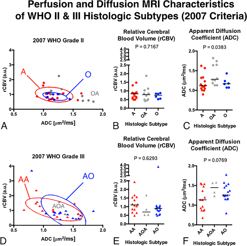
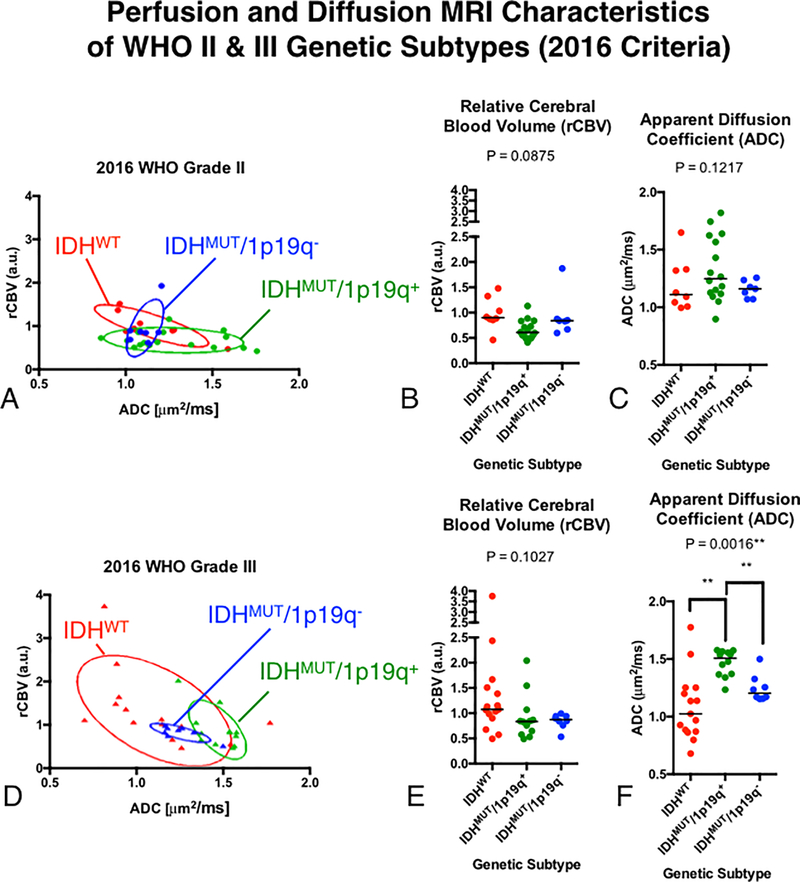
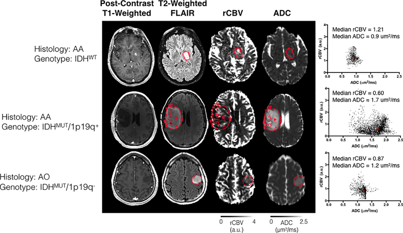
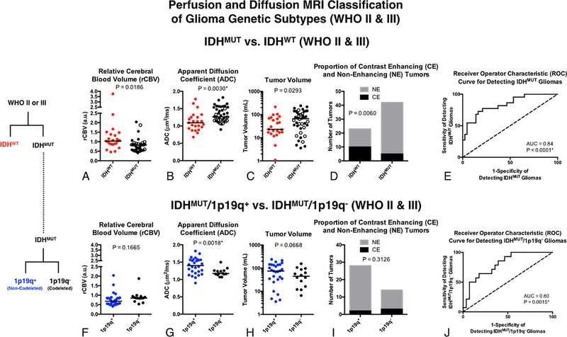
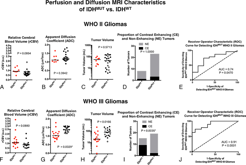
The value of perfusion and diffusion-weighted MRI in differentiating histological subtypes according to the 2007 WHO glioma classification scheme (i.e. astrocytoma vs. oligodendroglioma) and genetic subtypes according to the 2016 WHO reclassification (e.g. 1p/19q co-deletion and IDH1 mutation status) in WHO grade II and III diffuse gliomas remains controversial. In the current study, we describe unique perfusion and diffusion MR signatures between histological and genetic glioma subtypes. Sixty-five patients with 2007 histological designations (astrocytomas and oligodendrogliomas), 1p/19q status (+ = intact/- = co-deleted), and IDH1 mutation status (MUT/WT) were included in this study. In all patients, median relative cerebral blood volume (rCBV) and apparent diffusion coefficient (ADC) were estimated within T2 hyperintense lesions. Bootstrap hypothesis testing was used to compare subpopulations of gliomas, separated by WHO grade and 2007 or 2016 glioma classification schemes. A multivariable logistic regression model was also used to differentiate between 1p19q+ and 1p19q- WHO II-III gliomas. Neither rCBV nor ADC differed significantly between histological subtypes of pure astrocytomas and pure oligodendrogliomas. ADC was significantly different between molecular subtypes (p = 0.0016), particularly between IDHWT and IDHMUT/1p19q+ (p = 0.0013). IDHMUT/1p19q+ grade III gliomas had higher median ADC; IDHWT grade III gliomas had higher rCBV with lower ADC; and IDHMUT/1p19q- had intermediate rCBV and ADC values, similar to their grade II counterparts. A multivariable logistic regression model was able to differentiate between IDHWT and IDHMUT WHO II and III gliomas with an AUC of 0.84 (p < 0.0001, 74% sensitivity, 79% specificity). Within IDHMUT WHO II-III gliomas, a separate multivariable logistic regression model was able to differentiate between 1p19q+ and 1p19q- WHO II-III gliomas with an AUC of 0.80 (p = 0.0015, 64% sensitivity, 82% specificity). ADC better differentiated between genetic subtypes of gliomas according to the 2016 WHO guidelines compared to the classification scheme outlined in the 2007 WHO guidelines based on histological features of the tissue. Results suggest a combination of rCBV, ADC, T2 hyperintense volume, and presence of contrast enhancement together may aid in non-invasively identifying genetic subtypes of diffuse gliomas.

Keywords: Diffusion MRI; Glioma; IDH mutant; Perfusion MRI; WHO classification.


Figures

Fig 1.
Fig 1.. Diagram illustrating the 2007 and 2016 WHO classification criteria for gliomas.
(Top) Categories of grade II and III gliomas under the 2007 WHO criteria based on histological features using light microscopy and hematoxylin and eosin staining. (Bottom) Categories of grade II and III gliomas under the 2016 WHO criteria based on molecular genotype using IDH1 mutation status and 1p/19q co-deletion.
Fig 2.
Fig 2.. Comparisons of rCBV and ADC between histologic subtypes of WHO grade II and III gliomas using the 2007 WHO classification.
A) Scatter plot of median rCBV and ADC for populations of grade II glioma subtypes with elliptical error bars. B) Comparison of rCBV between grade II gliomas. C) Comparison of ADC between grade II gliomas. D) Scatter plot of median rCBV and ADC for populations of grade III glioma subtypes with elliptical error bars. E) Comparison of rCBV between grade III gliomas. F) Comparison of ADC between grade III gliomas.
Fig 3.
Fig 3.. Comparisons of rCBV and ADC between genetic subtypes of WHO grade II and III gliomas using the 2016 WHO classification.
A) Scatter plot of median rCBV and ADC for populations of grade II glioma subtypes with elliptical error bars. B) Comparison of rCBV between grade II gliomas. C) Comparison of ADC between grade II gliomas. D) Scatter plot of median rCBV and ADC for populations of grade III glioma subtypes with elliptical error bars. E) Comparison of rCBV between grade III gliomas. F) Comparison of ADC between grade III gliomas.
Fig 4.
Fig 4.. Imaging features extracted from three representative patients with different histologic and genetic subtypes.
Post-contrast T1-weighted anatomical, T2-weighted FLAIR anatomical, rCBV, and ADC maps are shown. The scatter plot shown on the right illustrates voxel-wise plots of rCBV versus ADC within the red region of interest outlining the T2 hyperintense lesion. Top Row: A patient with an AA subsequently characterized as IDHWT exhibited relatively high rCBV and low ADC. Middle Row: An AA patient subsequently characterized as IDHMUT/1p19q+ exhibiting a low rCBV and high ADC. Bottom Row: An AO oligodendroglioma characterized as IDHMUT/1p19q- exhibiting intermediary rCBV and ADC characteristics. (AA = Anaplastic Astrocytoma; AO = Anaplastic Oligodendroglioma).
Fig. 5.
Fig. 5.. Classification of glioma genetic subtypes in combined WHO II-III tumors using rCBV, ADC, T2 hyperintense lesion volume, and presence of contrast enhancement.
A) Comparison of rCBV between IDHWT and IDHMUT WHO II-III gliomas. B) Comparison of ADC between IDHWT and IDHMUT WHO II-III gliomas. C) Comparison of T2 hyperintense tumor volume between IDHWT and IDHMUT WHO II-III gliomas. D) Proportion of contrast enhancing (CE) and non-enhancing (NE) tumors between IDHWT and IDHMUT WHO II-III gliomas. E) ROC curve using all four biomarkers (rCBV, ADC, Volume, CE/NE) to differentiate IDHWT and IDHMUT WHO II-III gliomas. F) Comparison of rCBV between IDHMUT/1p19q+ and IDHMUT/1p19q- WHO II-III gliomas. G) Comparison of ADC between IDHMUT/1p19q+ and IDHMUT/1p19q- WHO II-III gliomas. H) Comparison of T2 hyperintense tumor volume between IDHMUT/1p19q+ and IDHMUT/1p19q- WHO II-III gliomas. I) Proportion of contrast enhancing (CE) and non-enhancing (NE) tumors between IDHMUT/1p19q+ and IDHMUT/1p19q- WHO II-III gliomas. J) ROC curve using all four biomarkers (rCBV, ADC, Volume, CE/NE) to differentiate IDHMUT/1p19q+ and IDHMUT/1p19q- WHO II-III gliomas.
Fig. 6.
Fig. 6.. Classification of IDH status in WHO II and III gliomas using rCBV, ADC, T2 hyperintense lesion volume, and presence of contrast enhancement.
A) Comparison of ADC between IDHWT and IDHMUT grade II gliomas. B) Comparison of rCBV between grade II gliomas. C) Comparison of tumor volume between grade II gliomas. D) Presence and absence of contrast enhancement among grade II gliomas. E) ROC curve analysis using all four biomarkers in grade II tumors. F) Comparison of ADC between IDHWT and IDHMUT grade III gliomas. B) Comparison of rCBV between grade III gliomas. C) Comparison of tumor volume between grade III gliomas. D) Presence and absence of contrast enhancement among grade III gliomas. E) ROC curve analysis using all four biomarkers in grade III tumors. The open circles within the IDHMUT groups indicate 1p/19q co-deletion.

Similar articles

Cited by

References

    1. Louis DN, Ohgaki H, Wiestler OD, Cavenee WK, Burger PC, Jouvet A, Scheithauer BW, Kleihues P: The 2007 WHO classification of tumours of the central nervous system. Acta Neuropathol 114: 97–109, 2007 - PMC - PubMed
    1. Ostrom QT, Gittleman H, Fulop J, Liu M, Blanda R, Kromer C, Wolinsky Y, Kruchko C, Barnholtz-Sloan JS: CBTRUS Statistical Report: Primary Brain and Central Nervous System Tumors Diagnosed in the United States in 2008–2012. Neuro Oncol 17 Suppl 4: iv1–iv62, 2015 - PMC - PubMed
    1. Sathornsumetee S, Rich JN, Reardon DA: Diagnosis and treatment of high-grade astrocytoma. Neurol Clin 25: 1111–1139, x, 2007 - PubMed
    1. Wen PY, Kesari S: Malignant gliomas in adults. N Engl J Med 359: 492–507, 2008 - PubMed
    1. Sharma S, Deb P: Intraoperative neurocytology of primary central nervous system neoplasia: A simplified and practical diagnostic approach. J Cytol 28: 147–158, 2011 - PMC - PubMed

MeSH terms

Substances

Supplementary concepts