. 2017 May;98(5):1080-1088.
doi: 10.1099/jgv.0.000763. Epub 2017 May 26.

Marek's disease virus infection of phagocytes: a de novo in vitro infection model

Affiliations

Marek's disease virus infection of phagocytes: a de novo in vitro infection model

Pankaj Chakraborty et al. J Gen Virol. 2017 May.

Abstract

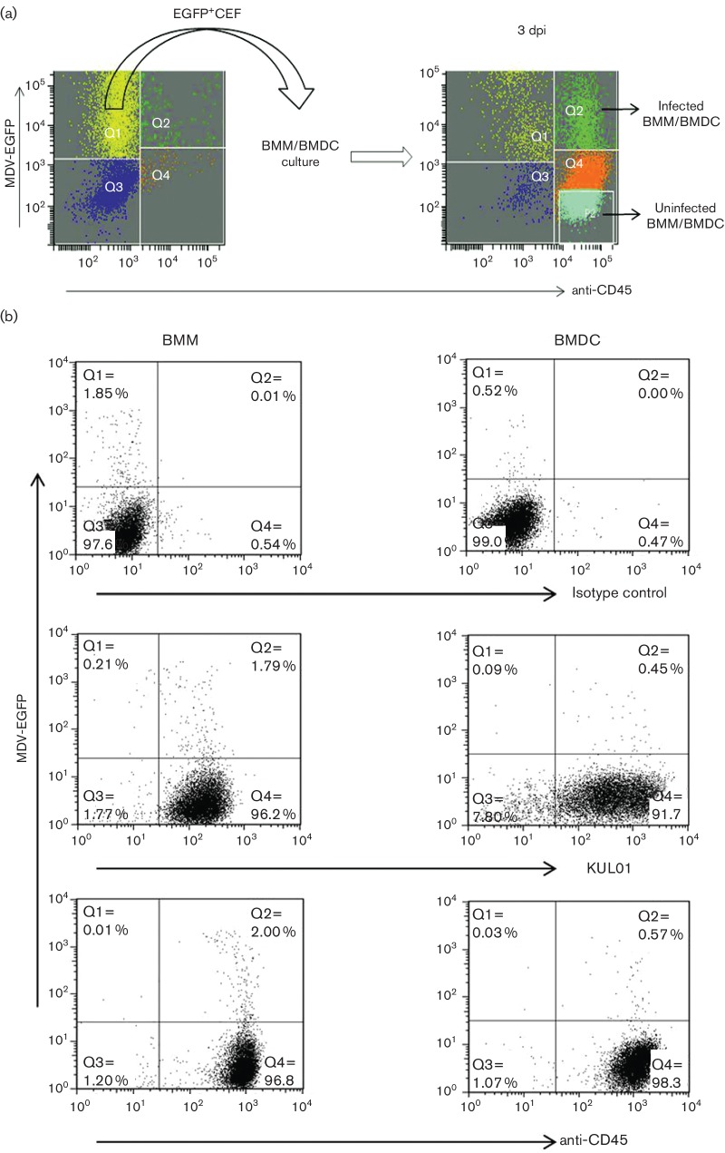
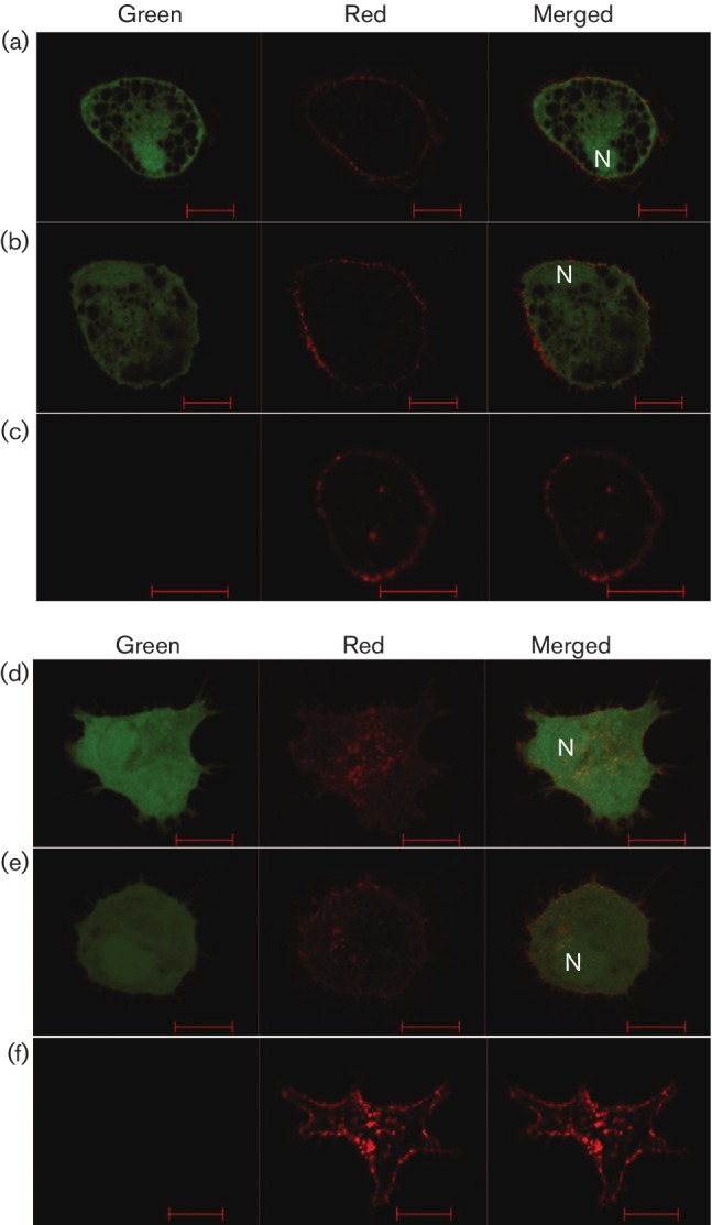
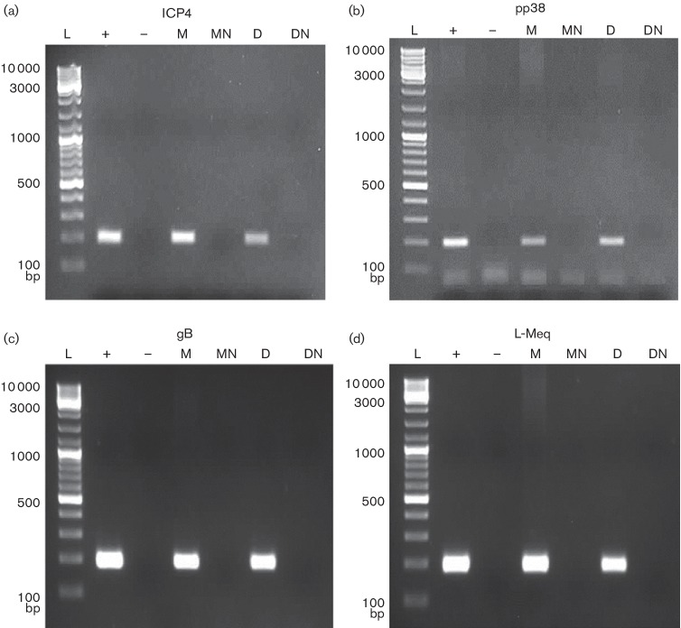
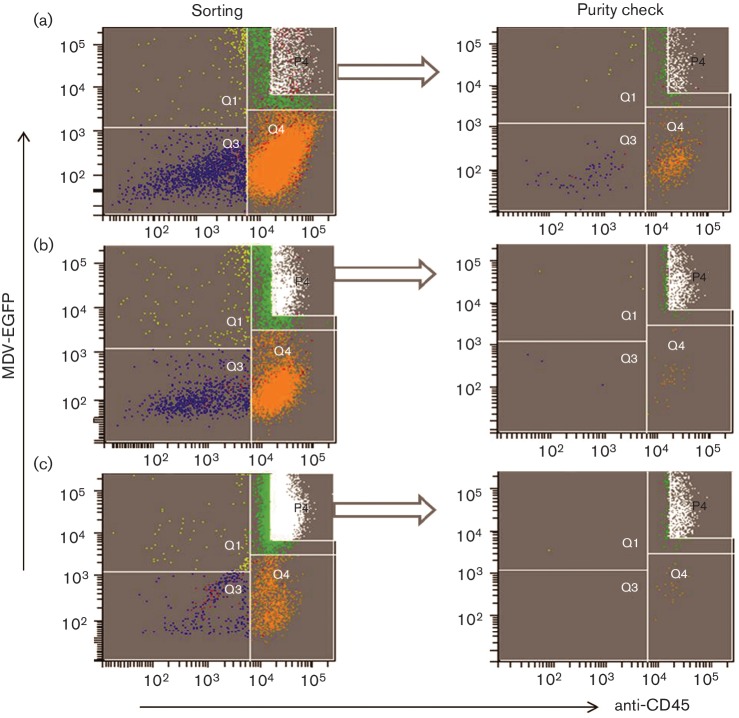
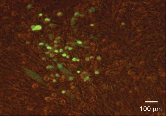
Marek's disease virus (MDV) is an alphaherpesvirus that induces T-cell lymphomas in chickens. Natural infections in vivo are caused by the inhalation of infected poultry house dust and it is presumed that MDV infection is initiated in the macrophages from where the infection is passed to B cells and activated T cells. Virus can be detected in B and T cells and macrophages in vivo, and both B and T cells can be infected in vitro. However, attempts to infect macrophages in vitro have not been successful. The aim of this study was to develop a model for infecting phagocytes [macrophages and dendritic cells (DCs)] with MDV in vitro and to characterize the infected cells. Chicken bone marrow cells were cultured with chicken CSF-1 or chicken IL-4 and chicken CSF-2 for 4 days to produce macrophages and DCs, respectively, and then co-cultured with FACS-sorted chicken embryo fibroblasts (CEFs) infected with recombinant MDV expressing EGFP. Infected phagocytes were identified and sorted by FACS using EGFP expression and phagocyte-specific mAbs. Detection of MDV-specific transcripts of ICP4 (immediate early), pp38 (early), gB (late) and Meq by RT-PCR provided evidence for MDV replication in the infected phagocytes. Time-lapse confocal microscopy was also used to demonstrate MDV spread in these cells. Subsequent co-culture of infected macrophages with CEFs suggests that productive virus infection may occur in these cell types. This is the first report of in vitro infection of phagocytic cells by MDV.


Figures

Fig. 1.
Fig. 1.
The in vitro infection of phagocytes with MDV. (a) The overall infection model. On the day of infection of phagocytes, MDV-infected CEFs were stained for CD45 expression to detect infected macrophages (Q2 in left plot) and EGFP+CD45-CEFs were sorted (left panel) and added to the bone marrow-derived macrophage (BMM) or bone marrow-derived DC (BMDC) culture. After 3 days in culture, infected and uninfected BMMs or BMDCs (EGFP+CD45+) were sorted (right panel). (b) Flow cytometric characterization of in vitro-infected BMMs and BMDCs. Chicken bone marrow-derived phagocytic cells were cultured with CSF-1 (for BMM) and with CSF-2 and IL-4 (for BMDC) for 4 days and then co-cultured with pre-sorted EGFP+CEF at a ratio of 1 : 5 (CEF:BMM/BMDC). Three days post-infection (p.i.), live cells were analysed for the surface expression of KUL01 and CD45 in BMM and BMDC. Gr 13.1 (class IgG1) was used as an isotype control antibody. Anti-CD45 antibody was used to detect the phagocytes via an AF647-tagged secondary antibody. Infected phagocytes were detected by double fluorescence of CD45 and EGFP (encoded with MDV). Data are shown as representative of two independent experiments for both BMM and BMDC. Distribution of cells: Q1, infected CEF; Q2, infected macrophage/DC; Q3, uninfected CEF; Q4, uninfected macrophage/DC; P2, sorting zone for uninfected macrophage/DC.
Fig. 2.
Fig. 2.
Visualization of infected and uninfected BMMs and BMDCs. Phagocytic cells were infected in vitro with EGFP+CEFs. Three days p.i., BMMs and BMDCs were sorted following staining with anti-CD45 and examined under confocal microscopy for (a, b) infected BMMs and (c) uninfected BMMs, as well as for (d, e) infected DCs and (f) uninfected DCs. Green channel: cells examined for the expression of EGFP-encoded MDV; red channel: cells examined for the expression of CD45 (AF647); merged channel: cells examined for combined expression of green and red. N, nucleus. Scale bar, 10 µm.
Fig. 3.
Fig. 3.
Detection of MDV transcripts in BMMs and BMDCs infected with MDV in vitro. BMMs and BMDCs were infected in vitro with EGFP-expressing MDV. After 3 days, EGFP-positive cells were sorted and RT-PCR was carried out for the detection of (a) immediate early ICP4 (200 bp), (b) early pp38 (198 bp), (c) late gB (193 bp) and (d) MDV-specific l-Meq (200 bp) transcripts. L, ladder; +, positive control MDV-infected CEFs; −, negative control, nuclease-free H2O; M, infected BMMs (cDNA); MN, infected BMMs no-RT control (DNase-treated RNA); D, infected BMDCs (cDNA); DN, infected BMDCs no-RT control (DNase-treated RNA).
Fig. 4.
Fig. 4.
Triple-sorting of MDV-infected BMMs with corresponding purity. (a) In the first sort, 1.28×10⁶ cells were sorted based on the gate P4 and the analysis revealed the presence of contaminant cells in Q1, Q3 and Q4. (b) Sorted cells (from P4) were re-sorted and contamination with infected CEFs (Q1); four events per 1000 infected macrophages were identified. (c) After sorting BMMs for the third time, only one infected CEF (Q1) per 1000 infected macrophages was detected. These triple-sorted infected BMMs were added to CEF cultures. The y-axis shows the fluorescence of intracellular EGFP-encoded MDV and the x-axis shows the fluorescence of AF 647-tagged anti-CD45. Distribution of cells in sorting plots: Q3, uninfected CEF; Q1, infected CEF; Q4, uninfected macrophage; Q2, infected macrophage; P4, sorting zone for infected macrophage.
Fig. 5.
Fig. 5.
Formation of plaques following infection of CEFs with MDV-infected BMMs. To investigate the productive or abortive nature of MDV-BMM infections, CEFs were re-infected with MDV-infected BMMs. In order to reduce the number of contaminant-infected CEFs, MDV-infected BMMs were sorted three times and freshly cultured CEFs were infected with these triple-sorted infected BMMs. Five days p.i., plaques were visualized based on EGFP (encoded with MDV) using a fluorescence microscope.

References

    1. Calnek BW. Pathogenesis of Marek's disease virus infection. Curr Top Microbiol Immunol. 2001;255:25–55. - PubMed
    1. Calnek BW, Schat KA, Ross LJ, Shek WR, Chen CL, et al. Further characterization of Marek's disease virus-infected lymphocytes. I. In vivo infection. Int J Cancer. 1984;33:389–398. doi: 10.1002/ijc.2910330318. - DOI - PubMed
    1. Calnek BW, Schat KA, Ross LJ, Chen CL. Further characterization of Marek's disease virus-infected lymphocytes. II. In vitro infection. Int J Cancer. 1984;33:399–406. doi: 10.1002/ijc.2910330319. - DOI - PubMed
    1. Schermuly J, Greco A, Härtle S, Osterrieder N, Kaufer BB, et al. In vitro model for lytic replication, latency, and transformation of an oncogenic alphaherpesvirus. Proc Natl Acad Sci USA. 2015;112:7279–7284. doi: 10.1073/pnas.1424420112. - DOI - PMC - PubMed
    1. McClean CM, Tobin DM. Macrophage form, function, and phenotype in mycobacterial infection: lessons from tuberculosis and other diseases. Pathog Dis. 2016;74:ftw068. doi: 10.1093/femspd/ftw068. - DOI - PMC - PubMed

LinkOut - more resources