. 2017 May 26:6:e27389.
doi: 10.7554/eLife.27389.

Asymmetric recognition of HIV-1 Envelope trimer by V1V2 loop-targeting antibodies

Affiliations

Asymmetric recognition of HIV-1 Envelope trimer by V1V2 loop-targeting antibodies

Haoqing Wang et al. Elife. .

Abstract

The HIV-1 envelope (Env) glycoprotein binds to host cell receptors to mediate membrane fusion. The prefusion Env trimer is stabilized by V1V2 loops that interact at the trimer apex. Broadly neutralizing antibodies (bNAbs) against V1V2 loops, exemplified by PG9, bind asymmetrically as a single Fab to the apex of the symmetric Env trimer using a protruding CDRH3 to penetrate the Env glycan shield. Here we characterized a distinct mode of V1V2 epitope recognition by the new bNAb BG1 in which two Fabs bind asymmetrically per Env trimer using a compact CDRH3. Comparisons between cryo-EM structures of Env trimer complexed with BG1 (6.2 Å resolution) and PG9 (11.5 Å resolution) revealed a new V1V2-targeting strategy by BG1. Analyses of the EM structures provided information relevant to vaccine design including molecular details for different modes of asymmetric recognition of Env trimer and a binding model for BG1 recognition of V1V2 involving glycan flexibility.

Keywords: Cryo-EM; Human; biophysics; broadly neutralizing antibodies; structural biology; virus.


Conflict of interest statement

MCN: Senior editor, eLife.

The other authors declare that no competing interests exist.

Figures
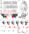
Figure 1.
Figure 1.. Comparison of BG1 and other V1V2 bNAbs.
(A) Sequences of the HCs of BG1 and representative other V1V2 bNAbs. Residue numbering (Kabat) refers to BG1, and the CDRs were defined using AbM (Swindells et al., 2017). (B) Crystal structure of BG1 Fab with highlighted CDRs. (C) Space-filling representations of antigen-binding site of BG1 in two orientations with CDRs highlighted. (D) Hydrogen bonds (dotted yellow lines) contributing to compact structure of CDRH3. Water molecules shown as red spheres. (E) Top: Space-filling representations of VH-VL domains of selected V1V2 bNAbs (BG1: this study; PG9: PDB 3U4E; PG16: PDB 3 MUG; PGT145: PDB 3U1S; CH03: PDB 5ESV; VRC26.03: PDB 4OD1). Residues within CDRs are highlighted. CDRH3 (middle) and CDRH2 (bottom) loops shown in ribbon (left) and space-filling (right) representations. Electrostatic potentials are shown on the space filling representations using blue and red for positive and negative electrostatic potentials, respectively. DOI: http://dx.doi.org/10.7554/eLife.27389.002
Figure 1—figure supplement 1.
Figure 1—figure supplement 1.. Electron densities for tyrosines within BG1 Fab CDRs.
(A) Tyrosine residues located within the antigen binding site are shown in stick representation together with their electron densities (contoured at 1.0 σ) from a 2Fo–Fc simulated annealing composite omit map. DOI: http://dx.doi.org/10.7554/eLife.27389.003
Figure 2.
Figure 2.. Fab-Env binding stoichiometries for V1V2 bNAbs.
(A) SEC-MALS profiles for BG505 SOSIP.664 Env trimer and complexes of Env trimer with BG1, PG16, and 8ANC195 Fabs. The absorbance at 280 nm (left y-axis) is plotted against elution volume from a Superdex 200 10/300 GL gel filtration column and overlaid with the molar mass determined for each peak (right y-axis). Predicted and calculated molecular masses are shown in the table. (B) Negative-stain single particle EM reconstructions BG1-Env-8ANC195, PG9-Env-8ANC195, and PG16-Env-8ANC195 complexes. EM density was fit with coordinates for the indicated Fabs and for BG505 Env trimer. (C) Density and coordinates from the BG1-Env portion of the BG1-Env-8ANC195 reconstruction (density for 8ANC195 Fabs removed) with coordinates for the indicated Fabs superimposed. The Env trimer portion from EM structures of complexes of PGT145-Env (PDB 5U1F), PG9-Env (panel B), or PG16-Env (panel B) were superimposed with the Env trimer portion of the BG1-Env-8ANC195 structure (panel B). DOI: http://dx.doi.org/10.7554/eLife.27389.005
Figure 2—figure supplement 1.
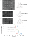
Figure 2—figure supplement 1.. Negative-stain EM structures of V1V2 bNAb-Env-8ANC195 complexes.
(A–C) 2D class averages and the final structures after refinement for BG1-Env-8ANC195 (panel A), PG16-Env-8ANC195 (panel B), and PG9-Env-8ANC195 (panel C). (D) Gold-standard FSC for each structure. DOI: http://dx.doi.org/10.7554/eLife.27389.007
Figure 3.
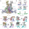
Figure 3.. ~6.2 Å cryo-EM reconstruction of a BG1-Env-8ANC195 complex.
(A) EM density fit by coordinates for BG1_A (HC in brown; LC in yellow), BG1_B (HC in dark gray; LC in light gray), gp120s associated with BG1_A and BG1_B Fabs in purple and cyan, respectively; gp120_C in green, gp41 in orange, and 8ANC195 Fabs in dark blue (HC) and light blue (LC). (B) Close-up views of densities contoured at 6.0σ (0.0378 e/Å3) for glycans attached to Asn156gp120 and Asn160gp120 in the three gp120 protomers of Env trimer. Glycan residues built into the cryo-EM map densities are shown as sticks with oxygen atoms in red and nitrogen atoms in blue. (C, D) Close-up views of densities contoured at 6.0σ (0.0378 e/Å3) surrounding CDRH3 (panel C) and CDRH2 (panel D) in BG1-gp120_A and BG1_B-gp120_B interactions. Asn156gp120 and Asn160gp120 glycans are shown as sticks. CDRH2 residue Trp54 (Figure 1D) is identified with an arrow. DOI: http://dx.doi.org/10.7554/eLife.27389.008
Figure 3—figure supplement 1.
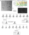
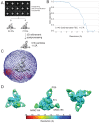
Figure 3—figure supplement 1.. Cryo-EM data processing for the BG1-Env-8ANC195 complex structure.
(A) Representative micrograph. Examples of complex particles are boxed. The accumulated dose and defocus values were ~80 e-/Å2 and ~1.8 µm underfocus. (B) CTF fitting of the left micrograph (generated using CTFFIND4) shows a good fit to 4.9 Å. (C) Data processing scheme. 2D classification of autopicked particles resulted in 76K particles that were 3D classified using a low-pass-filtered negative-stain 2:1 BG1-Env EM structure as a reference model. The particles from the best three 3D classes were refined to 6.18 Å resolution. One 3D class showed three BG1 bound to each Env trimer (see Figure 3—figure supplement 2). The particles in 6.18 Å reconstruction were further 3D classified with the Fab CHCL domains masked out. Then the best two 3D classes were refined to 6.18 Å resolution. (E) Gold-standard FSCs of reconstructions generated using different strategies (see Figure 3—figure supplement 2). (F) Local resolution estimation. The complex is shown from top (left), side (middle), and bottom (right) views. DOI: http://dx.doi.org/10.7554/eLife.27389.009
Figure 3—figure supplement 2.
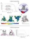
Figure 3—figure supplement 2.. Validation of the 2:1 and 3:1 BG1-Env structures.
(A) Gold-standard FSCs of 2:1 BG1-Env reconstructions generated using different strategies. (B) Orientation distribution of the 6.2 Å 2:1 BG1-Env reconstruction without masking out Fab CHCL domains. (C) Local resolution estimation for the 2:1 BG1-Env structure. The complex is shown from top (left), side (middle), and bottom (right) views. (D) Cryo-EM map contoured to show densities for α-helices (red); other secondary structures shown in grey. (E) 27 Å cryo-EM structure of a 3:1 BG1-Env complex. Cryo-EM map with fitted components are shown from side (left) and top (middle) views. Right: Gold-standard FSC indicating 27 Å resolution for the reconstruction. DOI: http://dx.doi.org/10.7554/eLife.27389.010
Figure 4.
Figure 4.. ~11.5 Å cryo-EM reconstruction of a PG9-Env-8ANC195 complex.
(A) EM density fit by coordinates for PG9 (HC in dark gray; LC in light gray), gp120 subunits in dark blue, cyan, and green, gp41 in orange, and 8ANC195 Fabs in dark blue (HC) and light blue (LC). (B) Density and coordinates from the PG9-Env portion of the PG9-Env-8ANC195 reconstruction (density for 8ANC195 Fabs removed) with coordinates for the PG9 Fab from the PG9-V1V2 scaffold structure (PDB 3U4E) superimposed. When the V1V2 scaffold from the PG9-V1V2 complex structure was superimposed with the V1V2 portion of the PG9-Env-8ANC195 structure (panel A), the angle of approach of the PG9 Fab differs from the angle observed in the PG9-Env structure. (C) Hypothetical model of three PG9 Fabs bound per Env trimer. One PG9 Fab (PG9_A) is shown its experimentally-determined position from the PG9-Env-8ANC195 structure interacting mainly with gp120_A on Env trimer (view looking down the trimer three-fold axis). The VH-VL domains for second and third PG9 Fabs (PG9 model) were positioned onto the gp120_B and gp120_C subunits assuming the interaction observed for PG9_A with gp120_A. Predicted van der Waals clashes (red dots) were calculated using the show_bumps module in Pymol (Schrödinger, 2011). DOI: http://dx.doi.org/10.7554/eLife.27389.013
Figure 4—figure supplement 1.
Figure 4—figure supplement 1.. Cryo-EM data processing for PG9-Env-8ANC195 complex structure.
(A) Data processing scheme. 2D classification of autopicked particles resulted in 12 K particles that were 3D classified using a low-pass-filtered negative-stain PG9-Env-8ANC195 EM structure as a reference model. The particles from the best class were refined to 11.5 Å resolution. (B) Orientation distribution of the reconstruction. (C) Local resolution estimation. The complex is shown from top (left), side (middle), and bottom (right) views. (D) Gold-standard FSC indicating 11.5 Å resolution for the reconstruction. DOI: http://dx.doi.org/10.7554/eLife.27389.014
Figure 5.
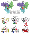
Figure 5.. Comparison of BG1 and PG9 epitopes and paratopes.
(A) Epitopes on Env trimer (top view surface representations) were defined as protein or glycan residues whose Cα (for protein) or C1 (for glycans) atom was within 7 Å of the bound Fab. Contacts on Env are color-coded to indicate which CDR loop made the contact (CDRH3 in red, CDRH2 in green, and CDRH1 in dark blue). Glycans are shown as sticks with a nearby label to identify the glycan as attached to either Asn156gp120 or Asn160gp120, and locations of residues of interest (Lys168gp120, Lys169gp120, and Gln130gp120) are indicated by black dots. Center box compares geometric mean IC50 values for BG1 and PG9 IgGs evaluated against HIV-1 strains either containing or not containing a PNGS at position 130 (number of HIV-1 strains indicated in the parentheses). IC50values > 50 µg/mL set to 50 µg/mL for geometric mean calculations. (B) Paratopes on BG1_A, BG1_B, and PG9 Fabs indicated by spheres on ribbon representations of VH-VL domains shown in two orientations related by a 90˚ rotation. CDRs are color coded as in panel A. DOI: http://dx.doi.org/10.7554/eLife.27389.015
Figure 5—figure supplement 1.
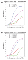
Figure 5—figure supplement 1.. Coverage curves for results of in vitro neutralization across panels of viruses that do or do not include a lysine at position 169gp120 (panel A) or a threonine at position 132gp120 (panel B).
The number of strains used to calculate each curve is shown in parentheses in the key inside each graph. DOI: http://dx.doi.org/10.7554/eLife.27389.017
Figure 6.
Figure 6.. BG1 and PG9 interactions with V1V2.
(A) BG1 interactions with N-linked glycans attached to Asn156gp120 and Asn160gp120 shown for BG1_A (left) and BG1_B (right). (B) PG9 interactions with N-linked glycans attached to Asn156gp120 and Asn160gp120 from PG9-Env cryo-EM structure reported here (left) and from the crystal structure of PG9-V1V2 scaffold (PDB 3U4E) (right). The position of the CDRH3 loop in the PG9-Env cryo-EM structure is shown by a dashed line representing the position of CDRH3 in the PG9-V1V2 scaffold structure. DOI: http://dx.doi.org/10.7554/eLife.27389.018
Figure 7.
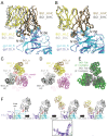
Figure 7.. Asymmetry of BG1 interactions with Env trimer.
(A,B) Superposition of the two BG1 binding sites in the 2:1 BG1-Env structure by aligning either the V1V2 regions of gp120_A and gp120_B (panel A) or the Asn156gp120 and Asn160gp120 glycans within gp120_A and gp120_B (panel B). (C) Hypothetical model of three BG1 Fabs bound per Env trimer based on Fab orientations in the 2:1 BG1-Env structure. The BG1_A and BG_1 B Fabs are shown in their respective positions on the gp120_A and gp120_B subunits of Env trimer (view looking down the trimer three-fold axis). A third BG1 Fab (BG1 model) was positioned onto the gp120_C subunit assuming that it would interact with the Asn156gp120 and Asn160gp120 glycans in the common interaction observed for BG1_A and BG1_B (panel B). Predicted van der Waals clashes (red dots) were calculated using the show_bumps module in Pymol (Schrödinger, 2011). (D) Positions of three BG1 Fabs in the 27 Å 3:1 BG1-Env structure (no predicted van der Waals clashes). (E) Comparison of BG1 Fab positions in the 2:1 BG1-Env structure (gray; the third Fab is from the model shown in panel C) and the 3:1 BG1-Env structure (green) after superimposing the BG1_A Fabs. (F) Model for binding of two BG1 Fabs to form the 2:1 BG1-Env structure. Panels 1 and 2: BG1_B binds first to the V1V2 epitope on gp120_B such that its CDRL1 interacts with the Asn160gp120 glycan from gp120_A. Panels 3 and 4: BG1_A recognizes the Asn156gp120 and Asn160gp120 glycans from gp120_A. The flexible glycans are shifted away from the trimer 3-fold axis (indicated by arrows in the close-up view of the third panel) such that a potential clash with BG1_B is avoided. DOI: http://dx.doi.org/10.7554/eLife.27389.019
Author response image 1.
DOI: http://dx.doi.org/10.7554/eLife.27389.020

Similar articles

Cited by

References

    1. Adams PD, Afonine PV, Bunkóczi G, Chen VB, Davis IW, Echols N, Headd JJ, Hung LW, Kapral GJ, Grosse-Kunstleve RW, McCoy AJ, Moriarty NW, Oeffner R, Read RJ, Richardson DC, Richardson JS, Terwilliger TC, Zwart PH. PHENIX: a comprehensive Python-based system for macromolecular structure solution. Acta Crystallographica Section D Biological Crystallography. 2010;66:213–221. doi: 10.1107/S0907444909052925. - DOI - PMC - PubMed
    1. Agirre J, Iglesias-Fernández J, Rovira C, Davies GJ, Wilson KS, Cowtan KD. Privateer: software for the conformational validation of carbohydrate structures. Nature Structural & Molecular Biology. 2015;22:833–834. doi: 10.1038/nsmb.3115. - DOI - PubMed
    1. Barouch DH, Whitney JB, Moldt B, Klein F, Oliveira TY, Liu J, Stephenson KE, Chang HW, Shekhar K, Gupta S, Nkolola JP, Seaman MS, Smith KM, Borducchi EN, Cabral C, Smith JY, Blackmore S, Sanisetty S, Perry JR, Beck M, Lewis MG, Rinaldi W, Chakraborty AK, Poignard P, Nussenzweig MC, Burton DR. Therapeutic efficacy of potent neutralizing HIV-1-specific monoclonal antibodies in SHIV-infected rhesus monkeys. Nature. 2013;503:224–228. doi: 10.1038/nature12744. - DOI - PMC - PubMed
    1. Bonsignori M, Hwang KK, Chen X, Tsao CY, Morris L, Gray E, Marshall DJ, Crump JA, Kapiga SH, Sam NE, Sinangil F, Pancera M, Yongping Y, Zhang B, Zhu J, Kwong PD, O'Dell S, Mascola JR, Wu L, Nabel GJ, Phogat S, Seaman MS, Whitesides JF, Moody MA, Kelsoe G, Yang X, Sodroski J, Shaw GM, Montefiori DC, Kepler TB, Tomaras GD, Alam SM, Liao HX, Haynes BF. Analysis of a clonal lineage of HIV-1 envelope V2/V3 conformational epitope-specific broadly neutralizing antibodies and their inferred unmutated common ancestors. Journal of Virology. 2011;85:9998–10009. doi: 10.1128/JVI.05045-11. - DOI - PMC - PubMed
    1. Brilot AF, Chen JZ, Cheng A, Pan J, Harrison SC, Potter CS, Carragher B, Henderson R, Grigorieff N. Beam-induced motion of vitrified specimen on holey carbon film. Journal of Structural Biology. 2012;177:630–637. doi: 10.1016/j.jsb.2012.02.003. - DOI - PMC - PubMed

Publication types

MeSH terms

Substances