. 2017 May 26;7(5):e565.
doi: 10.1038/bcj.2017.40.

Waldenstrom macroglobulinemia cells devoid of BTKC481S or CXCR4WHIM-like mutations acquire resistance to ibrutinib through upregulation of Bcl-2 and AKT resulting in vulnerability towards venetoclax or MK2206 treatment

Affiliations

Waldenstrom macroglobulinemia cells devoid of BTKC481S or CXCR4WHIM-like mutations acquire resistance to ibrutinib through upregulation of Bcl-2 and AKT resulting in vulnerability towards venetoclax or MK2206 treatment

A Paulus et al. Blood Cancer J. .

Abstract

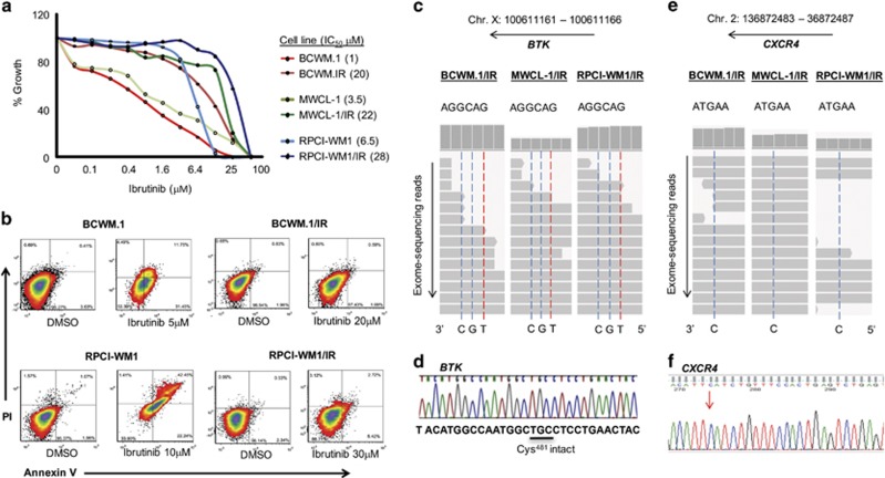
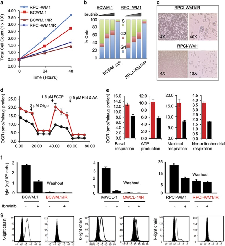
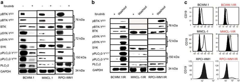
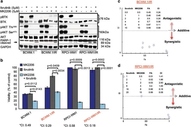
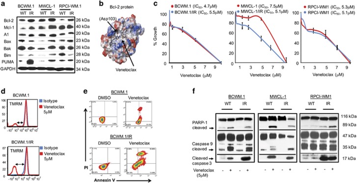
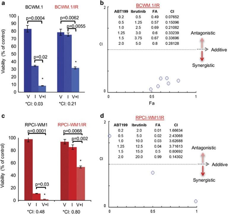
Although ibrutinib is highly effective in Waldenstrom macroglobulinemia (WM), no complete remissions in WM patients treated with ibrutinib have been reported to date. Moreover, ibrutinib-resistant disease is being steadily reported and is associated with dismal clinical outcome (overall survival of 2.9-3.1 months). To understand mechanisms of ibrutinib resistance in WM, we established ibrutinib-resistant in vitro models using validated WM cell lines. Characterization of these models revealed the absence of BTKC481S and CXCR4WHIM-like mutations. BTK-mediated signaling was found to be highly attenuated accompanied by a shift in PI3K/AKT and apoptosis regulation-associated genes/proteins. Cytotoxicity studies using the AKT inhibitor, MK2206±ibrutinib, and the Bcl-2-specific inhibitor, venetoclax±ibrutinib, demonstrated synergistic loss of cell viability when either MK22016 or venetoclax were used in combination with ibrutinib. Our findings demonstrate that induction of ibrutinib resistance in WM cells can arise independent of BTKC481S and CXCR4WHIM-like mutations and sustained pressure from ibrutinib appears to activate compensatory AKT signaling as well as reshuffling of Bcl-2 family proteins for maintenance of cell survival. Combination treatment demonstrated greater (and synergistic) antitumor effect and provides rationale for development of therapeutic strategies encompassing venetoclax+ibrutinib or PI3K/AKT inhibitors+ibrutinib in ibrutinib-resistant WM.


Conflict of interest statement

The authors declare no conflict of interest.

Figures

Figure 1
Figure 1
Establishment and characterization of ibrutinib-resistant cell lines demonstrates growth and apoptosis resistance without acquisition of BTKC481S or CXCR4WHIM-like mutations. Ibrutinib-resistant clones of established WM cell lines (n=3) were developed through chronic ibrutinib exposure over a period of 6 months. (a) A 72 h MTS assay was conducted to determine resistance of ibrutinib-resistant (/IR) WM cells vs WT parental cells. Median IC50 of ibrutinib-resistant cells was 22 μm (range, 20–28 μm), whereas median IC50 of WT WM cells was 3.5 μm (range, 1–6.5 μm). (b) All WM cell lines (WT and ibrutinib-resistant, n=6) were treated with DMSO or ibrutinib at 5, 10, 20 and 30 μm concentrations for 24 h and stained with annexin-V and propidium iodide, followed by flow cytometry to examine apoptosis. Representative heat density plots from two ibrutinib-resistant models demonstrate apoptosis resistance in BCWM.1/IR and RPCI-WM1/IR cells (~0–11% annexin-V positivity) relative to WT counterparts (~43–62% annexin-V positivity) after ibrutinib treatment. (c) Whole-exome sequencing (WES) analysis of all ibrutinib-resistant WM cell lines (n=3) revealed no mutation in the DNA region encoding for the Cys481 residue of the BTK gene as illustrated through integrative genomics viewer (IGV) visualization of nucleotide read alignments of BTK on chromosome X (Chr. X): position 100 611 161–100 611 166. (d) Conservation of the BTK Cys481 region was validated by Sanger sequencing (representative chromatogram from BCWM.1/IR cells shown). (e) In a similar manner, WES revealed no WHIM-like mutations in the CXCR4 gene on Chr. 2 (4 regions analyzed). One representative region shown by IGV read alignment visualization on Chr. 2, position 136 872 485, which if altered from C>A or C>G, results in a S338X mutation. (f) Sanger sequencing of the corresponding region region confirmed no mutation present in S338 (representative chromatogram from BCWM.1/IR cells). Additional regions corresponding to other WHIM-like mutations shown in Supplementary Figure S1.
Figure 2
Figure 2
Ibrutinib-resistant WM clones show altered growth kinetics, reduced IgM secretion and Ig-light-chain expression. (a) WT and ibrutinib-resistant WM clones (two representative isogenic pairs shown) were seeded at 0.5 million/ml, followed by measurement of total cell numbers and viability at 24 and 48 h. (b) WM cells and ibrutinib-resistant derivatives (two representative isogenic pairs shown) were treated with DMSO or increasing concentrations of ibrutinib (1 or 5 μm for 24 h), followed by flow cytometry and cell cycle analysis. Ibrutinib treatment resulted in accumulation of WM tumor cells in the G1 phase of the cell cycle. By comparison, ibrutinib-resistant WM clones (BCWM.1/IR and RPCI-WM1/IR) were noted be G1 arrested. (c) Light microscopy showed ibrutinib-resistant WM cells to be more irregular in shape and larger in size as compared with their WT counterparts (one isogenic pair shown as representative). (d) Mitochondrial respiration was measured side by side in parental cells (BCWM.1) and ibrutinib-resistant WM cells (BCWM.1/IR) at baseline condition, followed by inhibition of mitochondrial ATP production (introduction of oligomycin), uncoupling oxidative phosphorylation (OP) and ATP production causing maximal mitochondrial respiration (introduction of carbonyl cyanide-4-phenylhydrazone (FCCP)) and finally complete inhibition of the OP by the combination of rotenone and antimycin A. oxygen consumption rate (OCR) is displayed in values normalized to total protein content. (e) Compared with parental cells, BCWM.1/IR cells showed increased values in all analyzed parameters: basal respiration (left), ATP production (middle left) and maximal respiration (middle right) and non-mitochondrial respiration (right). (f) IgM secretion was quantified in the supernatant of WT and ibrutinib-resistant WM clones (±ibrutinib 1μm, 24 h exposure). In WT WM cells, secretion of IgM decreased after exposure to ibrutinib. In ibrutinib-resistant clones, IgM secretion was measured in cells after a washout period (cells cultured in ibrutinib-free media for 1 week) as well as after ibrutinib treatment and was noted to be markedly reduced in washout cells compared to WT counterparts, regardless of exposure to ibrutinib thereafter. (g) In a similar manner, all WM cell lines were incubated with anti-λ-light-chain or anti-k-light-chain antibody and analyzed by flow cytometry, which demonstrated decreased surface expression of native Ig-light-chain on ibrutinib-resistant WM cells.
Figure 3
Figure 3
BCR signaling is dowregulated in ibrutinib-resistant WM cells. (a) WT WM cell lines were treated with DMSO or ibrutinib (1 μm) for 12 h and subjected to immunoblotting with anti-BTK/pBTK, anti-SYK/pSYK, anti-PLCγ2/pPLCγ2 and GAPDH antibodies. Ibrutinib treatment effectively reduced pBTK, pSYK and pPLCγ2 protein levels in all WT WM cell lines, indicating attenuation of BCR signaling. (b) In a similar manner, ibrutinib-resistant WM clones were also probed with anti-BTK/pBTK, anti-SYK/pSYK, anti-PLCγ2/pPLCγ2 and GAPDH antibodies. While immunobot analysis showed very low pBTK, pSYK or pPLCγ2 protein levels in ibrutinib-resistant clones (kept in ibrutinib-containing media at >90% viability), protein levels increased after washout of cells (after 48 h in ibrutinib-free media). Notably, viability of ibrutinib-resistant clones- before or after washout, was not impacted. (c) All WM cell lines were incubated with anti-CD19 antibody followed by flow cytometry. Compared to WT parental cells, BCWM.1/IR and MWCL-1/IR cells showed reduced surface expression of CD19. RPCI-WM1 is known to be CD19- and this was also noted in RPCI-WM1/IR cells.
Figure 4
Figure 4
Phospho-AKT is increased in ibrutinib-resistant WM cells and its pharmacologic inhibition results in decreased tumor cell survival and apoptosis. (a) WM cell lines were treated with DMSO, ibrutinb or MK2206 (allosteric pan-AKT inhibitor) at indicated concentrations for 24 h and immunoblot analysis was performed probing for BTK, pBTK AKT, pAKT (Thr308), pAKT (Ser473) and PARP-1 protein expression. pAKT was noted to be decreased in all WM cells (alone or in combination with ibrutinib). PARP-1 cleavage in WM cells was most markedly observed after combination ibrutinib and MK2206 treatment. (b) Two isogenic cell line pairs (BCWM.1 and BCWM.1IR; RPCI-WM1 and RPCI-WM1/IR) were treated with DMSO or various concentrations of MK2206, ibrutinib or ibrutinib+MK2206 for 48 h and assessed for viability by the CellTiter Glo Luminescent Cell Assay to determine pharmacologic synergy. A combination index (CI) value of 1 signifies additivity, whereas a CI<1 signifies synergism and a CI>1 signifies antagonism between the drugs at particular concentrations. Bar graph shows representative results of combination experiments where drug combination synergy was noted. In BCWM.1 cells, ibrutinib (2 μm)+MK2206 (2 μm) reduced cell viability to 19% and in BCWM.1/IR cells, a similar trend was noted at similar concentrations, where the combination of both agents caused significant reduction in cell viability (40% viable cells) compared with either agent alone. (c) Representative isobologram analysis of BCWM.1/IR cells shows synergistic cytotoxic activity of ibrutinib+MK2206 in 6/6 drug combinations tested (represented by blue circles). (d) In RPCI-WM1 and RPCI-WM1/IR cells, ibrutinib or MK2206 alone did not significantly reduce cell viability; however, the combination of the two agents (ibrutinib 4 μm+MK2206 4 μm in RPCI-WM1 cells; ibrutinib 16 μm+MK2206 8 μm in RPCI-WM1/IR cells) significantly reduced cell viability to 16 and 11% in RPCI-WM1 and RPCI-WM1/IR cells, respectively. (e) Representative isobologram analysis of RPCI-WM1/IR cells showed synergistic cytotoxic activity in 2/6 combinations (represented by blue circles).
Figure 5
Figure 5
Inhibition of Bcl-2 with venetoclax results in loss of viability and mitochondrial-mediated apoptosis of ibrutinib-resistant WM cells. (a) WM cell lines were treated with DMSO or ibrutinib (1 μm) for 12 h and immunoblot analysis was performed probing for anti- and proapoptotic Bcl-2 family proteins. Compared with WT cells, ibrutinib-resistant clones showed increased expression of Bcl-2, with more variability in proapoptotic protein (Bax, Bak, Bim and PUMA) expression among the different ibrutinib-resistant models. (b) Three-dimensional (3D) rendering of venetoclax, a BH3 mimetic (black colored ligand) that selectively binds Bcl-2, occupying the P2 and P4 pockets in the BH3 groove of the Bcl-2 protein. (c) A 48 h MTS assay was conducted to assess sensitivity of all WM cell lines towards venetoclax, which demonstrated comparable activity in WT and ibrutinib-resistant cells alike, with the exception of MWCL-1/IR cells, which showed greater growth inhibition compared with MWCL-1 parental cells. (d) WM cell lines were treated with DMSO or venetoclax (5 μm) for 24 h and stained with annexin-V and propidium iodide, followed by flow cytometry to examine apoptosis. Venetoclax induced significant apoptosis in all WM cell lines tested; heat density plots from a representative isogenic pair model, BCWM.1 and BCWM.1/IR, show 60% and 63% annexin-V positivity, respectively, after treatment with the drug. (e) WM cells were treated with DMSO or venetoclax (5 μm) for 12 h and MOMP was measured in relation to TMRM fluorescence; TMRM-negative cells were calculated to represent (%) MOMP. MOMP was more increased in ibrutinib-resistant clones after ibrutinib exposure as compared with WT WM cells. Representative histograms are shown for BCWM.1 and BCWM.1/IR cells, where the blue line represents isotype control and the red line indicates shift in TMRM fluorescence after venetoclax treatment. (f) Mitochondrial-mediated apoptosis was validated in WM cells treated with DMSO or venetoclax by immunoblotting for PARP-1, caspase-9 and caspase-3 protein products, which demonstrated cleavage of these proteins in all cells treated with the drug.
Figure 6
Figure 6
Venetoclax and ibrutinib display synergistic anti-WM activity. Two isogenic cell line pairs (BCWM.1 and BCWM.1IR; RPCI-WM1 and RPCI-WM1/IR) were treated with DMSO or various concentrations of venetoclax (V), ibrutinib (I) or venetoclax+ibrutinib (V+I) for 48 h and assessed for viability by the CellTiter Glo Luminescent Cell Assay. A combination index (CI) value of 1 signifies additivity, whereas a CI<1 signifies synergism and a CI>1 signifies antagonism between the drugs at particular concentrations. Bar graph shows representative results from combination experiments where drug combination synergy was noted. (a) In BCWM.1 and BCWM.1/IR cells, the combination of V (1 μm)+I (2.5 μm) synergistically reduced tumor cell viability; significantly more so than either V or I alone at a CI of 0.03 and 0.21, respectively. (b) Representative isobologram analysis of BCWM.1/IR cells demonstrated synergistic cytotoxic activity of V+I in 6/6 combinations (represented by blue circles). (c) In the RPCI-WM1 cell line and its ibrutinib-resistant derivative, the combination of V (1.5 μm)+I (15 μm) synergistically reduced tumor cell viability (CI of 0.48 and 0.80, respectively), an effect more notable than when cells were treated with V or I alone. (d) Representative isobologram analysis of RPCI-WM1/IR cells showed synergistic cytotoxic activity in 2/6 combinations (represented by blue circles).

Similar articles

Cited by

References

    1. Owen RG, Treon SP, Al-Katib A, Fonseca R, Greipp PR, McMaster ML et al. Clinicopathological definition of Waldenstrom's macroglobulinemia: consensus panel recommendations from the Second International Workshop on Waldenstrom's macroglobulinemia. Semin Oncol 2003; 30: 110–115. - PubMed
    1. Oza A, Rajkumar SV. Waldenstrom macroglobulinemia: prognosis and management. Blood Cancer J 2015; 5: e296. - PMC - PubMed
    1. Treon SP, Tripsas CK, Meid K, Warren D, Varma G, Green R et al. Ibrutinib in previously treated Waldenstrom's macroglobulinemia. N Engl J Med 2015; 372: 1430–1440. - PubMed
    1. Cao Y, Hunter ZR, Liu X, Xu L, Yang G, Chen J et al. The WHIM-like CXCR4(S338X) somatic mutation activates AKT and ERK, and promotes resistance to ibrutinib and other agents used in the treatment of Waldenstrom's macroglobulinemia. Leukemia 2015; 29: 169–176. - PubMed
    1. Young RM, Staudt LM. Ibrutinib treatment of CLL: the cancer fights back. Cancer Cell 2014; 26: 11–13. - PMC - PubMed

MeSH terms