. 2018 Feb;233(2):1468-1480.
doi: 10.1002/jcp.26033. Epub 2017 Aug 4.

Microtubules regulate brush border formation

Affiliations

Microtubules regulate brush border formation

Facundo M Tonucci et al. J Cell Physiol. 2018 Feb.

Abstract

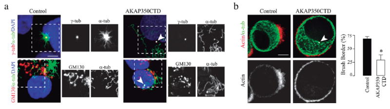
Most epithelial cells contain apical membrane structures associated to bundles of actin filaments, which constitute the brush border. Whereas microtubule participation in the maintenance of the brush border identity has been characterized, their contribution to de novo microvilli organization remained elusive. Hereby, using a cell model of individual enterocyte polarization, we found that nocodazole induced microtubule depolymerization prevented the de novo brush border formation. Microtubule participation in brush border actin organization was confirmed in polarized kidney tubule MDCK cells. We also found that centrosome, but not Golgi derived microtubules, were essential for the initial stages of brush border development. During this process, microtubule plus ends acquired an early asymmetric orientation toward the apical membrane, which clearly differs from their predominant basal orientation in mature epithelia. In addition, overexpression of the microtubule plus ends associated protein CLIP170, which regulate actin nucleation in different cell contexts, facilitated brush border formation. In combination, the present results support the participation of centrosomal microtubule plus ends in the activation of the polarized actin organization associated to brush border formation, unveiling a novel mechanism of microtubule regulation of epithelial polarity.

Keywords: MTOC; actin; brush border; epithelial polarity; microtubules.


Figures

FIGURE 1
FIGURE 1
Ls174T-W4 polarized cells organize their microtubule cytoskeleton as typical columnar epithelial cells. (a,b) Ls174T-W4 cells stably expressing Utrophin-GFP (UtrGFP) as actin marker (red) were activated with doxycycline for 6 hr and fixed with glutaraldehyde. Cells were stained with anti α-tubulin antibody for microtubule visualization (green) and analyzed by confocal microscopy (a) or structural illumination microscopy (b). (b) Arrowheads indicate microtubules with random distribution; arrows, microtubules oriented perpendicular to the brush border. Microtubule directionality toward the brush border was analyzed using an ImageJ plugin. The chart shows microtubule distribution according to their direction, corresponding to the cell shown in the image. These are typical results, representative of 10 different cells. (c) Cells were stained with anti-GM130 (blue) and anti-γ-tubulin (γ-tub, red) antibodies for Golgi and centrosome visualization, respectively. Bars show the percentage of cells which contains the Golgi apparatus or the centrosomes oriented toward the brush border and are representative of three independent experiments. At least 80 cells were analyzed in each experiment. Scale bars, 2.5 μm. *p < 0.05
FIGURE 2
FIGURE 2
Microtubules are essential for de novo brush border formation in Ls174T-W4 cells. Cells were treated with nocodazole 17 μM for 30 min previous to and during activation with doxycycline. (a) Images show actin staining in control cells and in cells pretreated with nocodazole. Bars represent the percentage of control and nocodazole treated cells which developed brush border in the presence or absence of doxycycline, for at least 80 cells counted in three independent experiments. (b) Typical western blot showing pEzrin expression in control and nocodazole treated cells subjected to induction with doxycycline. α-tubulin was used as loading control. Bars show the densitometric analysis of the western blot. (c) The images show pEzrin (green) and actin (red) localization in activated cells subjected or not to nocodazole treatment. Bars represent total cell pEzrin-specific fluorescence intensity for 30 cells. Scale bars, 10 μm (a) or 5 μm (c). *p < 0.05
FIGURE 3
FIGURE 3
Microtubules play a central role in actin organization at the brush border in MDCK cells. MDCK cells were seeded on filters at confluence and grown for 7 days. One group of cells was fixed to analyze actin localization at the brush border in control conditions. In order to induce microtubule depolymerization, cells were pretreated with nocodazole 33 μM on ice during 60 min and maintained with nocodazole 33 μM throughout the experiment. Non-treated and nocodazole treated cells were exposed to cytochalasin D 20 μM for 60 min. One group of cytochalasin D treated cells was fixed after this treatment (Cytochalasin) to verify the effect of cytochalasin D treatment on actin integrity at the brush border. The other groups of cells treated with cytochalasin D, in the absence or presence of nocodazole, were washed with PBS and then incubated in cytochalasin free media for 2 hr (Cytochalasin washout and Cytochalasin washout + nocodazole, respectively). Cells were fixed and stained with anti α-tubulin, phalloidin, and DAPI, and analyzed by confocal microscopy. The figure shows three-dimensional reconstructions of F-actin (red), nuclear (blue) and microtubule (green) staining representative of each condition (first row) and orthogonal views of merged channels (second row) or actin staining (third row) corresponding to the same fields. Bars represent the F-actin present at the top 2 μm of the cell, which corresponds to the brush border area, expressed as percentage of total F-actin, for at least 120 cells, representative of three independent experiments. Scale bars, 10 μm. *p < 0.05
FIGURE 4
FIGURE 4
The impairment in Golgi function does not affect brush border development. (a) Western blot showing AKAP350 expression in control and AKAP350KD cells. Calreticulin was used as loading control. Molecular weight markers are indicated. (b) Control and AKAP350KD cells were subjected to ice recovery assays, and microtubule nucleation at centrosomes and Golgi apparatus, quantified. Images show centrosome (red, first row) or Golgi (red, second row) derived microtubules (green) 7 min after cell rewarming. The inset images show views of microtubule nucleation sites (boxed areas). (c) Images show the organization of the actin cytoskeleton (green), the Golgi apparatus (blue), and the centrosomes (red) in AKAP350KD activated cells. Bars represent the percentage of cells that developed brush borders and, among these, the percentage of cells that had polarized localization of the centrosomes or Golgi apparatus. (d) Cells were treated as indicated in the schema. As positive control, a group of cells received nocodazole treatment the whole period (Noc). Images show actin (red) and Golgi (green) organization in BFA treated cells. Bars represent the fraction of activated cells which developed brush borders. (e) The RGB image shows brush border development (red) in a cell which expressed AKAP350NTD (green), and in a control cell. The gray scale image shows the channel corresponding to AKAP350 staining. Bars represent the fraction of activated cells which developed brush border. Data are representative of 40 (b) or 80 (c–e) cells. Scale bars, 5 μm. *p < 0.05
FIGURE 5
FIGURE 5
Centrosome-derived microtubules are essential for actin organization at the brush border. Cells were transfected with AKAP350CTD fused to GFP (AKAP350CTD), or with the empty plasmid (control). (a) Cells were subjected to ice recovery assays. Images show centrosome (red, first row) or Golgi (red, second row) derived microtubules (green) in control cells and in cells expressing AKAP350CTD (gray, arrowhead). The inset images show views of centrosome or Golgi microtubule nucleation sites (boxed areas). (b) AKAP350CTD and control cells were treated with nocodazole, activated with doxycycline, and brush border formation analyzed 2 hr after nocodazole removal. Images show AKAP350CTD-GFP expression (arrowhead, white), actin (red), and microtubule (green) organization. Bars represent the fraction of cells that developed brush borders. Data are representative of 80 cells. Scale bars, 5 μm. *p < 0.05
FIGURE 6
FIGURE 6
Microtubule plus ends have an early apical orientation during de novo brush border formation. UtrGFP Ls174T-W4 cells were transiently transfected with EB3-Cherry. Cells were activated with doxycycline, and visualized by live imaging. Frames were obtained every 30 s. (a) Images show actin (red) and EB3 (green) distribution. (b) EB3 accumulation in the anterior pole (BB), measured in a 120° angle toward the brush border, or media EB3 accumulation in the other 120° sections of the cell (other) were analyzed using ImageJ tools. Data are representative of three independent experiments. Scale bar, 5 μm
FIGURE 7
FIGURE 7
CLIP170 promotes, whereas CLIP170Δhead inhibits, actin organization at the brush border. Ls174T-W4 cells were transfected with CLIP170 (a) or CLIP170Δhead (b) fused to GFP. Cells were activated with doxycycline, and brush border formation analyzed 4 hr after activation. Images show F-actin staining (red) and CLIP170 (a) or CLIP170Δhead (b) (green) expression. The inset images show magnified views of CLIP170 (a) or CLIP170Δhead (b) expression (boxed areas). Bars represent the fraction of transfected or non-transfected (control) cells which developed brush borders representative of, at least, 80 cells counted per experiment. Stars indicate transfected cells. Scale bars, 20 μm. *p < 0.05

References

    1. Achler C, Filmer D, Merte C, Drenckhahn D. Role of microtubules in polarized delivery of apical membrane proteins to the brush border of the intestinal epithelium. The Journal of Cell Biology. 1989;109:179–189. - PMC - PubMed
    1. Akhtar N, Streuli CH. An integrin-ILK-microtubule network orients cell polarity and lumen formation in glandular epithelium. Nature Cell Biology. 2013;15:17–27. - PMC - PubMed
    1. Baas AF, Kuipers J, van der Wel NN, Batlle E, Koerten HK, Peters PJ, Clevers HC. Complete polarization of single intestinal epithelial cells upon activation of LKB1 by STRAD. Cell. 2004;116:457–466. - PubMed
    1. Bornens M. The centrosome in cells and organisms. Science. 2012;335:422–426. - PubMed
    1. Buck KB, Zheng JQ. Growth cone turning induced by direct local modification of microtubule dynamics. The Journal of Neuroscience: The Official Journal of the Society for Neuroscience. 2002;22:9358–9367. - PMC - PubMed

MeSH terms

LinkOut - more resources