. 2017 Jul 3;13(7):1130-1144.
doi: 10.1080/15548627.2017.1319544. Epub 2017 May 26.

The constitutive lipid droplet protein PLIN2 regulates autophagy in liver

Affiliations

The constitutive lipid droplet protein PLIN2 regulates autophagy in liver

Tsung-Huang Tsai et al. Autophagy. .

Abstract

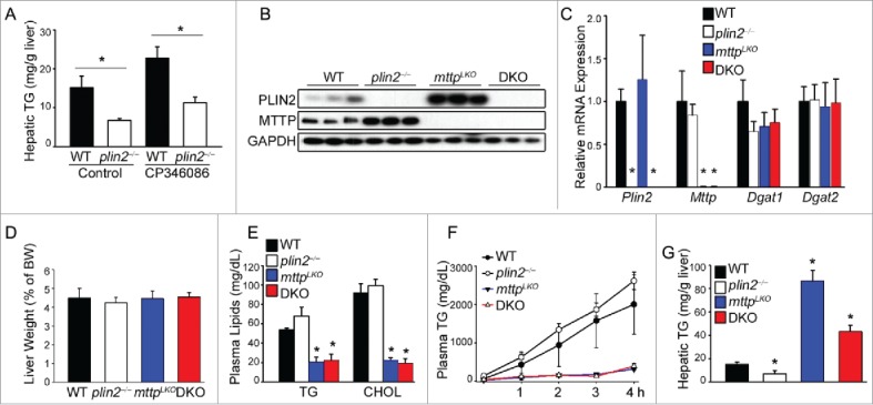
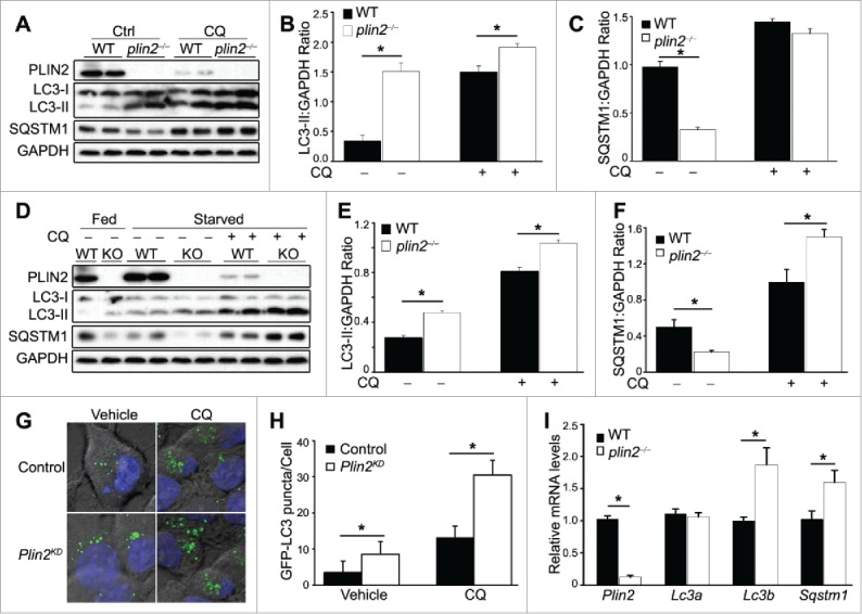
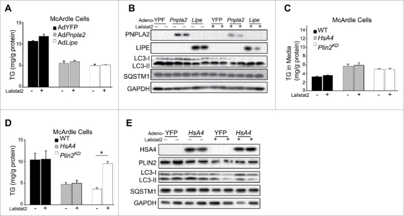
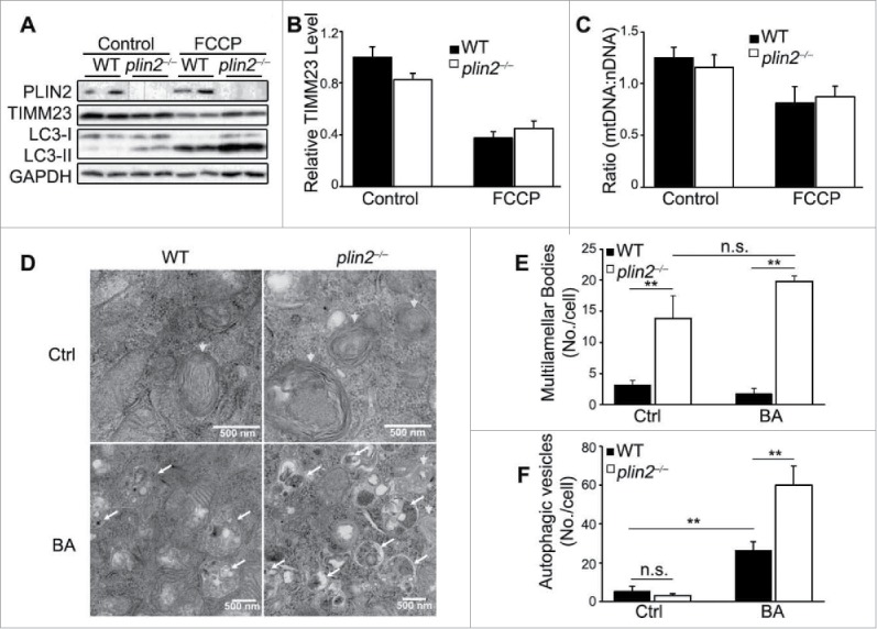
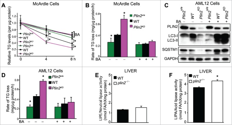
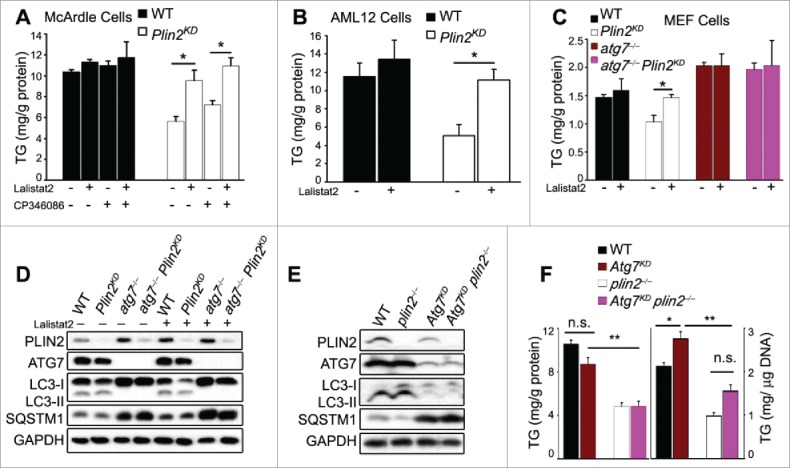
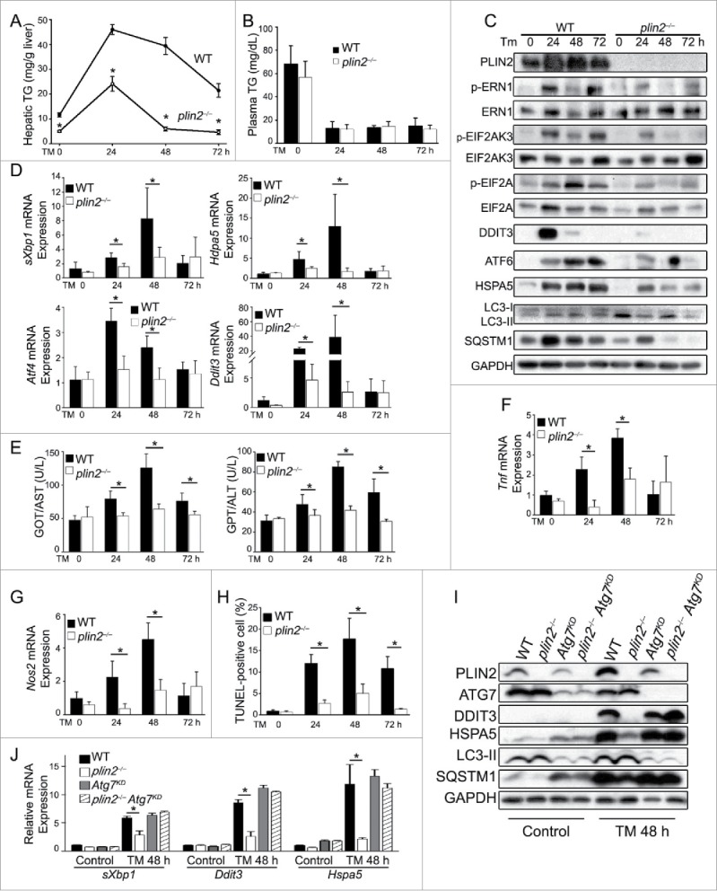
Excess triglyceride (TG) accumulation in the liver underlies fatty liver disease, a highly prevalent ailment. TG occurs in the liver sequestered in lipid droplets, the major lipid storage organelle. Lipid droplets are home to the lipid droplet proteins, the most abundant of which are the perilipins (PLINs), encoded by 5 different genes, Plin1 to Plin5. Of the corresponding gene products, PLIN2 is the only constitutive and ubiquitously expressed lipid droplet protein that has been used as a protein marker for lipid droplets. We and others reported that plin2-/- mice have an ∼60% reduction in TG content, and are protected against fatty liver disease. Here we show that PLIN2 overexpression protects lipid droplets against macroautophagy/autophagy, whereas PLIN2 deficiency enhances autophagy and depletes hepatic TG. The enhanced autophagy in plin2-/- mice protects against severe ER stress-induced hepatosteatosis and hepatocyte apoptosis. In contrast, hepatic TG depletion resulting from other genetic and pharmacological manipulations has no effect on autophagy. Importantly, PLIN2 deficiency lowers cellular TG content in wild-type mouse embryonic fibroblasts (MEFs) via enhanced autophagy, but does not affect cellular TG content in atg7-/- MEFs that are devoid of autophagic function. Conversely, adenovirus-shAtg7-mediated hepatic Atg7 knockdown per se does not alter the hepatic TG level, suggesting a more complex regulation in vivo. In sum, PLIN2 guards its own house, the lipid droplet. PLIN2 overexpression protects against autophagy, and its downregulation stimulates TG catabolism via autophagy.

Keywords: ADRP; PLIN2; autophagy; hepatic TG; lipophagy; neutral lipase.


Figures

Figure 1.
Figure 1.
Plin2 regulates hepatic TG independently of MTTP (microsomal triglyceride transfer protein). (A) Mice were given CP346086 (10 mg/kg body weight) or vehicle once daily for 5 d. Mice (n = 4) were killed 2 h after the last dose and liver removed for TG measurement by thin-layer chromatography. (B) Immunoblot of liver homogenates of WT, plin2−/−, Mttp liver-specific knockout (mttpLKO), and plin2 and mttp double knockout (DKO) mice. (C) Expression of hepatic Plin2, Mttp, Dgat1, and Dgat2 assessed by real-time RT-PCR using liver RNA from WT, plin2−/−, mttpLKO, and DKO mice (n = 6). Liver weight (D), plasma TG and cholesterol (CHOL) (E), hepatic VLDL secretion rate (F), and hepatic TG content (G) of the indicated mouse genotypes (n = 6). VLDL secretion was deduced by injecting Pluronic F127 (an inhibitor of VLDL catabolism) i.p. to mice and measuring plasma TG before (0 h) and hourly after injection (n = 6). *, p < 0.05 compared with the WT control.
Figure 2.
Figure 2.
plin2−/− mice display elevated autophagy in the liver. (A) Immunoblot of liver homogenates of WT and plin2−/− mice (2 representatives shown) with or without chloroquine (CQ) treatment (CQ 100 µM). Quantification of ratios of LC3-II:GAPDH (B) and SQSTM1:GAPDH (C) using immunoblots shown in (A) (n = 6). (D) Immunoblot of primary hepatocytes isolated from WT and plin2−/− mice that were grown in normal medium (Fed) or serum-deprived media for 6 h (Starved) with or without chloroquine (CQ 100 μM) (1 to 2 representatives shown). Quantification of ratios of LC3-II:GAPDH (E) and SQSTM1:GAPDH (F) using immunoblots shown in (D) (n = 6). Representative images (G) and quantification (H) of GFP-LC3 puncta in McArdle RH-7777 control or Plin2 knockdown (Plin2KD) cells transiently transfected with GFP-LC3 plasmid, and treated with vehicle or 100 μM CQ for 6 h. (n = 5). *, p < 0.05 between WT and plin2−/− mice. Quantitative RT-PCR analysis of Plin2, Lc3a, Lc3b and Sqstm1 mRNA expression in the liver tissues (I) isolated from WT or plin2−/− mice (n = 4), with or without Plin2 knockdown or overexpression (n = 4).
Figure 3.
Figure 3.
Changes in cellular TG content per se do not modulate autophagy (A) Cellular TG levels of McArdle cells overexpressing YFP control (Ad-YFP) or PNPLA2 (AdPnpla2), or LIPE (AdLipe) transduced with the respective adenovirus vectors (MOI = 10) for 48 h +/− Lalistat2. (B) Immunoblot of McArdle RH7777 cells studied as outlined in (A). TG secreted into the medium (C) or cellular TG retained in the McArdle RH-7777 cells (D) that were either control (WT), or Plin2 knockdown (Plin2KD), or overexpressing human APOA4 (HsA4) using an adenoviral vector (MOI = 10) for 48 h +/−Lalistat2. (E) Immunoblot of McArdle RH7777 cells studied as outlined in ((C)and D). TG levels were analyzed by enzymatic kits. *, p < 0.05.
Figure 4.
Figure 4.
Plin2 deficiency does not alter mitophagy, but elevates autophagic flux. (A) Immunoblot of primary hepatocytes isolated from WT and plin2−/− mice that were treated with or without FCCP (10 μM) for 6 h. (B) Quantification of ratios of TIMM23:GAPDH using immunoblots shown in (A) (n = 6). (C) Real-time PCR of mitochondrial (mt-Rnr2/16S rRNA) to nuclear DNA (Hk/hexokinase) ratio (n = 6). (D) Electron microscopy imaging of control or plin2−/− AML12 generated by CRISPR technology that are treated with bafilomycin A1 (BA) or with vehicle (Ctrl). Arrowheads, multilamellar bodies (MLB), a special kind of autophagosomes; arrows, autophagic vesicles. Quantitative analysis of MLB (E) and Autophagic vesicles (F) (n = 6). n.s., not significant.
Figure 5.
Figure 5.
Plin2 regulates lipophagy in rat and mouse hepatic cells. (A) TG turnover in McArdle cells transfected with empty vector (WT) or Plin2 plasmid (Plin2o/e for overexpression) or transduced with Lenti-shPlin2 (Plin2KD for knockdown). Cellular lipid accumulation was induced by overnight incubation with medium supplemented with 400 μM oleate complexed to BSA. At time zero, cells were washed and replaced with regular medium with or without bafilomycin A1 (BA) and then harvested immediately or allowed to grow for 4 or 8 h. TG was measured at 0, 4 and 8 h with enzymatic kits (n = 6). (B) Rates of TG turnover in cells under different conditions calculated from (A). (C) Immunoblot of autophagy markers in AML-12 cells transfected with empty vector (WT), Plin2 overexpression vector (Plin2o/e), or transduced with Lenti-shPlin2 (Plin2KD) with or without BA (one representative shown). (D) The rate of TG turnover in WT, Plin2o/e and Plin2KD AML-12 cells treated as outlined in (A). Neutral lipase (E) and LIPA/acid lipase (F) activities measured from liver homogenates of WT and plin2−/− mice (n = 4). *, p < 0.05. FA, fatty acid.
Figure 6.
Figure 6.
Plin2 deficiency-mediated TG reduction requires LIPA. Intracellular TG levels of control (WT) and Plin2KD McArdle (A) and AML-12 cells (B) grown in the presence or absence of Lalistat2 (20 μM; a LIPA inhibitor) and the presence or absence of CP346086 (10 μM; an MTTP inhibitor). (C) Cellular TG levels of MEF cells grown in the presence or absence of Lalistat2. (D) Immunoblot of MEF cell extracts as outlined in (D). (E) Immunoblot of autophagy markers in liver tissue from WT and plin2−/− mice treated with Ad-shAtg7. (F) Total hepatic TG content as outlined in (E) expressed as mg/g protein (left panel) or mg/g DNA (right panel). n.s., not significant.
Figure 7.
Figure 7.
Loss of Plin2 protects against tunicamycin (TM)-induced ER stress via augmented autophagy. TM was injected i.p. (0.5 mg/kg body weight) at 0 h. Hepatic TG (A) and plasma TG (B) were measured in WT and plin2−/− mice before (0 h) or 24, 48, and 72 h after TM (n = 6). (C) Immunoblots of ER stress and autophagy markers in the liver before and after TM injection (one representative shown). (D) Quantitative real-time RT-PCR analyses of ER stress markers sXbp1, Atf4, Ddit3, and Hspa5 on liver RNA isolated before (0 h) or at the indicated times after TM. (E) Plasma GPT/ALT and GOT/AST (n = 6). Quantitative real-time RT-PCR analysis of hepatic Tnf (F) and Nos2 (G) (n = 6). (H) TUNEL-positive cells were identified in liver sections of WT and plin2−/− mice (n = 6). (I) Immunoblots of ER stress and autophagy markers in the livers of WT and plin2−/− mice injected with Ad-GFP (WT and plin2−/−) or Ad-shAtg7 (Atg7KD and plin2−/− Atg7KD) before (Control)and 48 h (TM 48 h) after TM injection (one representative shown). (J) Quantitative real-time RT-PCR analyses of ER stress markers sXbp1, Ddit3, and Hspa5 on liver RNA isolated before (Control) or 48 h after TM (TM 48 h). *, p < 0.05 between WT and plin2−/− mice.

References

    1. Kawano Y, Cohen DE. Mechanisms of hepatic triglyceride accumulation in non-alcoholic fatty liver disease. J Gastroenterol 2013; 48(4):434-41; https://doi.org/10.1007/s00535-013-0758-5 - DOI - PMC - PubMed
    1. Quiroga AD, Lehner R. Liver triacylglycerol lipases. Biochim Biophys Acta 2012; 1821(5):762-9; https://doi.org/10.1016/j.bbalip.2011.09.007 - DOI - PubMed
    1. Czaja MJ, Ding WX, Donohue TM Jr., Friedman SL, Kim JS, Komatsu M, Lemasters JJ, Lemoine A, Lin JD, Ou JH, et al.. Functions of autophagy in normal and diseased liver. Autophagy 2013; 9(8):1131-58. - PMC - PubMed
    1. Sekiya M, Osuga J, Yahagi N, Okazaki H, Tamura Y, Igarashi M, Takase S, Harada K, Okazaki S, Iizuka Y, et al.. Hormone-sensitive lipase is involved in hepatic cholesteryl ester hydrolysis. J Lipid Res 2008; 49(8):1829-38; https://doi.org/10.1194/jlr.M800198-JLR200 - DOI - PubMed
    1. Wu JW, Wang SP, Alvarez F, Casavant S, Gauthier N, Abed L, Soni KG, Yang G, Mitchell GA. Deficiency of liver adipose triglyceride lipase in mice causes progressive hepatic steatosis. Hepatology 2011; 54(1):122-32; https://doi.org/10.1002/hep.24338 - DOI - PubMed