

Microenvironment of a tumor-organoid system enhances hepatocellular carcinoma malignancy-related hallmarks
- PMID: 28548903
- PMCID: PMC5654820
- DOI: 10.1080/15476278.2017.1322243
Microenvironment of a tumor-organoid system enhances hepatocellular carcinoma malignancy-related hallmarks
Abstract
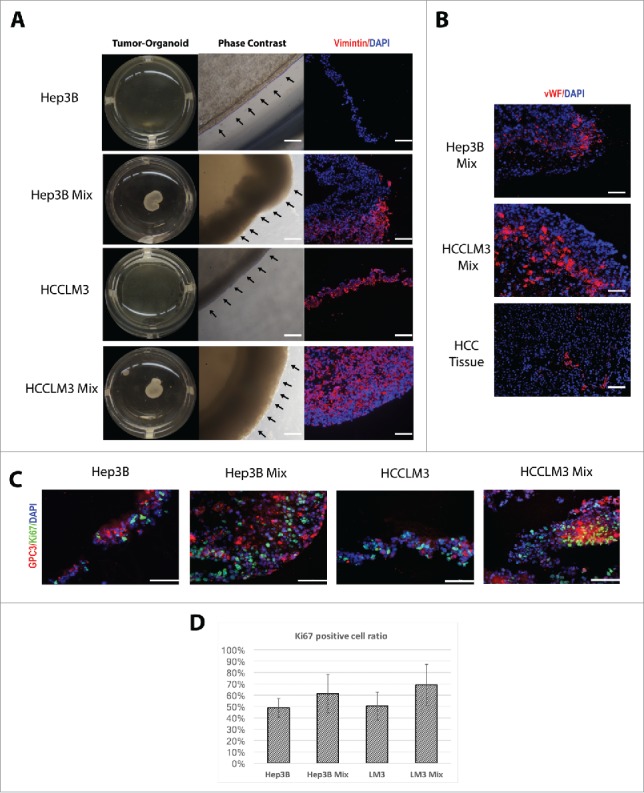
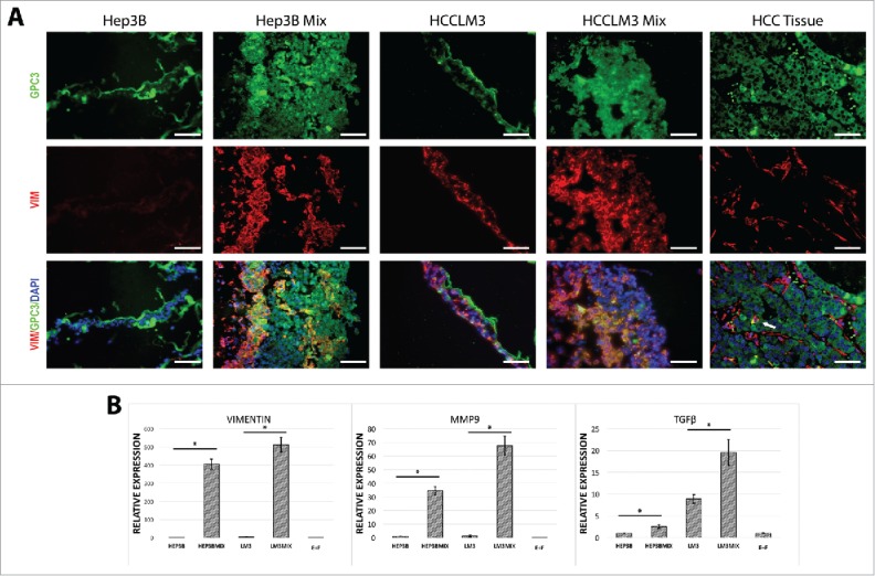
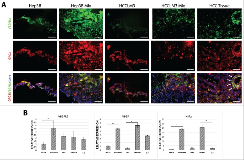
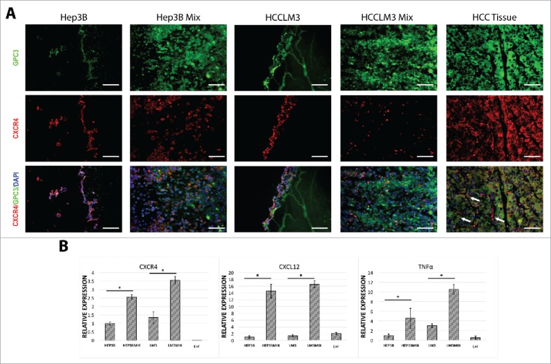
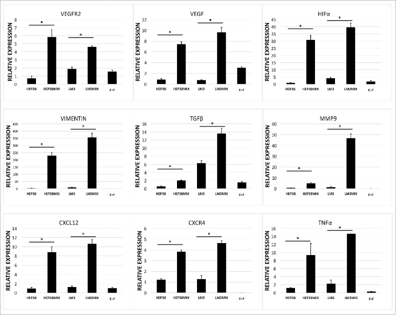
Organ-like microenviroment and 3-dimensional (3D) cell culture conformations have been suggested as promising approaches to mimic in a micro-scale a whole organ cellular functions and interactions present in vivo. We have used this approach to examine biologic features of hepatocellular carcinoma (HCC) cells. In this study, we demonstrate that hepatocellular carcinoma (HCC) cells, fibroblasts, endothelial cells and extracellular matrix can generate organoid-like spheroids that enhanced numerous features of human HCC observed in vivo. We show that the addition of non-parenchymal cells such as fibroblast and endothelial cells is required for spheroid formation as well as the maintenance of the tissue-like structure. Furthermore, HCC cells cultured as spheroids with non-parenchymal cells express more neo-angiogenesis-related markers (VEGFR2, VEGF, HIF-α), tumor-related inflammatory factors (CXCR4, CXCL12, TNF-α) and molecules-related to induced epithelial-mesenchymal transition (TGFβ, Vimentin, MMP9) compared with organoids containing only HCC cells. These results demonstrate the importance of non-parenchymal cells in the cellular composition of HCC organoids. The novelty of the multicellular-based organotypic culture system strongly supports the integration of this approach in a high throughput approach to identified patient-specific HCC malignancy and accurate anti-tumor therapy screening after surgery.
Keywords: endothelial cell; fibroblast; hepatocellular carcinoma; organoid; tumor microenvironment.
Figures
Similar articles
-
Plasma-derived extracellular matrix for xenofree and cost-effective organoid modeling for hepatocellular carcinoma.J Transl Med. 2024 May 21;22(1):487. doi: 10.1186/s12967-024-05230-7. J Transl Med. 2024. PMID: 38773585 Free PMC article.
-
HIF-1α promoted vasculogenic mimicry formation in hepatocellular carcinoma through LOXL2 up-regulation in hypoxic tumor microenvironment.J Exp Clin Cancer Res. 2017 Apr 27;36(1):60. doi: 10.1186/s13046-017-0533-1. J Exp Clin Cancer Res. 2017. PMID: 28449718 Free PMC article.
-
EphA1 activation promotes the homing of endothelial progenitor cells to hepatocellular carcinoma for tumor neovascularization through the SDF-1/CXCR4 signaling pathway.J Exp Clin Cancer Res. 2016 Apr 11;35:65. doi: 10.1186/s13046-016-0339-6. J Exp Clin Cancer Res. 2016. PMID: 27066828 Free PMC article.
-
Exosome-mediated communication in the tumor microenvironment contributes to hepatocellular carcinoma development and progression.J Hematol Oncol. 2019 May 29;12(1):53. doi: 10.1186/s13045-019-0739-0. J Hematol Oncol. 2019. PMID: 31142326 Free PMC article. Review.
-
Components of the Hepatocellular Carcinoma Microenvironment and Their Role in Tumor Progression.Biochemistry (Mosc). 2017 Aug;82(8):861-873. doi: 10.1134/S0006297917080016. Biochemistry (Mosc). 2017. PMID: 28941454 Review.
Cited by
-
Tumoroids, a valid preclinical screening platform for monitoring cancer angiogenesis.Stem Cell Res Ther. 2024 Aug 26;15(1):267. doi: 10.1186/s13287-024-03880-4. Stem Cell Res Ther. 2024. PMID: 39183337 Free PMC article. Review.
-
Organoids of liver diseases: From bench to bedside.World J Gastroenterol. 2019 Apr 28;25(16):1913-1927. doi: 10.3748/wjg.v25.i16.1913. World J Gastroenterol. 2019. PMID: 31086460 Free PMC article.
-
Liver organoids: a promising three-dimensional model for insights and innovations in tumor progression and precision medicine of liver cancer.Front Immunol. 2023 Jun 2;14:1180184. doi: 10.3389/fimmu.2023.1180184. eCollection 2023. Front Immunol. 2023. PMID: 37334366 Free PMC article. Review.
-
Promising Applications of Tumor Spheroids and Organoids for Personalized Medicine.Cancers (Basel). 2020 Sep 23;12(10):2727. doi: 10.3390/cancers12102727. Cancers (Basel). 2020. PMID: 32977530 Free PMC article. Review.
-
Organoid as a promising tool for primary liver cancer research: a comprehensive review.Cell Biosci. 2024 Aug 27;14(1):107. doi: 10.1186/s13578-024-01287-5. Cell Biosci. 2024. PMID: 39192365 Free PMC article. Review.
References
-
- Organization WH. World Cancer Report 2014. World Health Organization: International Agency for Research on Cancer; 2014.
-
- Wang FS, Fan JG, Zhang Z, Gao B, Wang HY. The global burden of liver disease: the major impact of China. Hepatology 2014; 60(6):2099-108; PMID:25164003; https://doi.org/10.1002/hep.27406 - DOI - PMC - PubMed
-
- Forner A, Llovet JM, Bruix J. Hepatocellular carcinoma. Lancet 2012; 379(9822):1245-55; PMID:22353262; https://doi.org/10.1016/S0140-6736(11)61347-0 - DOI - PubMed
-
- Shibata T, Aburatani H. Exploration of liver cancer genomes. Nat Rev Gastroenterol Hepatol 2014; 11(6):340-9; PMID:24473361; https://doi.org/10.1038/nrgastro.2014.6 - DOI - PubMed
-
- Tao J, Xu E, Zhao Y, Singh S, Li X, Couchy G, Chen X, Zucman-Rossi J, Chikina M, Monga SP. Modeling a human hepatocellular carcinoma subset in mice through coexpression of met and point-mutant beta-catenin. Hepatology 2016; 64(5):1587-605; PMID:27097116; https://doi.org/10.1002/hep.28601 - DOI - PMC - PubMed
Publication types
MeSH terms
Substances
LinkOut - more resources
Full Text Sources
Other Literature Sources
Medical
Miscellaneous