. 2017 May 1;58(5):2785-2795.
doi: 10.1167/iovs.17-21826.

KLF4 Plays an Essential Role in Corneal Epithelial Homeostasis by Promoting Epithelial Cell Fate and Suppressing Epithelial-Mesenchymal Transition

Affiliations

KLF4 Plays an Essential Role in Corneal Epithelial Homeostasis by Promoting Epithelial Cell Fate and Suppressing Epithelial-Mesenchymal Transition

Anil Tiwari et al. Invest Ophthalmol Vis Sci. .

Abstract

Purpose: The purpose of this study was to test the hypothesis that KLF4 promotes corneal epithelial (CE) cell fate by suppressing the epithelial-mesenchymal transition (EMT), using spatiotemporally regulated CE-specific ablation of Klf4 in Klf4Δ/ΔCE (Klf4LoxP/LoxP/Krt12rtTA/rtTA/Tet-O-Cre) mice.

Methods: CE-specific ablation of Klf4 was achieved by feeding Klf4Δ/ΔCE mice with doxycycline chow. The wild-type (WT; normal chow-fed littermates) and the Klf4Δ/ΔCE histology was compared by hematoxylin and eosin-stained sections; EMT marker expression was quantified by quantitative PCR, immunoblots, and immunofluorescent staining; and wound healing rate was measured by CE debridement using Algerbrush. KLF4 and EMT markers were quantified in human corneal limbal epithelial (HCLE) cells undergoing TGF-β1-induced EMT by quantitative PCR, immunoblots, and immunofluorescent staining.

Results: The epithelial markers E-cadherin, Krt12, claudin-3, and claudin-4 were down-regulated, whereas the mesenchymal markers vimentin, β-catenin, survivin, and cyclin-D1 and the EMT transcription factors Snail, Slug, Twist1, Twist2, Zeb1, and Zeb2 were up-regulated in the Klf4Δ/ΔCE corneas. The Klf4Δ/ΔCE cells migrated faster, filling 93% of the debrided area within 16 hours compared with 61% in the WT. After 7 days of wounding, the Klf4Δ/ΔCE cells that filled the gap failed to regain epithelial characteristics, as they displayed abnormal stratification; down-regulation of E-cadherin and Krt12; up-regulation of β-catenin, survivin, and cyclin-D1; and a 2.5-fold increase in the number of proliferative Ki67+ cells. WT CE cells at the migrating edge and the HCLE cells undergoing TGF-β1-induced EMT displayed significant down-regulation of KLF4.

Conclusions: Collectively, these results reveal that KLF4 plays an essential role in CE homeostasis by promoting epithelial cell fate and suppressing EMT.


Figures
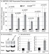
Figure 1
Figure 1
Up-regulation of EMT-associated transcription factors in the Klf4Δ/ΔCE CE. (A) qRT-PCR for EMT transcription factors. qPCR was performed in duplicate using three different pools of WT and Klf4Δ/ΔCE corneal cDNA, each generated using total RNA from two corneas of different mice. Results shown are mean ± SEM. P ≤ 0.05 was considered statistically significant. The sequence of oligonucleotide primers used is shown in Supplementary Table S1. (B) Immunoblots for representative EMT-transcription factors Slug and Twist1. The blot was stripped of the antibody and reprobed with anti-actin antibody for normalization. (C) Densitometric scan from three independent replicates using actin as a loading control. Results shown are mean ± SEM.
Figure 2
Figure 2
Down-regulation of epithelial markers and up-regulation of mesenchymal marker vimentin in the Klf4Δ/ΔCE CE. (A) qRT-PCR for EMT markers. qPCR was performed in duplicate using three different pools of WT and Klf4Δ/ΔCE corneal cDNA, each generated using total RNA from two corneas of different mice. (B) Immunoblot shows increased expression of vimentin in the Klf4Δ/ΔCE CE compared with the WT. (C) Histogram showing densitometric quantitation from three independent replicates, using actin as a loading control. Results shown are mean ± SEM; P ≤ 0.05 was considered statistically significant. (D) Immunofluorescent stain shows robust expression of vimentin in the Klf4Δ/ΔCE (arrows) but not the WT CE (arrowheads). No primary antibody control is shown.
Figure 3
Figure 3
E-cadherin is down-regulated in the Klf4Δ/ΔCE CE. (A) Immunoblot shows decreased expression of E-cadherin in the Klf4Δ/ΔCE CE compared with the WT. (B) Histogram showing densitometric quantitation from three independent replicates, using actin as a loading control. Results shown are mean ± SEM; P ≤ 0.05 was considered statistically significant. (C) Immunofluorescent stain shows decreased expression of E-cadherin in the Klf4Δ/ΔCE compared with the WT CE. Note that E-cadherin is localized predominantly on the cell membranes in the WT but not the Klf4Δ/ΔCE CE.
Figure 4
Figure 4
Increased expression and nuclear translocation of β-catenin in Klf4Δ/ΔCE CE. (A) Immunoblot shows increased expression of β-catenin in the Klf4Δ/ΔCE CE compared with the WT. (B) Histogram showing densitometric quantitation from three independent replicates, using actin as a loading control. Results are mean ± SEM; P ≤ 0.05 was considered statistically significant. (C) Immunofluorescent stain shows increased expression and nuclear translocation of β-catenin in Klf4Δ/ΔCE (arrows) compared with the WT CE, where the base level expression of β-catenin is mostly localized to the cell membrane and not the nucleus (arrowheads). The two panels in the bottom show enlarged images of the corresponding inset regions from Cii and Ciii.
Figure 5
Figure 5
Altered expression of survivin and cyclin-D in the Klf4Δ/ΔCE CE. (AC) Increased number of survivin-positive cells in the Klf4Δ/ΔCE compared with the WT CE, revealed by immunofluorescent staining. Corresponding histogram shows the mean number of survivin-positive cells per unit area, using data from three independent replicates. (DF) Increased expression of cyclin-D1 in the Klf4Δ/ΔCE cells compared with the WT. Corresponding histograms show relative fluorescence intensities measured throughout the CE. Results are mean ± SEM; P ≤ 0.05 was considered statistically significant.
Figure 6
Figure 6
KLF4 is down-regulated in HCLE cells undergoing TGF-β1–induced EMT. (A) Phase contrast images of control HCLE cells and those treated for 48 hours with TGF-β1. Note that the TGF-β1–treated HCLE cells are elongated and more spindle shaped compared with the untreated control. (B) Immunofluorescent stain reveals decreased expression of epithelial marker E-cadherin and increased expression, as well as nuclear localization of mesenchymal marker β-catenin in TGF-β1–treated HCLE cells (arrows) compared with the vehicle-treated control cells (arrowheads). (C.i) qPCR showing the relative KLF4 mRNA levels in the control and TGF-β1–treated HCLE cells. (C.ii) Immunoblot probed with anti-KLF4 antibody, showing the decreased expression of KLF4 in TGF-β1–treated HCLE cells compared with the control. (C.iii) Histogram showing densitometric quantitation from three independent immunoblots. Results are mean ± SEM; P ≤ 0.05 was considered statistically significant. (C.iv) Immunofluorescent stain for KLF4 in HCLE cells treated with and without (control) TGF-β1. Arrows in C.iv.a point to nuclear expression of KLF4 in control vehicle-treated HCLE cells. Arrowheads in C.iv.b point to the nuclei of TGF-β1–treated HCLE cells that lack KLF4 expression.
Figure 7
Figure 7
After debridement, Klf4Δ/ΔCE cells migrate faster and display abnormal morphology. (A) Representative images showing the wounded area detected by fluorescein staining at 0 and 16 hours after debridement in the WT and Klf4Δ/ΔCE CE. (B) Histogram showing the relative wound area (fluorescein-stained area) at 0 and 16 hours after debridement in the WT and Klf4Δ/ΔCE CE. (C) Histology of debrided WT and Klf4Δ/ΔCE corneas at 16 hours and 7 days after debridement. (D) Immunofluorescent stain with anti-laminin antibody at 16 hours after debridement reveals intact basement membrane in debrided WT and Klf4Δ/ΔCE. Note that the debrided gap remains in the WT, but not the Klf4Δ/ΔCE CE at 16 hours after debridement. (E) Immunofluorescent stain with anti-Klf4 antibody at 16 hours after debridement reveals markedly decreased expression of Klf4 in the WT CE cells at the migrating edge (arrowheads), unlike the cells away from the wound (arrows) where it is abundantly expressed.
Figure 8
Figure 8
Enhanced signs of EMT in Klf4Δ/ΔCE CE after 7 days of wounding. Immunofluorescent stain and corresponding histogram showing the relative expression of EMT-associated proteins (AC) Ki67, (DF) survivin, and (GI) cyclin-D1 in the WT and Klf4Δ/ΔCE cornea. Results presented in histograms are mean ± SEM. P ≤ 0.05 was considered statistically significant.
Figure 9
Figure 9
Migrated Klf4Δ/ΔCE CE cells fail to revert to epithelial phenotype. Immunofluorescent staining and histograms of corresponding fluorescence intensities of re-epithelialized WT and Klf4Δ/ΔCE cells 7 days after corneal debridement with (AC) anti-KLF4 antibody and (DF) CE-specific marker anti–keratin-12. Results presented in histograms are mean ± SEM. P ≤ 0.05 was considered statistically significant.
Figure 10
Figure 10
Migrated Klf4Δ/ΔCE CE cells maintain mesenchymal characteristics. Images showing immunofluorescent stain and histogram of corresponding fluorescence intensities for epithelial marker E-cadherin (AC) and mesenchymal marker β-catenin (DF) in the WT and Klf4Δ/ΔCE corneas 7 days after wounding. Results presented in histograms are mean ± SEM. P ≤ 0.05 was considered statistically significant.
Figure 11
Figure 11
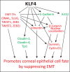
Schematic representation of a model for the role of Klf4 in promoting CE cell fate by suppressing EMT. The results presented in this manuscript demonstrate that Klf4 suppresses the expression of EMT transcription factors, vimentin, cyclin-D1, and survivin (indicated in red and connected by blunt-ended lines) and activates the expression of tight junction proteins Tjp1, claudin-3, and claudin-4 and CE markers Krt12 and E-cadherin (indicated in green and connected by pointed arrows). Collectively, these results reveal that KLF4 promotes corneal epithelial cell fate by suppressing EMT.

Similar articles

Cited by

References

    1. McDade SS,, Henry AE,, Pivato GP,, et al. Genome-wide analysis of p63 binding sites identifies AP-2 factors as co-regulators of epidermal differentiation. Nucleic Acids Res. 2012; 40: 7190–7206. - PMC - PubMed
    1. Stephens DN,, Klein RH,, Salmans ML,, Gordon W,, Ho H,, Andersen B. The Ets transcription factor EHF as a regulator of cornea epithelial cell identity. J Biol Chem. 2013; 288: 34304–34324. - PMC - PubMed
    1. Kouwenhoven EN,, Oti M,, Niehues H,, et al. Transcription factor p63 bookmarks and regulates dynamic enhancers during epidermal differentiation. EMBO Rep. 2015; 16: 863–878. - PMC - PubMed
    1. Flandez M,, Guilmeau S,, Blache P,, Augenlicht LH. KLF4 regulation in intestinal epithelial cell maturation. Exp Cell Res. 2008; 314: 3712–3723. - PMC - PubMed
    1. Thiery JP,, Sleeman JP. Complex networks orchestrate epithelial-mesenchymal transitions. Nat Rev Mol Cell Biol. 2006; 7: 131–142. - PubMed