

Kimura's disease affecting the axillary lymph nodes: a case report
- PMID: 28549475
- PMCID: PMC5446720
- DOI: 10.1186/s12893-017-0260-8
Kimura's disease affecting the axillary lymph nodes: a case report
Abstract
Background: Kimura's disease (KD; eosinophilic granuloma of soft tissue) is an inflammatory granulomatous disorder of unknown cause with eosinophilic infiltration that occurs mainly in soft tissue. KD occurs mainly in the head and neck, but development in the axillary region is very rare.
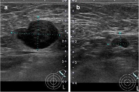
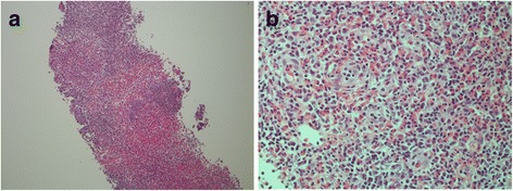
Case presentation: A 74-year-old Japanese woman was evaluated for a mass that she noted in the left axillary region. On physical examination, there was a palpable, thumb-sized, hard, elastic, freely movable mass in the left axilla. Blood tests showed elevated soluble interleukin-2 receptor (sIL-2R), normal serum immunoglobulin (Ig) G4, and elevated serum IgE. Ultrasonography of the left axilla showed multiple lymph nodes (LNs) with irregular margins in which central hyperechogenicity was lost. A systemic search by computed tomography (CT) showed no systemic lymphadenopathy or other mass-like lesions suspicious for a primary tumour other than in the left axillary LNs. Biopsy of an excised LN was performed under local anaesthesia for a definitive diagnosis. Histopathology showed various-sized lymphoid follicles, large nodular lesions with an enlarged mantle zone, multiple various-sized germinal centres in single nodules, and eosinophilic infiltration between the nodes. Immunohistochemical (IHC) staining of the germinal centres was positive for cluster of differentiation (CD) 10, positive for B-cell lymphoma (bcl)-6, and negative for bcl-2. These findings led to a diagnosis of KD. Ultrasound after 3 months of follow-up showed disappearance of the axillary lymphadenopathy.
Conclusions: A very rare case of KD in the axillary LNs was described. KD has the potential to occur in any region.
Keywords: Axillary lymph nodes; Biopsy; Eosinophilic granuloma; IgE-RIST; Kimura’s disease.
Figures


References
-
- Abuel-Haija M, Hurford MT. Kimura disease. Arch Pathol Lab Med. 2007;131:650–651. - PubMed
-
- Kimura T, Yoshimura S, Ishikawa E. On the unusual granula- tion combined with hyperplastic changes of lymphatic tissue. Trans Soc Pathol Jpn. 1948;37:179–180.
Publication types
MeSH terms
Substances
LinkOut - more resources
Full Text Sources
Other Literature Sources
