Review
. 2018 Feb 16;430(4):424-437.
doi: 10.1016/j.jmb.2017.05.021. Epub 2017 May 23.

Applications of Single-Molecule Methods to Membrane Protein Folding Studies

Affiliations
Review

Applications of Single-Molecule Methods to Membrane Protein Folding Studies

Robert E Jefferson et al. J Mol Biol. .

Abstract

Protein folding is a fundamental life process with many implications throughout biology and medicine. Consequently, there have been enormous efforts to understand how proteins fold. Almost all of this effort has focused on water-soluble proteins, however, leaving membrane proteins largely wandering in the wilderness. The neglect has occurred not because membrane proteins are unimportant but rather because they present many theoretical and technical complications. Indeed, quantitative membrane protein folding studies are generally restricted to a handful of well-behaved proteins. Single-molecule methods may greatly alter this picture, however, because the ability to work at or near infinite dilution removes aggregation problems, one of the main technical challenges of membrane protein folding studies.

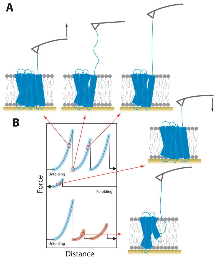
Keywords: atomic force spectroscopy; fluorescence; forced unfolding; magnetic tweezer.


Figures

Figure 1
Figure 1. Single-molecule fluorescence measurements of membrane protein oligomerization
(A) Measuring membrane protein oligomerization in isolated lipid vesicles immobilized on a surface via DNA-biotin-avidin tethers. Expected fluorescence signals from a line across the confocal plane are plotted from aggregated protein (i), empty vesicles (ii), homo-oligomers (iii), monomers (iv), and hetero-oligomers (v). (B) Large multilamellar vesicles containing fluorescently labeled protein (i) are converted to small unilammelar vesicles (ii) by extrusion, preserving the oligomeric equilibrium distribution in the large vesicles. Photobleaching events are counted in extruded vesicles, from which the fraction of dimeric complexes can be calculated as a function of protein concentrations (iii).
Figure 2
Figure 2. Single-molecule FRET measurements of membrane protein folding
(A) Example trajectory of a membrane protein undergoing folding and unfolding transitions through the confocal excitation volume. (B) Mistic in a micelle environment unfolded with urea (triangles). (C) Histogram of FRET efficiencies from recorded single-molecule bursts. Low FRET efficiencies represent the fraction of unfolded molecules (green), while high FRET efficiencies represent folded molecules (red). The mid-range FRET efficiencies arise from molecules undergoing fast folding and unfolding transitions during single bursts, reflecting the unfolding and folding rates, ku and kf.
Figure 3
Figure 3
Unfolding of a membrane protein using atomic force spectroscopy. (A) Schematic of forced unfolding of a membrane protein. The AFM stylus is shown in gray and the black arrows show the direction of tip movement. (B) Illustration of representative force-distance curves from unfolding, refolding, and a second cycle of unfolding. Force peaks matching the unfolding segments in the first pull are highlighted in blue. Misfolded segments whose force peaks do not match are highlighted in red.
Figure 4
Figure 4
Membrane protein unfolding domains from AFM-based single-molecule force spectroscopy of (A) bacteriorhodopsin pulled from the C-terminus [69], (B) halorhodopsin pulled from the C-terminus [112] and (C) N-terminus [112], (D) LacY pulled from the C-terminus [87], (E) DtpA pulled from the N-terminus [86], (F) and C-terminus [86], (G) β2AR pulled from the N-terminus [88], (H) bovine rhodopsin pulled from the N-terminus [84], and (I) NhaA pulled from the C-terminus [72] and (J) N-terminus [72]. Unfolding domains are highlighted by color and the start of each segment is labeled with the contour length of the corresponding force peak. Black arrows show position of the applied pulling force.
Figure 5
Figure 5
Forced unfolding of GlpG using magnetic tweezers. (A) Schematic of the experimental set up for unfolding a membrane protein in a bicelle using magnetic tweezers. (B) Force-extension curves for repeated unfolding and refolding of GlpG. (C) The energy landscape for second-stage folding of GlpG in bicelles determined from magnetic tweezer experiments [95].

Similar articles

Cited by

References

    1. Bowie JU. Membrane proteins: A new method enters the fold. Proc Natl Acad Sci. 2004;101:3995–3996. doi: 10.1073/pnas.0400671101. - DOI - PMC - PubMed
    1. Dalbey RE, Wang P, Kuhn A. Assembly of Bacterial Inner Membrane Proteins. Annu Rev Biochem. 2011;80:161–187. doi: 10.1146/annurev-biochem-060409-092524. - DOI - PubMed
    1. Knowles TJ, Scott-Tucker A, Overduin M, Henderson IR. Membrane protein architects: the role of the BAM complex in outer membrane protein assembly. Nat Rev Microbiol. 2009;7:206–214. doi: 10.1038/nrmicro2069. - DOI - PubMed
    1. White SH, von Heijne G. The machinery of membrane protein assembly. Curr Opin Struct Biol. 2004;14:397–404. doi: 10.1016/j.sbi.2004.07.003. - DOI - PubMed
    1. White SH, Wimley WC. Membrane protein folding and stability: physical principles. Annu Rev Biophys Biomol Struct. 1999;28:319–365. - PubMed

Publication types

MeSH terms