Review
. 2017 Jun 7;12(6):983-997.
doi: 10.2215/CJN.11761116. Epub 2017 May 26.

Primary Membranous Nephropathy

Affiliations
Review

Primary Membranous Nephropathy

William G Couser. Clin J Am Soc Nephrol. .

Erratum in

  • Correction.
    [No authors listed] [No authors listed] Clin J Am Soc Nephrol. 2017 Sep 7;12(9):1528. doi: 10.2215/CJN.07190717. Epub 2017 Aug 3. Clin J Am Soc Nephrol. 2017. PMID: 28775127 Free PMC article. No abstract available.

Abstract

Membranous nephropathy (MN) is a unique glomerular lesion that is the most common cause of idiopathic nephrotic syndrome in nondiabetic white adults. About 80% of cases are renal limited (primary MN, PMN) and 20% are associated with other systemic diseases or exposures (secondary MN). This review focuses only on PMN. Most cases of PMN have circulating IgG4 autoantibody to the podocyte membrane antigen PLA2R (70%), biopsy evidence PLA2R staining indicating recent immunologic disease activity despite negative serum antibody levels (15%), or serum anti-THSD7A (3%-5%). The remaining 10% without demonstrable anti-PLA2R/THSd7A antibody or antigen likely have PMN probably secondary to a different, still unidentified, anti-podocyte antibody. Considerable clinical and experimental data now suggests these antibodies are pathogenic. Clinically, 80% of patients with PMN present with nephrotic syndrome and 20% with non-nephrotic proteinuria. Untreated, about one third undergo spontaneous remission, especially those with absent or low anti-PLA2R levels, one-third progress to ESRD over 10 years, and the remainder develop nonprogressive CKD. Proteinuria can persist for months after circulating anti-PLA2R/THSD7A antibody is no longer detectable (immunologic remission). All patients with PMN should be treated with supportive care from the time of diagnosis to minimize protein excretion. Patients with elevated anti-PLA2R/THSD7A levels and proteinuria >3.5 g/d at diagnosis, and those who fail to reduce proteinuria to <3.5 g after 6 months of supportive care or have complications of nephrotic syndrome, should be considered for immunosuppressive therapy. Accepted regimens include steroids/cyclophosphamide, calcineurin inhibitors, and B cell depletion. With proper management, only 10% or less will develop ESRD over the subsequent 10 years.

Keywords: Adult; B-Lymphocytes; Biopsy; Calcineurin Inhibitors; Chronic; Cyclophosphamide; Glomerulonephritis; Humans; Immunoglobulin G; Kidney Failure; Kidney Glomerulus; Lipoid; Membranous; Nephrosis; PLA2R; PLA2R1 protein; Phospholipase A2; Podocytes; Receptors; Remission; Renal Insufficiency; Spontaneous; Staining and Labeling; THSD7A; congenital; human; membranous nephropathy; nephrotic syndrome; proteinuria.


Figures

Figure 1.
Figure 1.
Glomerulus from a patient with primary membranous nephropathy showing the pathognomonic “spikes” of basement membrane projecting from the outer surface of the glomerular basement membrane (arrows) when stained with silver-methenamine (original magnification, ×40). (Provided by Dr. Charles Alpers, Department of Pathology, University of Washington, Seattle, WA.)
Figure 2.
Figure 2.
Immunofluorescence microscopy in primary membranous nephropathy (PMN). (A) Finely granular staining for IgG, predominately IgG4, present uniformly in a subepithelial distribution in all glomeruli in a patient with PLA2R-associated PMN (original magnification, ×40) (generously provided by Dr. Charles Alpers, Department of Pathology, University of Washington, Seattle, WA). (B) Finely granular staining for PLA2R antigen that colocalizes with IgG4 in a patient with PMN. The presence of PLA2R indicates that anti-PLA2R antibody was present and forming deposits in glomeruli at the time of biopsy or within the past several weeks. (original magnification, ×40) (generously provided by Dr. Charles Alpers, Department of Pathology, University of Washington, Seattle, WA). (C) Finely granular staining for the complement membrane attack complex, C5b-9, in a patient with active PMN (original magnification, ×40). Reprinted from reference , with permission.
Figure 3.
Figure 3.
Electron micrograph of chronic primary membranous nephropathy showing discontinuous, electron-dense deposits representing aggregates of PLA2R–anti-PLA2R immune complexes formed in situ along the outer surface of the glomerular capillary wall beneath a layer of effaced podocyte foot processes (arrows). BM, basement membrane; CL, capillary lumen. Original photomicrograph generously provided by Dr. Charles Alpers, Department of Pathology, University of Washington, Seattle, WA.
Figure 4.
Figure 4.
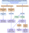
Antibody-guided diagnosis and treatment algorithm for primary membranous nephropathy (PMN). Patients who undergo biopsies for proteinuria of uncertain cause who have MN should initially be classified as anti-PLA2R/THSD7A–positive (active disease) or negative (Table 2). Patients who are antibody-negative should have the absence of a PLA2R/THSD7A-related mechanism further excluded by the absence of PLA2R/THSD7A staining in glomeruli and usually the dominance of IgG1–3 in the biopsy sample. Most of these latter patients have secondary MN and require other tests to identify the cause. They are treated with supportive care and therapy for their underlying systemic disease. Patients who are negative for anti-PLA2R/THSD7A in the serum but have PLA2R or THSD7A antigen staining in the biopsy sample, and usually predominately IgG4 deposition in glomeruli, have inactive anti-PLA2R/THSD7A–mediated PMN and should be treated with supportive care while monitoring anti-PLA2R levels for 4–6 months. They would be considered for IST only if anti-PLA2R becomes positive or proteinuria>3.5 g/d persists after 6 months of supportive care. Patients with elevated anti-PLA2R levels (with positive PLA2R staining and [usually] predominantly IgG4 in the biopsy sample) and proteinuria <3.5 g/d have active anti-PLA2R/THSD7A–mediated PMN but would receive supportive care with monthly anti-PLA2R monitoring because most of these patients will undergo spontaneous remission. If patients have, or develop, elevated anti-PLA2R levels and >3.5 g/d of proteinuria they have active, anti-PLA2R/THSD7A–mediated PMN and would be considered for immediate IST. IST options would be selected on the basis of characteristics of individual patients with dose and duration of therapy (Table 6) guided by regular monitoring of anti-PLA2R levels. About 10% of patients with anti-PLA2R/THSD7A–negative antibody and glomerular staining have PMN presumably mediated by a different anti-podocyte antibody rather than secondary MN and would be treated with traditional restrictive care (4,52,53). Occasional patients with negative anti-PLA2R antibody but dominant IgG4 in the biopsy sample have also been reported and should be monitored for later development of positive circulating anti-PLA2R antibody. ANA, anti-nuclear antibody; HBV, hepatitis B; HCV, hepatitis C; IST, immunosuppressive therapy; MN, membranous nephropathy. Modified from other schemas in references and , with permission.

Comment in

References

    1. Cattran DC, Brenchley PE: Membranous nephropathy: Integrating basic science into improved clinical management. Kidney Int 91: 566–574, 2017 - PubMed
    1. Salant DJ, Cattran DC: Membranous nephropathy. Chapter 20. In: Comprehensive Clinical Nephrology, 5th Ed., edited by Floege J, Johnson RJ, Feehally J, St. Louis, MI, Saunders, an imprint of Elsevier Inc., 2015, pp 239–251
    1. Kumar V, Ramachandran R, Kumar A, Nada R, Suri D, Gupta A, Kohli HS, Gupta KL, Jha V: Antibodies to m-type phospholipase A2 receptor in children with idiopathic membranous nephropathy. Nephrology (Carlton) 20: 572–575, 2015 - PubMed
    1. De Vriese AS, Glassock RJ, Nath KA, Sethi S, Fervenza FC: A proposal for a serology-based approach to membranous nephropathy. J Am Soc Nephrol 28: 421–430, 2016 - PMC - PubMed
    1. Francis JM, Beck LH Jr., Salant DJ: Membranous nephropathy: A journey from bench to bedside. Am J Kidney Dis 68: 138–147, 2016 - PMC - PubMed

MeSH terms