. 2017 Aug 1;10(8):1015-1025.
doi: 10.1242/dmm.028852. Epub 2017 May 26.

A novel experimental rat model of peripheral nerve scarring that reliably mimics post-surgical complications and recurring adhesions

Affiliations

A novel experimental rat model of peripheral nerve scarring that reliably mimics post-surgical complications and recurring adhesions

Angela Lemke et al. Dis Model Mech. .

Abstract

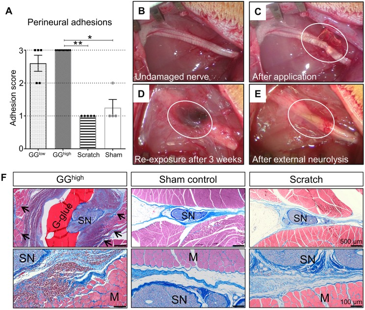
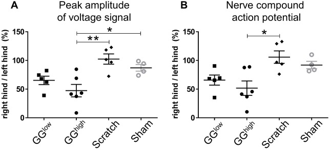
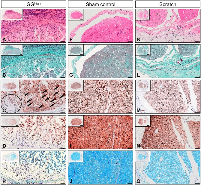
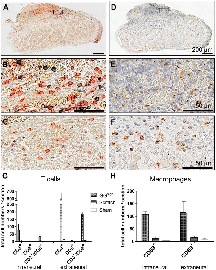
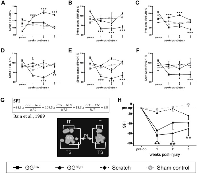
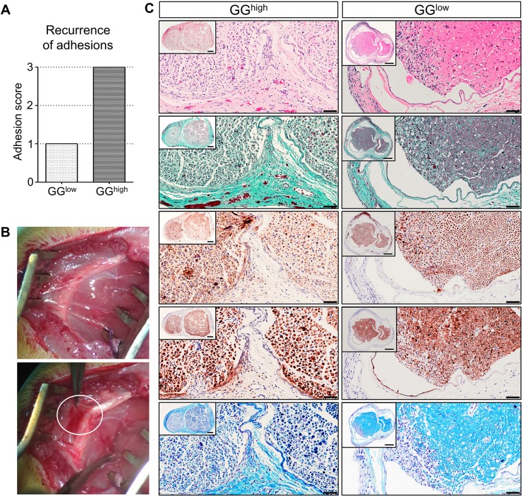
Inflammation, fibrosis and perineural adhesions with the surrounding tissue are common pathological processes following nerve injury and surgical interventions on peripheral nerves in human patients. These features can reoccur following external neurolysis, currently the most common surgical treatment for peripheral nerve scarring, thus leading to renewed nerve function impairment and chronic pain. To enable a successful evaluation of new therapeutic approaches, it is crucial to use a reproducible animal model that mimics the main clinical symptoms occurring in human patients. However, a clinically relevant model combining both histological and functional alterations has not been published to date. We therefore developed a reliable rat model that exhibits the essential pathological processes of peripheral nerve scarring. In our study, we present a novel method for the induction of nerve scarring by applying glutaraldehyde-containing glue that is known to cause nerve injury in humans. After a 3-week contact period with the sciatic nerve in female Sprague Dawley rats, we could demonstrate severe intra- and perineural scarring that resulted in grade 3 adhesions and major impairments in the electrophysiological peak amplitude compared with sham control (P=0.0478). Immunohistochemical analysis of the nerve structure revealed vigorous nerve inflammation and recruitment of T cells and macrophages. Also, distinct nerve degeneration was determined by immunostaining. These pathological alterations were further reflected in significant functional deficiencies, as determined by the analysis of relevant gait parameters as well as the quantification of the sciatic functional index starting at week 1 post-operation (P<0.01). Moreover, with this model we could, for the first time, demonstrate not only the primary formation, but also the recurrence, of severe adhesions 1 week after glue removal, imitating a major clinical challenge. As a comparison, we tested a published model for generating perineural fibrotic adhesions, which did not result in significant pathological changes. Taken together, we established an easily reproducible and reliable rat model for peripheral nerve scarring that allows for the effective testing of new therapeutic strategies.

Keywords: Nerve fibrosis; Nerve inflammation; Nerve scarring; Perineural adhesions; Peripheral nerve adhesions.


Conflict of interest statement

Competing interestsThe authors declare no competing or financial interests.

Figures

Fig. 1.
Fig. 1.
Analysis of perineural adhesions between the sciatic nerve and the surrounding tissue 3 weeks after primary surgery. (A) Rats belonging to the GGhigh group (n=8) exhibited distinct adhesive fibrotic tissue requiring sharp dissection during external neurolysis, whereas sham (n=4, P=0.0125) and scratch (n=5, P=0.0012) groups had no or mild adherence . Rats of the GGlow group (n=5) also showed predominantly severe adhesions. The difference between the GGlow group and the other groups was, however, not statistically significant (P=0.2129 and P=0.0562). *P<0.05, **P<0.01. (B-E) Morphological gross evaluation of the sciatic nerve. Circled area shows the sciatic nerve immediately after glue application (C), 3 weeks afterwards during re-exposure (D) and after removal of the glue and the developed adhesions (E; external neurolysis). (F) Histological en bloc cross sections of GGhigh, sham control and scratch groups stained with CAB, showing the sciatic nerves and surrounding tissue 3 weeks following primary surgery. The application of glutaraldehyde glue induced strong inflammatory mononuclear cell infiltration of both nerve and muscles, as well as severe growth of dense collagenized matrix infiltrating muscle fibres (arrows). Although, in the scratch group, a slight increase of loose connective tissue could be observed, the surrounding muscles were not affected, similar to sham controls, which showed only mild formation of collagen. G-glue, glutaraldehyde glue; SN, sciatic nerve; M, muscle fibres.
Fig. 2.
Fig. 2.
Peak amplitude of voltage signal and nerve compound action potential in experimental groups at 3 weeks post-operation. The data for peak amplitude (A) and nerve compound action potential (B) are presented as the ratio of the right hind limb signal to the left hind limb signal, shown as a percentage (×100). GGlow: n=5, GGhigh: n=6, scratch: n=5, sham-operated control: n=4. *P<0.05; **P<0.01.
Fig. 3.
Fig. 3.
Representative photomicrographs showing cross sections of the right sciatic nerve of GGhigh, sham control and scratch group at 3 weeks after primary surgery. Sections were stained with (A,F,K) H&E, (B,G,L) Masson's trichrome, (C,H,M) anti-neurofilament protein, (D,I,N) anti-S100 and (E,J,O) Luxol fast blue. The nerves treated with GGhigh exhibited (A) severe inflammatory cell infiltration and (B) intraneural and perineural increase of collagen-rich fibrotic tissue compared to (F,G) sham control and (K,L) scratch, for which only minor cell recruitment and loose perineural connective tissue were observed. Intraneural damage of the GGhigh group was visible by (C) swelling of axons (arrows) and reduced amount thereof (circle) and (D) an overall decrease of Schwann cells as well as (E) demyelination. (M-O) In contrast, nerves treated with the cotton swab exhibited normal intraneural structure, similar to (H-J) sham control. Scale bars: 100 µm (high magnification) and 200 µm (inset).
Fig. 4.
Fig. 4.
T-cell and macrophage recruitment at the sciatic nerve 3 weeks after glutaraldehyde glue application. (A-C) Cells were co-stained with antibodies against CD3 (red) and CD8 (blue) to detect T cells and (D-F) against CD68 (brown) for macrophages. Nuclei in D-F were counterstained with hematoxylin (blue). Quantification of cell numbers of representative cross sections revealed (G) high amounts of T cells and (H) macrophages inside and surrounding the sciatic nerve of glutaraldehyde-glue-treated rats in comparison to scratch and sham groups. Displayed are mean cell numbers with s.e.m. of respective histological sections of two rats of each experimental group.
Fig. 5.
Fig. 5.
Functional analysis of the sciatic nerve in the different experimental groups. (A-F) Quantitative Catwalk™ gait analysis of motor function, including (A) swing, (B) swing speed, (C) print area, (D) stand, (E) single stance and (F) duty cycle over 3 weeks following primary surgery revealed gait impairment in the GGhigh group compared to sham control, in contrast to rats of GGlow and scratch groups. *P<0.05, **P<0.01, ***P<0.001. Illustrated are the ratios as percentages between right (RH) and left hind limb (LH). n=6: scratch and sham control; n=8: GGlow; n=11: GGhigh. (G) Formula for the calculation of the SFI according to Bain et al. (1989), and representative paw prints of the sham control (left) and GGhigh group (right) 3 weeks after operation acquired with CatWalk™. E, experimental; N, normal; IT, intermediate toe spread; TS, toe spread; PL, print length. (H) Analysis of the SFI exhibited severe functional diminution of the sciatic nerve in the GGhigh group compared to sham control starting 1 week after surgery. No severe functional deficiencies were detectable in the GGlow and scratch group; **P<0.01 GGhigh compared with sham; n=6: scratch and sham control; n=8: GGlow; n=11: GGhigh.
Fig. 6.
Fig. 6.
Morphology of the sciatic nerve and secondary perineural adhesions 1 week after external neurolysis and glutaraldehyde glue removal. (A) Comparison of recurrence of perineural scar tissue demonstrated severe adhesions within the GGhigh group (right bar), in contrast to the GGlow group (left bar); n=2. (B) Gross morphological analysis revealed the formation of connective tissue (upper image) and regrowth of strong fibrotic fibers (lower image, encircled) 1 week after removal of 50 µl applied glutaraldehyde glue. (C) Representative cross sections of sciatic nerves of GGhigh (left column) and GGlow (right column) were stained with H&E, Masson's trichrome, anti-neurofilament protein, anti-S100 and Luxol fast blue (from top to bottom). Comparison of both experimental groups revealed major differences in growth of perineural collagen-rich connective tissue and intraneural damage. Scale bars: 50 µm (high magnification) and 200 µm (inset).

References

    1. Abe Y., Doi K. and Kawai S. (2005). An experimental model of peripheral nerve adhesion in rabbits. Br. J. Plast. Surg. 58, 533-540. 10.1016/j.bjps.2004.05.012 - DOI - PubMed
    1. Albayrak B. S., Ismailoglu O., Ilbay K., Yaka U., Tanriover G., Gorgulu A. and Demir N. (2010). Doxorubicin for prevention of epineurial fibrosis in a rat sciatic nerve model: outcome based on gross postsurgical, histopathological, and ultrastructural findings Laboratory investigation. J. Neurosurg. Spine 12, 327-333. 10.3171/2009.9.SPINE09407 - DOI - PubMed
    1. Atkins S., Smith K. G., Loescher A. R., Boissonade F. M., O'Kane S., Ferguson M. W. J. and Robinson P. P. (2006). Scarring impedes regeneration at sites of peripheral nerve repair. Neuroreport 17, 1245-1249. 10.1097/01.wnr.0000230519.39456.ea - DOI - PubMed
    1. Avellino A. M. M., Hart D., Dailey A. T., MacKinnon M., Ellegala D. and Kliot M. (1995). Differential macrophage responses in the peripheral and central nervous system during Wallerian degeneration of axons. Neurol. Exp. 136, 183-198. 10.1006/exnr.1995.1095 - DOI - PubMed
    1. Bain J. R., Mackinnon S. E. and Hunter D. A. (1989). Functional evaluation of complete sciatic, peroneal, and posterior tibial nerve lesions in the rat. Plast. Reconstr. Surg. 83, 129-136. 10.1097/00006534-198901000-00024 - DOI - PubMed

Publication types

MeSH terms