. 2017 Jul 1;199(1):348-362.
doi: 10.4049/jimmunol.1601494. Epub 2017 May 26.

Comprehensive Approach for Identifying the T Cell Subset Origin of CD3 and CD28 Antibody-Activated Chimeric Antigen Receptor-Modified T Cells

Affiliations

Comprehensive Approach for Identifying the T Cell Subset Origin of CD3 and CD28 Antibody-Activated Chimeric Antigen Receptor-Modified T Cells

Michael Schmueck-Henneresse et al. J Immunol. .

Abstract

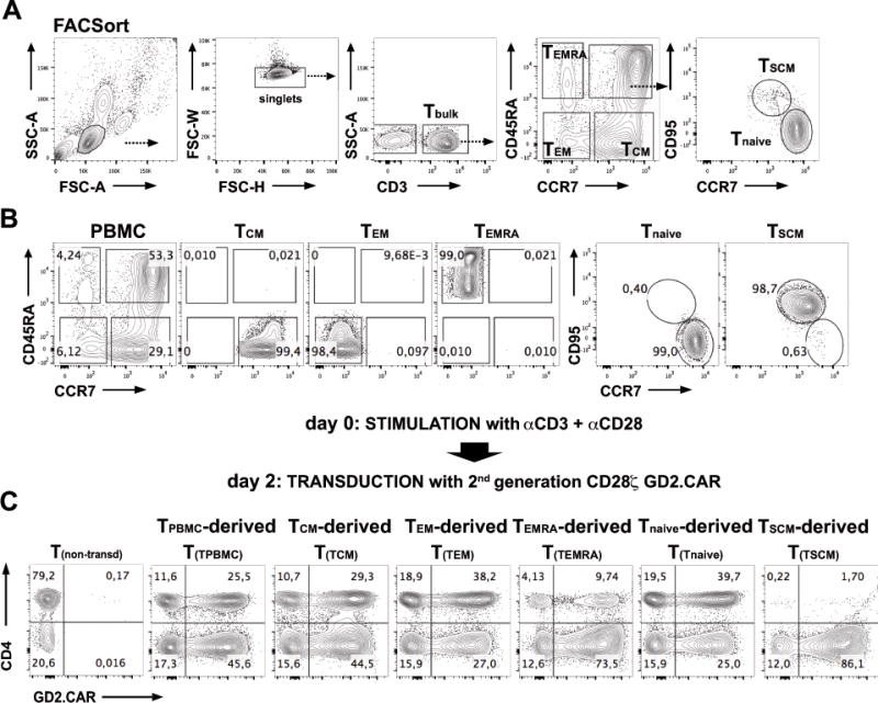
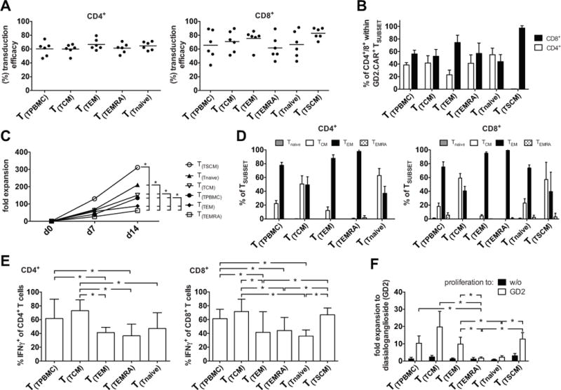
The outcome of therapy with chimeric Ag receptor (CAR)-modified T cells is strongly influenced by the subset origin of the infused T cells. However, because polyclonally activated T cells acquire a largely CD45RO+CCR7- effector memory phenotype after expansion, regardless of subset origin, it is impossible to know which subsets contribute to the final T cell product. To determine the contribution of naive T cell, memory stem T cell, central memory T cell, effector memory T cell, and terminally differentiated effector T cell populations to the CD3 and CD28-activated CAR-modified T cells that we use for therapy, we followed the fate and function of individually sorted CAR-modified T cell subsets after activation with CD3 and CD28 Abs (CD3/28), transduction and culture alone, or after reconstitution into the relevant subset-depleted population. We show that all subsets are sensitive to CAR transduction, and each developed a distinct T cell functional profile during culture. Naive-derived T cells showed the greatest rate of proliferation but had more limited effector functions and reduced killing compared with memory-derived populations. When cultured in the presence of memory T cells, naive-derived T cells show increased differentiation, reduced effector cytokine production, and a reduced reproliferative response to CAR stimulation. CD3/28-activated T cells expanded in IL-7 and IL-15 produced greater expansion of memory stem T cells and central memory T cell-derived T cells compared with IL-2. Our strategy provides a powerful tool to elucidate the characteristics of CAR-modified T cells, regardless of the protocol used for expansion, reveals the functional properties of each expanded T cell subset, and paves the way for a more detailed evaluation of the effects of manufacturing changes on the subset contribution to in vitro-expanded T cells.


Figures

FIGURE 1
FIGURE 1. Strategy for T-cell subset sorting, activation and transduction
(A) Illustrates the strategy for sorting T-cell subsets from PBMC according to their expression of CD3+, CD45RA+, CCR7+ and CD95+. Prior to FACSorting, CD3+ T-cells were enriched from PBMC by untouched MACS separation. They were then sorted into the following subsets: TCM (CCR7+CD45RA), TEM (CCR7CD45RA), TEMRA (CCR7CD45RA+), TSCM (CD45RA+CCR7+→CD95+) and Tnaive (CD45RA+CCR7+→CD95). (B) FACSort purified T-cell populations were isolated to greater than 98% purity. (C) Strategy for polyclonal activation and retroviral transduction of PBMC-derived T(TPBMC), TCM-derived (T(TCM)), TEM-derived (T(TEM)), TEMRA-derived (T(TEMRA)), TSCM-derived (T(TSCM)) and Tnaive-derived (T(Tnaive)) cells. FACSort purified TSUBSET were activated with CD3 and CD28 antibodies and cultured in the presence of IL-7 and IL-15 at 10ng/mL each and transduced on day 2 with a 2nd generation GD2.CAR (14g2a.CD28-ζ CAR). Shown are examples of GD2.CAR transduction efficacies in separately cultured PBMC-, TCM-, TEM-, TEMRA-, TSCM- and Tnaive-derived populations on day 7 post activation. Transduction efficacies were assessed by fluorescence staining for the GD2.CAR expression on the T-cell surface using the 1A7 antibody targeting 14g2a.
FIGURE 2
FIGURE 2. Fold Expansion and phenotype of separately cultured GD2.CAR-modified TSUBSETS in response to GD2 stimulation
(A) FACSort purified TSUBSET were cultured according to Fig. 1. GD2 transduction efficacies from 6 donors: left graph: CD4+; right graph: CD8+ TSUBSET-derived (T(TSUBSET)) populations on day 14 after CD3/28-activation. (B) CD4+/CD8+ proportion within GD2.CAR+ TSUBSETS. (C) TSUBSET expansion in response to CD3/28 stimulation (measured as fold increase) analyzed for each TSUBSET on days 7 and 14. (D) Quantification of CCR7 and CD45RO expression 14 days after CD3/28-activation in each TSUBSET-derived population: TCM (CCR7+CD45RO+), TEM (CCR7CD45RO+), TEMRA (CCR7CD45RO) and Tnaive (CCR7+CD45RO). Left graph: CD4+; right graph: CD8+ TSUBSET-derived populations. (E) CD3/28-expanded TSUBSET-derived populations were re-stimulated via the CAR using a GD2-positive neuroblastoma cell line, JF at a stimulator to responder ratio of 10:1 14 days after the initial CD3/28 activation and a non-CAR control stimulation (GD2 Hodgkin lymphoma: HDLM-2). Shown is background-subtracted (negative control: HDLM-2) IFNγ production to JF by CD4+ (left graph) and CD8+ (right graph) TSUBSET-derived populations and was determined by intracellular staining 6 hrs after co-culture. Cells were permeabilized and stained for IFNγ, CD3, CD4 and CD8. (F) TSUBSET expansion (measured as fold increase) was analyzed for each cell subset between days 14 and 21 after stimulation with plate-bound GD2 on day 14. Mean data from 6 healthy donors are presented, and error bars represent SEM. Data were analyzed using repeated measures 1-way ANOVA, after verifying Gaussian distribution with the Kolmogorov-Smirnov test. p-values ≤ 0.05 were considered significant: *p<0.05.
FIGURE 3
FIGURE 3. Experimental design to assess the fate of TSUBSETS polyclonally expanded within PBMC
(A) Illustrates the strategy for FACSort-purification of T-cell populations: TCM, TEM, TEMRA, Tnaive and TSCM were FACSort-depleted from PBMC (B: TSUBSETS; C: PBMC-TCM, PBMC-TEM, PBMC-TEMRA, PBMC-Tnaive, PBMC-TSCM). After isolation each TSUBSET and each single subset-depleted PBMC population was activated with CD3 and CD28 antibodies in the presence of IL-7 and IL-15, then transduced on day 2 with a CD19.CAR construct encoding a marker gene (NGFR), while TSUBSET–depleted cells were transduced with the CD19.CAR alone (CD19.CD28ζ CAR). Each activated and transduced TSUBSET was reconstituted into the appropriate activated and transduced TSUBSET –depleted PBMC at their initial frequencies one day after transduction to allow tracking of the subset within the bulk population after reconstitution (details in methods). (D) Representative FACS plots are shown for the flow cytometric detection of each reconstituted CD19.CAR-NGFR+ TSUBSET-derived (T(TSUBSET)) population (CD4+ T(TSUBSET) indicated in red dots; CD8+ T(TSUBSET) indicated in blue dots) and CD19.CAR+ PBMC-TSUBSET (P-TSUBSET, gray dots) by the assessment of the CD19.CAR and the CD19.CAR-NGFR+, respectively. (E; upper graph) CD19.CAR-NGFR transduction efficacies for CD4+ and CD8+ TCM, TEM, TEMRA, TSCM and Tnaive-derived (T(TSUBSET)) population and (E; lower graph) CD19.CAR transduction efficacies for CD4+ and CD8+ PBMC-TSUBSET (P-TSUBSET).
FIGURE 4
FIGURE 4. Fate of TSUBSETS polyclonally expanded within PBMC: naïve-derived T-cells expand preferentially within polyclonally activated PBMC
CAR-modified TSUBSET were reconstituted into CD19.CAR modified, subset-depleted PBMC according to Figure 3. (A) Fold expansion of CD19.CAR-NGFR+ TSUBSET-derived (T(TSUBSET)) population cultured alone or after reconstitution into the PBMC-TSUBSET: CD4+ (upper graph), CD8+ (middle graph) and whole CD3+ (lower graph). (B) Analysis on day 14 post initial polyclonal activation of each T(TSUBSET) (CD19.CAR-NGFR+) within PBMC-TSUBSET expanded T-cells (CD19.CAR+) showing higher frequencies of CD4+ (red, upper graph), CD8+ (blue, middle graph) and whole CD3+ (lower graph) T(Tnaive) and T(TCM) cells compared to other T(TSUBSET). Data were analyzed using repeated measures 1-way ANOVA, after verifying Gaussian distribution with the Kolmogorov-Smirnov test. p-values ≤ 0.05 were considered significant: *p<0.05. (C) Frequency of each TSUBSET within CD3+ PBMC (ex vivo) and frequency at day 14 of CD4+ (red, upper graph) and CD8+ (blue, lower graph) CD19.CAR-NGFR+ T(TSUBSET) within PBMC-TSUBSET expanded T-cells. Mean data from 6 healthy donors are presented, and error bars represent SEM.
FIGURE 5
FIGURE 5. Cytokine production by reconstituted T-cell subsets in response to CAR-stimulation
CAR-modified T-cell subsets were reconstituted into CD19.CAR modified, subset-depleted PBMC according to Figure 3. On day 14, cultures were stimulated with a CD19 (HDLM-2) or CD19+ JEKO lymphoma cells for 6 hrs at a stimulator to responder ratio of 10:1. Cytokine production was measured by intracellular cytokine staining. (A) Shows a representative FACSplot in which IFNγ and TNFα production by Tnaive–derived cells in response to HDLM-2 (upper plots) or JEKO (lower plots) is shown. TSUBSETS could be distinguished from the depleted fraction by their expression of NGFR (B). The fraction of all subsets producing IFNγ, TNFα, IL-2 is shown for CD4+ and CD8+ T-cells in C, D and E. Cytokine production by transduced and non-transduced PBMC is also shown. Black bars represent PBMC-TSUBSET–derived CD4+ T-cells, red bars represent TSUBSET–derived CD4+ T-cells, white bars represent PBMC-TSUBSET–derived CD8+ T-cells, blue bars represent TSUBSET–derived CD8+ T-cells. Data from TSUBSET–derived (CD19.CAR-NGFR+) and PBMC-TSUBSET–derived (CD19.CAR+) populations within one approach were analyzed using paired t-test, after verifying Gaussian distribution with the Kolmogorov-Smirnov test. p-values ≤ 0.05 were considered significant: *p<0.05. Mean data from 3 healthy donors are presented, and error bars represent SEM.
FIGURE 6
FIGURE 6. Cytotoxic killing capacity of each TSUBSET–derived population cultured alone and its respective TSUBSET-depleted and TSUBSET-reconstituted PBMC cell preparation
Assessment of cytotoxic activity by flow cytometric vital assay. CAR-modified T-cell subsets were cultured alone or reconstituted into CD19.CAR modified, subset-depleted PBMC according to Figure 3. (A) CFDA-SE+ CD19+ JEKO cell lines (serving as stimulator APC for 2nd generation CD19.CAR T-cells) were used as targets, whereas DDOA+ CD19 HDLM-2 cells lack CD19 were used as control targets. Cells were co-cultured for 16 hrs in T-cell / target cell ratio of 1:1 and 10:1. Samples without T-cells, containing only APC (CD19+ JEKO cell lines or CD19 HDLM-2) displayed as internal control. The mean percent survival of CD19+ JEKO incubated targets was calculated relative to CD19 HDLM-2 controls. (B) Shows the killing capacity of each TSUBSET-depleted bulk population (upper panel), TSUBSET-reconstituted bulk population (middle panel) and TSUBSET cultured alone (lower panel). Data from TSUBSET–derived population cultured alone, TSUBSET-depleted and TSUBSET-reconstituted PBMC cell preparations were analyzed using paired t-test, after verifying Gaussian distribution with the Kolmogorov-Smirnov test. p-values ≤ 0.05 were considered significant: *p<0.05. Mean data from 3 healthy donors are presented, and error bars represent SEM.
FIGURE 7
FIGURE 7. T-cells with TCM and TEM phenotypes dominate all TSUBSET–derived populations during culture
CAR-modified T-cell subsets were cultured alone or reconstituted into CD19.CAR modified, subset-depleted PBMC according to Figure 3. (A) Representative FACS plots illustrating flow cytometric detection of CCR7 and CD45RO expression on days 7, 14 and 21 post initial activation of TSUBSET-derived (T(TSUBSET)) populations (cultured alone) and of TSUBSET-derived (T(TSUBSET)) populations within the relevant PBMC-TSUBSET preparation (cultured within PBMC): (B) Results for TCM phenotype (CCR7+CD45RO+) at indicated time-points (days 7, 14 and 21) for all subsets of CD4+CAR+ and CD8+CAR+ T-cells. Data from TSUBSET–derived (CD4+CD19.CAR-NGFR+ cultured alone: black bars; CD8+CD19.CAR-NGFR+ cultured alone: white bars versus CD4+CD19.CAR+ cultured within PBMC preparations: red bars; CD8+CD19.CAR+ cultured within PBMC preparations: blue bars) were analyzed using paired t-test, after verifying Gaussian distribution with the Kolmogorov-Smirnov test. p-values ≤ 0.05 were considered significant: *p<0.05. Mean data from 3 healthy donors are presented, and error bars represent SEM.
FIGURE 8
FIGURE 8. Proliferation of CD19.CAR-modified TSUBSETS cell cultured alone or expanded within bulk PBMC in response to CAR stimulation
CAR-modified TSUBSET were cultured alone or reconstituted into CD19.CAR modified, subset-depleted PBMC according to Figure 3. (A) Each CD3/28-expanded transduced TSUBSET-derived population cultured alone or within bulk PBMC were stimulated via the CAR using vital CD19+ JEKO cells in the presence or absence of exogenous cytokines (IL-7 and IL-15) at a stimulator to responder ratio of 10:1 14 days after the initial CD3/28 activation. The frequency of each subset was measured by co-staining with antibodies to NGFR and to the spacer/hinge region of the CD19.CAR at assay initiation and post 7 days of consecutive expansion. (B and C) TSUBSET expansion (measured as fold increase) was analyzed for each cell subset between days 14 and 21 after stimulation with CD19+ JEKO cells on day 14 in the absence (B) or presence (C) of exogenous cytokines (IL-7 and IL-15). Data from TSUBSET–derived (CD19.CAR-NGFR+ cultured alone versus cultured within PBMC preparations) were analyzed using paired t-test, after verifying Gaussian distribution with the Kolmogorov-Smirnov test. p-values ≤ 0.05 were considered significant: *p<0.05. Mean data from 3 healthy donors are presented, and error bars represent SEM.
FIGURE 9
FIGURE 9. A combination of IL-7 and IL-15 increases the yield of TSCM and TCM derived T-cells compared to IL-2
TCM and TSCM were FACSort-depleted from PBMC, activated with CD3 and CD28 antibodies in the presence of either IL-2 (50IU/mL) or IL-7 and IL-15 (each at 10ng/mL) and transduced on day 2 with a retroviral vector encoding mOrange. mOrange+ TCM and TSCM were reconstituted on day 3 to the respective TSUBSET-depleted PBMC. The medium and cytokines were changed every 3 days during culture or when passaging the T-cells for splitting during expansion. (A) Shows the frequency of TSCM and TCM in PBMC. (B) Representative plot showing the frequency of mOrange-positive TCM-derived (T(TCM)) and TSCM-derived (T(TSCM)) populations in reconstituted cultures on day 11. Cells were stained for CD3, CD4 and CD8. (C) Frequencies of reconstituted CD4+ TCM (upper graph), CD8+ TCM (middle graph) and CD8+ TSCM (lower graph) during expansion from day 2 to 35 of culture in either IL-2 or IL-7 and IL-15 (TCM-derived: T(TCM); TSCM-derived: T(TSCM)). (D) Expansion within the bulk population of reconstituted CD4+ TCM (upper graph), CD8+ TCM (middle graph) and CD8+ TSCM (lower graph) measured as fold increase of mOrange+ T-cells over 35 days of culture in IL-2 or IL-7/15 (TCM-derived: T(TCM); TSCM-derived: T(TSCM)). Data were analyzed using repeated measures 2-way ANOVA, after verifying Gaussian distribution with the Kolmogorov-Smirnov test. p-values ≤ 0.05 were considered significant: *p<0.05. Mean data from 4 healthy donors are presented.

References

    1. Rosenberg Sa, Restifo NP, Yang JC, Morgan Ra, Dudley ME. Adoptive cell transfer: a clinical path to effective cancer immunotherapy. Nat Rev Cancer. 2008;8:299–308. - PMC - PubMed
    1. Porter DL, Levine BL, Kalos M, Bagg A, June CH. Chimeric antigen receptor-modified T cells in chronic lymphoid leukemia. N Engl J Med. 2011;365:725–33. - PMC - PubMed
    1. Kalos M, Levine BL, Porter DL, Katz S, Grupp Sa, Bagg A, June CH. T cells with chimeric antigen receptors have potent antitumor effects and can establish memory in patients with advanced leukemia. Sci Transl Med. 2011;3:95ra73. - PMC - PubMed
    1. Grupp Sa, Kalos M, Barrett D, Aplenc R, Porter DL, Rheingold SR, Teachey DT, Chew A, Hauck B, Wright JF, Milone MC, Levine BL, June CH. Chimeric antigen receptor-modified T cells for acute lymphoid leukemia. N Engl J Med. 2013;368:1509–18. - PMC - PubMed
    1. Savoldo B, Ramos CA, Liu E, Mims MP, Keating MJ, Carrum G, Kamble RT, Bollard CM, Gee AP, Mei Z, Liu H, Grilley B, Rooney CM, Heslop HE, Brenner MK, Dotti G. Brief report CD28 costimulation improves expansion and persistence of chimeric antigen receptor – modified T cells in lymphoma patients. 2011;121:1–5. - PMC - PubMed

Publication types

MeSH terms