. 2017 May 26;7(1):2444.
doi: 10.1038/s41598-017-02569-4.

Differences in selectivity to natural images in early visual areas (V1-V3)

Affiliations

Differences in selectivity to natural images in early visual areas (V1-V3)

David D Coggan et al. Sci Rep. .

Abstract

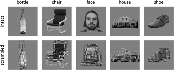
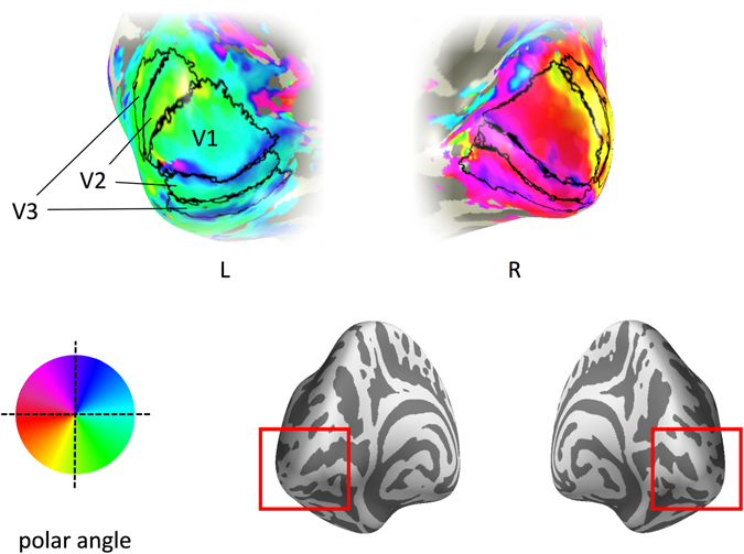
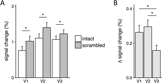
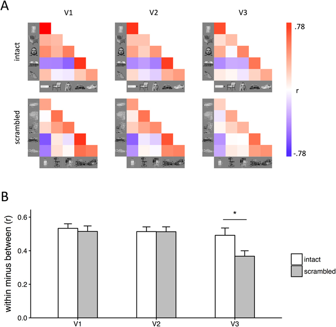
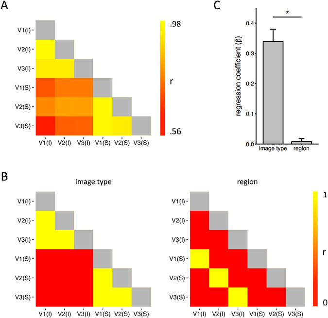
High-level regions of the ventral visual pathway respond more to intact objects compared to scrambled objects. The aim of this study was to determine if this selectivity for objects emerges at an earlier stage of processing. Visual areas (V1-V3) were defined for each participant using retinotopic mapping. Participants then viewed intact and scrambled images from different object categories (bottle, chair, face, house, shoe) while neural responses were measured using fMRI. Our rationale for using scrambled images is that they contain the same low-level properties as the intact objects, but lack the higher-order combinations of features that are characteristic of natural images. Neural responses were higher for scrambled than intact images in all regions. However, the difference between intact and scrambled images was smaller in V3 compared to V1 and V2. Next, we measured the spatial patterns of response to intact and scrambled images from different object categories. We found higher within-category compared to between category correlations for both intact and scrambled images demonstrating distinct patterns of response. Spatial patterns of response were more distinct for intact compared to scrambled images in V3, but not in V1 or V2. These findings demonstrate the emergence of selectivity to natural images in V3.


Conflict of interest statement

The authors declare that they have no competing interests.

Figures

Figure 1
Figure 1
Exemplars of intact and scrambled images from the different object categories.
Figure 2
Figure 2
Early visual cortical regions for a representative participant. Visual areas are superimposed onto the occipital lobe – see red insert on the posterior view of the inflated brain. Colour maps indicate the preferred polar angle.
Figure 3
Figure 3
(A) Magnitude of response to intact and scrambled images. Scrambled images evoked more activity than intact images in each visual area. (B) Differences in response to intact and scrambled images for each visual region. V3 showed a smaller difference in response to intact and scrambled images compared to V1 and V2. Error bars show ±1 SEM. *p < 0.05, FDR corrected.
Figure 4
Figure 4
(A) Similarity matrices showing the correlation in patterns of neural response to all within-category and between-category comparisons. Within-category comparisons (e.g. bottle-bottle) are shown on the diagonal. (B) Bar graph showing the mean within-category and between-category correlations for intact and scrambled images across participants. There was a significant interaction between Comparison, Image Type and Region, which was due to more distinct (within > between) patterns of neural response to intact relative to scrambled images in V3. Error bars show ±1 SEM. *p < 0.05, FDR corrected.
Figure 5
Figure 5
(A) Correlations between the similarity matrices shown in Fig. 4A. (B) Matrix predictions based on representations of image type and region. (C) Models were used in a regression analysis across participants. Performance was determined by the regression coefficients for each model. The results show that patterns of response were predicted significantly more by the image type than region. Error bars show ±1 SEM. *p < 0.05.

References

    1. Hubel DH, Wiesel TN. Receptive Fields and Functional Architecture of monkey striate cortex. J. Physiol. 1968;195:215–243. doi: 10.1113/jphysiol.1968.sp008455. - DOI - PMC - PubMed
    1. Tanaka K. Inferotemporal cortex and object vision. Annu. Rev. Neurosci. 1996;19:109–139. doi: 10.1146/annurev.ne.19.030196.000545. - DOI - PubMed
    1. Connor CE, Brincat SL, Pasupathy A. Transformation of shape information in the ventral pathway. Curr. Opin. Neurobiol. 2007;17:140–147. doi: 10.1016/j.conb.2007.03.002. - DOI - PubMed
    1. Kanwisher N. Functional specificity in the human brain: a window into the functional architecture of the mind. Proc. Natl. Acad. Sci. USA. 2010;107:11163–70. doi: 10.1073/pnas.1005062107. - DOI - PMC - PubMed
    1. Rolls ET. Neurophysiological mechanisms underlying face processing within and beyond the temporal cortical visual areas. Philos. Trans. R. Soc. Lond. B. Biol. Sci. 1992;335:11–21. doi: 10.1098/rstb.1992.0002. - DOI - PubMed

Publication types

LinkOut - more resources