. 2017 May 26;7(1):2446.
doi: 10.1038/s41598-017-02719-8.

Antigenicity of Bovine Pericardium Determined by a Novel Immunoproteomic Approach

Affiliations

Antigenicity of Bovine Pericardium Determined by a Novel Immunoproteomic Approach

Katherine V Gates et al. Sci Rep. .

Abstract

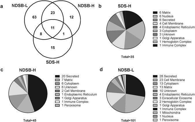
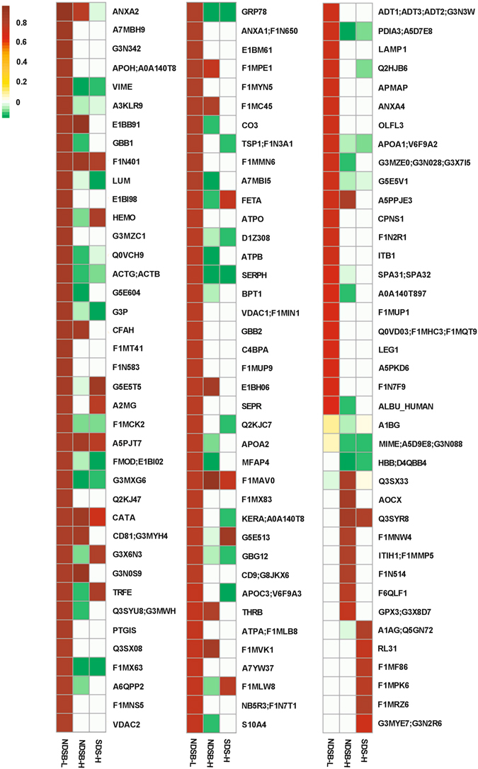
Despite bovine pericardium (BP) being the primary biomaterial used in heart valve bioprostheses, recipient graft-specific immune responses remain a significant cause of graft failure. Consequently, tissue antigenicity remains the principal barrier for expanding use of such biomaterials in clinical practice. We hypothesize that our understanding of BP antigenicity can be improved by application of a combined affinity chromatography shotgun immunoproteomic approach to identify antigens that have previously been overlooked. Liquid Chromatography Tandem Mass Spectrometry (LC-MS/MS) analysis of affinity chromatography purified antigens resulted in identification of 133 antigens. Most importantly, antigens were identified from all subcellular locations, including 18 integral membrane protein antigens. Critically, isoforms of several protein families were found to be antigenic suggesting the possibility that shared epitope domains may exist. Furthermore, proteins associated with immune, coagulation, and inflammatory pathways were over-represented, suggesting that these biological processes play a key role in antigenicity. This study brings to light important determinants of antigenicity in a clinically relevant xenogeneic biomaterial (i.e. BP) and further validates a rapid, high-throughput method for immunoproteomic antigen identification.


Conflict of interest statement

The authors declare that they have no competing interests.

Figures

Figure 1
Figure 1
Assessment of column IgG binding capacity and cross-linking efficiency. Serum protein electrophoresis (SPE) of native rabbit serum showing IgG levels (a). Column run-through following D84 anti-native BP rabbit serum loading on SpinTrap Column (b). Boxes indicate the gamma peak, containing IgG along with fibronectin and C-reactive protein. Gamma peak was reduced from 0.3 mg/dL in native serum to below 0.1 mg/dL in the run-through (n = 1). Calculated efficiency of rabbit IgG capture and cross-linking to the column determined by ELISA analysis of IgG content in native serum and column run-through (c). No difference in IgG capture or cross-linking efficiency was found between D0 versus D84 serum using a paired two-tailed Student’s t-test (n = 6 per timepoint, p = 0.4663, values represent the mean ± s.d.).
Figure 2
Figure 2
Diagram of workflow for affinity chromatography column generation and validation for antigen identification. Column IgG capture following serum loading was assessed using serum protein electrophoresis (SPE). Capture and cross-linking efficiency of IgG antibodies was determined using rabbit IgG specific ELISA. Specificity of antigen capture and non-specific protein binding were assessed using silver stained gels and Western blots with post-immunization rabbit serum (D84), for protein run-through from columns eluted at pH 5, 4 and 2.9. Antigen identifications were made using LC-MS/MS analysis of specifically bound proteins eluted from D0 versus D84 columns.
Figure 3
Figure 3
Western blot and silver stain of protein eluates. Western blot of total native protein extract (a1), protein extract run-through from D0 column (a2), and protein extract run-through from D84 column (a3). Both D0 and D84 columns demonstrate decreased antigenic protein content in column run-through, with greater removal of antigenic bands in D84 column. Silver stain for SDS-page gel of D84 pH 5, 4, and 2.9 eluates ((b1,b2 and b3) respectively) and corresponding D84 Western blot from pH 5, 4, and 2.9 eluates ((b4,b5 and b6) respectively), confirming that minimal non-specific binding is present and that all specifically bound proteins (pH 2.9 eluates) are antigenic. Comparison of D0 and D84 eluates at pH 2.9, demonstrating that more antigens are specifically captured from D84 columns (c2) than from D0 columns (c1). (n = 4 per extraction method and timepoint). Western blot and silver stained gel were cropped for easier visualization, uncropped images are available in the supplemental material.
Figure 4
Figure 4
Comparison of antigenic protein identifications depending on type of extraction solution, SDS-H, NDSB-H, and NDSB-L. Venn diagram showing the number of antigenic proteins identified using each protein extraction methods (a). Pie charts demonstrating subcellular location of proteins depending on the protein extraction method used (bd). Numbers associated with subcellular locations indicate number of antigens identified within that subcellular location. (n = 6 per extraction method).
Figure 5
Figure 5
Heatmap of the statistically significant antigenic protein identifications within each extraction method. Antigens were predominantly identified in NDSB-L, but alternative extraction methods are required to optimize total number of antigens identified. Scale is a Log of the fold change. Proteomic data was analyzed using Guassian linearized modeling to determine the differential abundance of proteins between the groups as previously reported. White squares indicate that the protein was not present in that sample. (n = 6 per extraction method). Groups were considered significantly different when p < 0.05.

References

    1. Platt J, DiSesa V, Gail D, Massicot-Fisher J. Recommendations of the National Heart, Lung, and Blood Institute Heart and Lung Xenotransplantation Working Group. Circulation. 2002;106:1043–1047. doi: 10.1161/01.CIR.0000031064.67525.28. - DOI - PubMed
    1. Griesemer A, Yamada K, Sykes M. Xenotransplantation: immunological hurdles and progress toward tolerance. Immunol Rev. 2014;258:241–258. doi: 10.1111/imr.12152. - DOI - PMC - PubMed
    1. Manji, R. A., Lee, W. & Cooper, D. K. Xenograft bioprosthetic heart valves: Past, present and future. Int J Surg, doi:10.1016/j.ijsu.2015.07.009 (2015). - PubMed
    1. Zilla P, Human P, Bezuidenhout D. Bioprosthetic heart valves: the need for a quantum leap. Biotechnol Appl Bioc. 2004;40:57–66. doi: 10.1042/BA20030211. - DOI - PubMed
    1. Simon, P. et al. Early failure of the tissue engineered porcine heart valve SYNERGRAFT in pediatric patients. Eur J Cardiothorac Surg23, 1002–1006, discussion 1006 (2003). - PubMed

Publication types

LinkOut - more resources