Diagnostic accuracy of post-mortem CT with targeted coronary angiography versus autopsy for coroner-requested post-mortem investigations: a prospective, masked, comparison study
- PMID: 28551075
- PMCID: PMC5506259
- DOI: 10.1016/S0140-6736(17)30333-1
Diagnostic accuracy of post-mortem CT with targeted coronary angiography versus autopsy for coroner-requested post-mortem investigations: a prospective, masked, comparison study
Abstract
Background: England and Wales have one of the highest frequencies of autopsy in the world. Implementation of post-mortem CT (PMCT), enhanced with targeted coronary angiography (PMCTA), in adults to avoid invasive autopsy would have cultural, religious, and potential economic benefits. We aimed to assess the diagnostic accuracy of PMCTA as a first-line technique in post-mortem investigations.
Methods: In this single-centre (Leicester, UK), prospective, controlled study, we selected cases of natural and non-suspicious unnatural death referred to Her Majesty's (HM) Coroners. We excluded cases younger than 18 years, known to have had a transmittable disease, or who weighed more than 125 kg. Each case was assessed by PMCTA, followed by autopsy. Pathologists were masked to the PMCTA findings, unless a potential risk was shown. The primary endpoint was the accuracy of the cause of death diagnosis from PMCTA against a gold standard of autopsy findings, modified by PMCTA findings only if additional substantially incontrovertible findings were identified.
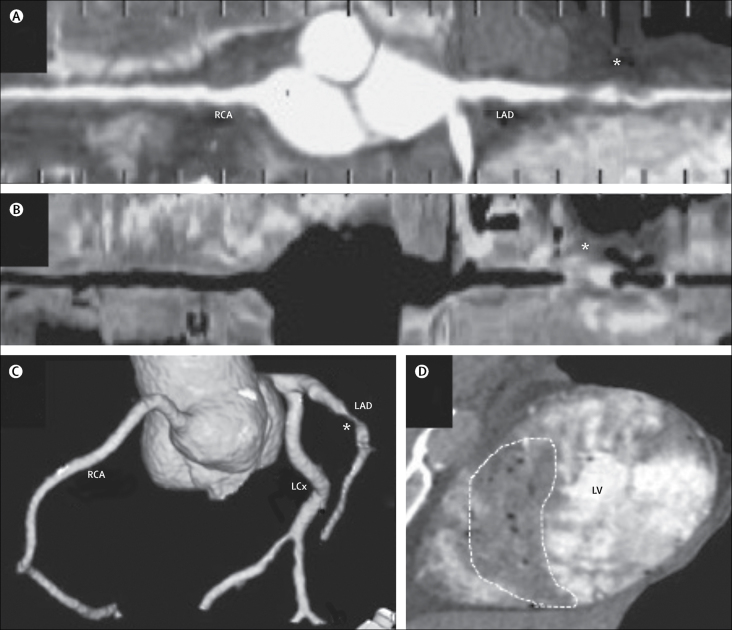
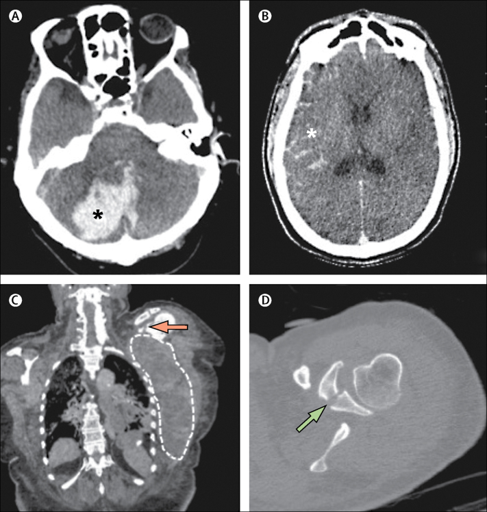
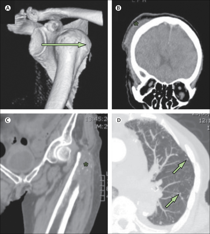
Findings: Between Jan 20, 2010, and Sept 13, 2012, we selected 241 cases, for which PMCTA was successful in 204 (85%). Seven cases were excluded from the analysis because of procedural unmasking or no autopsy data, as were 24 cases with a clear diagnosis of traumatic death before investigation; 210 cases were included. In 40 (19%) cases, predictable toxicology or histology testing accessible by PMCT informed the result. PMCTA provided a cause of death in 193 (92%) cases. A major discrepancy with the gold standard was noted in 12 (6%) cases identified by PMCTA, and in nine (5%) cases identified by autopsy (because of specific findings on PMCTA). The frequency of autopsy and PMCTA discrepancies were not significantly different (p=0·65 for major discrepancies and p=0·21 for minor discrepancies). Cause of death given by PMCTA did not overlook clinically significant trauma, occupational lung disease, or reportable disease, and did not significantly affect the overall population data for cause of death (p≥0·31). PMCTA was better at identifying trauma and haemorrhage (p=0·008), whereas autopsy was better at identifying pulmonary thromboembolism (p=0·004).
Interpretation: For most sudden natural adult deaths investigated by HM Coroners, PMCTA could be used to avoid invasive autopsy. The gold standard of post-mortem investigations should include both PMCT and invasive autopsy.
Funding: National Institute for Health Research.
Copyright © 2017 The Author(s). Published by Elsevier Ltd. This is an Open Access article under the CC BY 4.0 licence. Published by Elsevier Ltd.. All rights reserved.
Figures
Comment in
-
Targeted coronary post-mortem CT angiography, straight to the heart.Lancet. 2017 Jul 8;390(10090):100-101. doi: 10.1016/S0140-6736(17)31260-6. Epub 2017 May 24. Lancet. 2017. PMID: 28551070 No abstract available.
-
Can post-mortem CT and angiography provide all the answers?Lancet. 2017 Aug 12;390(10095):646-647. doi: 10.1016/S0140-6736(17)31828-7. Lancet. 2017. PMID: 28816131 No abstract available.
References
-
- Rutty GN, Brogdon G, Dedouit F. Terminology used in publications for post-mortem cross-sectional imaging. Int J Legal Med. 2013;127:465–466. - PubMed
-
- Westphal SE, Apitzsch J, Penzkofer T, Mahnken AH, Knuchel R. Virtual CT autopsy in clinical pathology: feasibility in clinical autopsies. Virchows Arch. 2012;461:211–219. - PubMed
-
- Leth PM, Struckmann H, Lauritsen J. Interobserver agreement of the injury diagnoses obtained by postmortem computed tomography of traffic fatality victims and a comparison with autopsy results. Forensic Sci Int. 2013;225:15–19. - PubMed
-
- Wichmann D, Obbelode F, Vogel H. Virtual autopsy as an alternative to traditional medical autopsy in the intensive care unit: a prospective cohort study. Ann Intern Med. 2012;156:123–130. - PubMed
-
- Donchin Y, Rivkind AI, Bar-Ziv J, Hiss J, Almog J, Drescher M. Utility of postmortem computed tomography in trauma victims. J Trauma. 1994;37:552–556. - PubMed
Publication types
MeSH terms
Grants and funding
LinkOut - more resources
Full Text Sources
Other Literature Sources
Medical