. 2017 Jun:20:39-49.
doi: 10.1016/j.ebiom.2017.04.022. Epub 2017 Apr 13.

A Specific Mutational Signature Associated with DNA 8-Oxoguanine Persistence in MUTYH-defective Colorectal Cancer

Affiliations

A Specific Mutational Signature Associated with DNA 8-Oxoguanine Persistence in MUTYH-defective Colorectal Cancer

Alessandra Viel et al. EBioMedicine. 2017 Jun.

Abstract

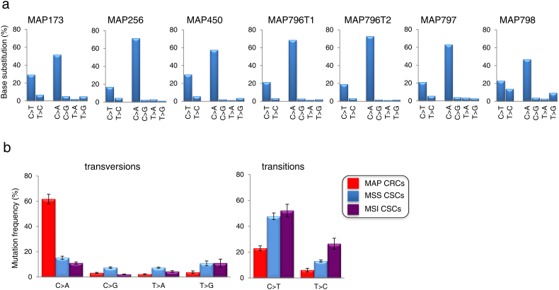
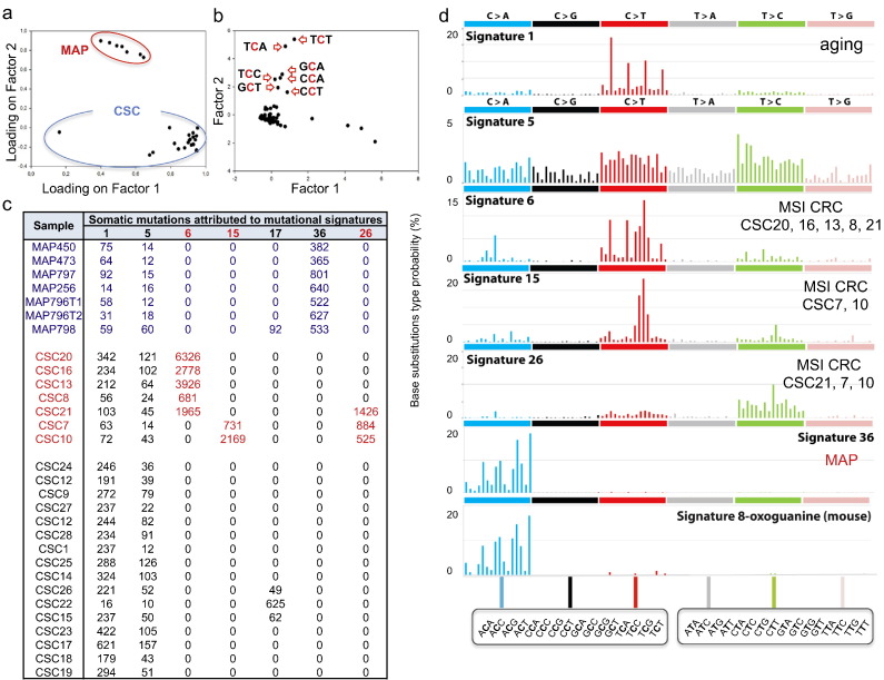
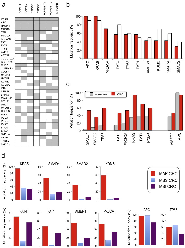
8-Oxoguanine, a common mutagenic DNA lesion, generates G:C>T:A transversions via mispairing with adenine during DNA replication. When operating normally, the MUTYH DNA glycosylase prevents 8-oxoguanine-related mutagenesis by excising the incorporated adenine. Biallelic MUTYH mutations impair this enzymatic function and are associated with colorectal cancer (CRC) in MUTYH-Associated Polyposis (MAP) syndrome. Here, we perform whole-exome sequencing that reveals a modest mutator phenotype in MAP CRCs compared to sporadic CRC stem cell lines or bulk tumours. The excess G:C>T:A transversion mutations in MAP CRCs exhibits a novel mutational signature, termed Signature 36, with a strong sequence dependence. The MUTYH mutational signature reflecting persistent 8-oxoG:A mismatches occurs frequently in the APC, KRAS, PIK3CA, FAT4, TP53, FAT1, AMER1, KDM6A, SMAD4 and SMAD2 genes that are associated with CRC. The occurrence of Signature 36 in other types of human cancer indicates that DNA 8-oxoguanine-related mutations might contribute to the development of cancer in other organs.

Keywords: 8-Oxoguanine; Base excision repair; Colorectal cancer; Exome sequencing; MUTYH-associated polyposis; Mutational signature.


Figures

Image 1
Graphical abstract
Fig. 1
Fig. 1
Somatic mutation frequencies in MAP CRCs and colorectal CSCs. a) and b) Mutation frequencies per Mb in MAP CRCs (red), and MSS (blue) and MSI CSCs (purple). c) Mutation rates in MAP CRCs and MSS CSCs calculated by dividing the number of somatic mutations per tumour by the age of the patient at diagnosis. d) Distribution of indels (grey) and SNVs in MAP CRCs (red), and MSS (blue) and MSI (purple) CSCs. Data are mean ± SE.
Fig. 2
Fig. 2
Mutational spectra in MAP CRCs and colorectal CSCs. a) Mutational spectrum for base substitutions in each MAP CRC. b) Comparison of average values of transitions and transversions in MAP CRCs (red), MSS CSCs (blue) and MSI CSCs (purple). Data are mean ± SE. Each class of mutation in MAP CRC was statistically significant different from MSS and MSI CSCs with P values < 0.001 (Student's t-test).
Fig. 3
Fig. 3
Fingerprints of transitions and transversions in MAP CRCs. Mutational signatures are defined by the substitution class (in different colours) and the sequence context immediately 5′ and 3′ to the mutated base. The mutation types are on the horizontal axes, whereas vertical axes show the recurrence of mutations occurring in the specific sequence context.
Fig. 4
Fig. 4
Mutational signatures in MAP CRCs and MSS and MSI CSCs. a) Separation between MAP (red) and CSCs (blue) mutational fingerprints by PCA. Factor 1 and Factor 2 represent commonality and differences among profiles, respectively. b) G:C > T:A transversions at the indicated trinucleotides identify signatures with the greater discriminant power for MAP/CSCs separation. c) Somatic mutations in MAP CRCs (red) and MSI/MSS CSCs (black) attributed to reported mutational signatures (Alexandrov et al., 2013a, Alexandrov et al., 2013b). d) Graphical representations of signatures identified in MSI/MSS CSCs and MAP CRCs. Signature 8-oxoguanine derives from mice defective in Mutyh/Ogg1/Mth1 (Ohno et al., 2014).
Fig. 5
Fig. 5
Mutation analysis of target genes in CRCs and adenomas of MAP patients. a) Genes mutated in 7 MAP CRCs and identified by exome sequencing. Only genes mutated in ≥ 2 patients are shown. b) Comparison of the mutation frequency in 7 MAP CRCs analyzed by WES (red) and 10 CRCs analyzed by targeted sequencing (open bars) from independent MAP patients. c) Comparison of mutation frequency of the indicated genes in 26 adenomas (grey) and 17 CRCs (red) from MAP patients. d) Comparison of mutation frequency in the indicated genes in MAP CRCs (red), and MSS (blue) and MSI (purple) CRCs (data from a database of 376 samples).
Fig. 6
Fig. 6
KRAS sequence context and MUTYH adenine glycosylase activity. a) Duplex oligonucleotides containing a single 8-oxoG:A mispair at nt 34 or 35 of the KRAS sequence which are responsible for the G12C and G12V variants in MAP (top) and sporadic CRC (bottom). Codon 12 is in bold and trinucleotides containing 8-oxoG (G*) are underlined. b) Base substitution frequency of C > A transversions in MAP CRC. The black and red bars represent the GG*T “cold-spot” and TG*G “hot-spot” trinucleotides, respectively. c) Representative gels of single-turnover MUTYH assays using duplexes shown in panel A. DNA glycosylase activity of human MUTYH on single 8-oxoG:A mispairs was assayed using 10 nM duplex substrates and 20 nM active protein. Reaction products are indicated by an arrow. Lanes 1–9: 0, 0.5, 1, 2, 3, 4, 5, 10 and 20 min. d) Plots of MUTYH glycosylase assay data under single-turnover conditions. Data were derived from image analysis of gels by Image J and Kaleidagraph softwares. Values are the average from two independent experiments. G12C sequence (full black circle); G12 V (empty blue circle).

References

    1. Alexandrov L.B. Understanding the origins of human cancer. Science. 2015;350:1175. - PubMed
    1. Alexandrov L.B., Nik-Zainal S., Wedge D.C., Campbell P.J., Stratton M.R. Deciphering signatures of mutational processes operative in human cancer. Cell Rep. 2013;3:246–259. - PMC - PubMed
    1. Alexandrov L.B., Nik-Zainal S., Wedge D.C., Aparicio S.A., Behjati S., Biankin A.V., Bignell G.R., Bolli N., Borg A., Børresen-Dale A.L. Signatures of mutational processes in human cancer. Nature. 2013;500:415–421. - PMC - PubMed
    1. Alexandrov L.B., Jones P.H., Wedge D.C., Sale J.E., Campbell P.J., Nik-Zainal S., Stratton M.R. Clock-like mutational processes in human somatic cells. Nat. Genet. 2015;47:1402–1407. - PMC - PubMed
    1. Al-Tassan N., Chmiel N.H., Maynard J., Fleming N., Livingston A.L., Williams G.T., Hodges A.K., Davies D.R., David S.S., Sampson J.R. Inherited variants of MYH associated with somatic G:C > T:A mutations in colorectal tumors. Nat. Genet. 2002;30:227–232. - PubMed

MeSH terms