. 2017 Oct;74(19):3631-3645.
doi: 10.1007/s00018-017-2547-4. Epub 2017 May 27.

Necroptosis and ferroptosis are alternative cell death pathways that operate in acute kidney failure

Affiliations

Necroptosis and ferroptosis are alternative cell death pathways that operate in acute kidney failure

Tammo Müller et al. Cell Mol Life Sci. 2017 Oct.

Abstract

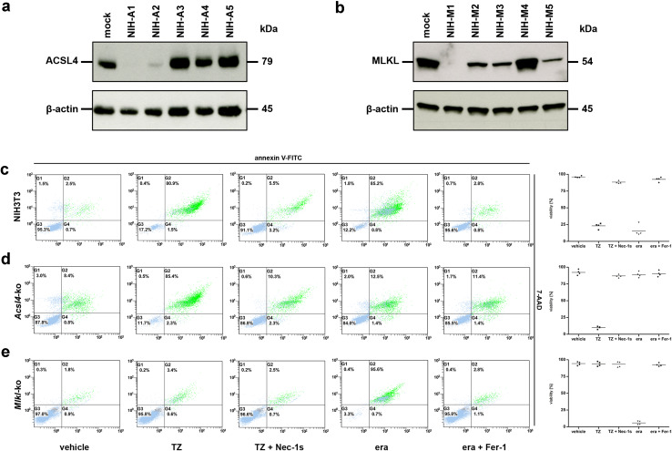
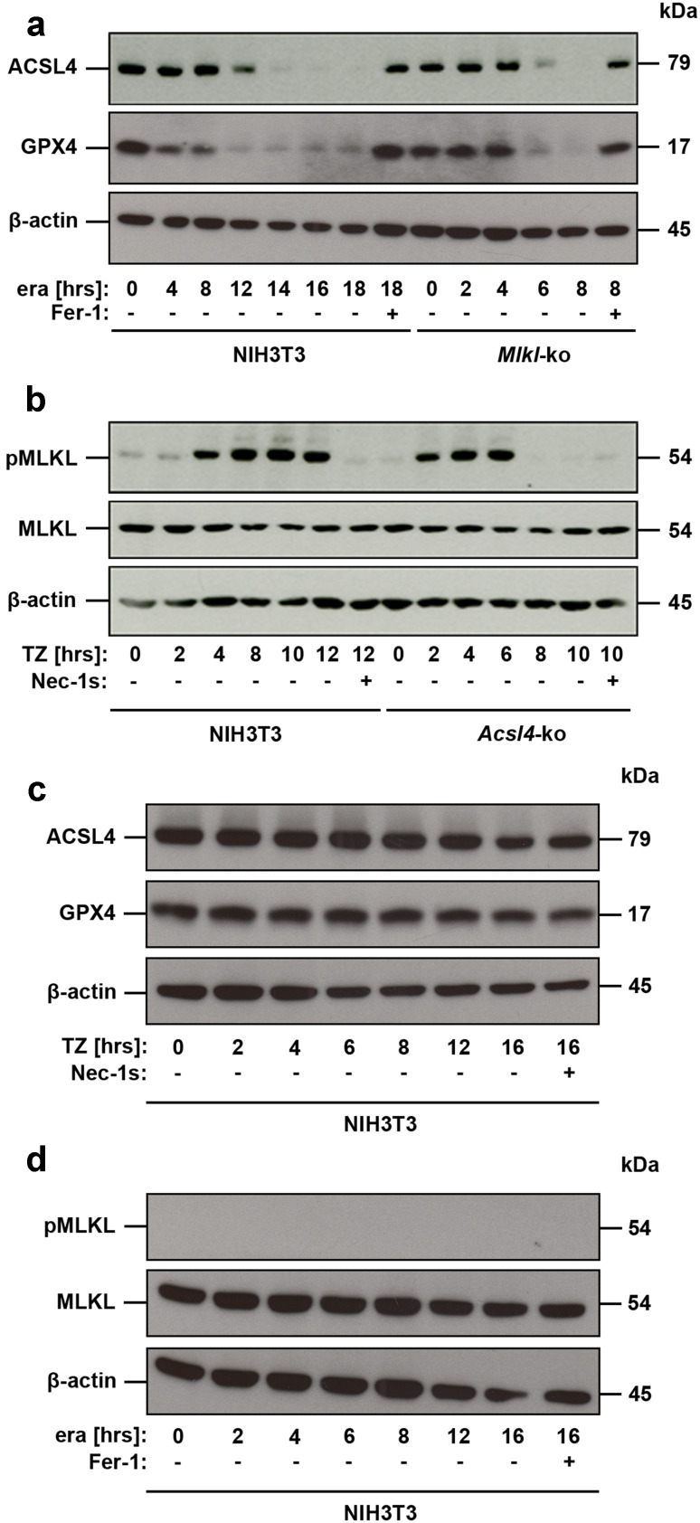
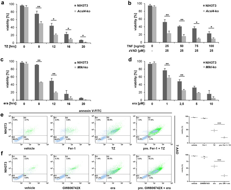
Ferroptosis is a recently recognized caspase-independent form of regulated cell death that is characterized by the accumulation of lethal lipid ROS produced through iron-dependent lipid peroxidation. Considering that regulation of fatty acid metabolism is responsible for the membrane-resident pool of oxidizable fatty acids that undergo lipid peroxidation in ferroptotic processes, we examined the contribution of the key fatty acid metabolism enzyme, acyl-CoA synthetase long-chain family member 4 (ACSL4), in regulating ferroptosis. By using CRISPR/Cas9 technology, we found that knockout of Acsl4 in ferroptosis-sensitive murine and human cells conferred protection from erastin- and RSL3-induced cell death. In the same cell types, deletion of mixed lineage kinase domain-like (Mlkl) blocked susceptibility to necroptosis, as expected. Surprisingly, these studies also revealed ferroptosis and necroptosis are alternative, in that resistance to one pathway sensitized cells to death via the other pathway. These data suggest a mechanism by which one regulated necrosis pathway compensates for another when either ferroptosis or necroptosis is compromised. We verified the synergistic contributions of ferroptosis and necroptosis to tissue damage during acute organ failure in vivo. Interestingly, in the course of pathophysiological acute ischemic kidney injury, ACSL4 was initially upregulated and its expression level correlated with the severity of tissue damage. Together, our findings reveal ACSL4 to be a reliable biomarker of the emerging cell death modality of ferroptosis, which may also serve as a novel therapeutic target in preventing pathological cell death processes.

Keywords: ACSL4; Ferroptosis; Ischemia-reperfusion injury; MLKL; Necroptosis.


Conflict of interest statement

The authors declare no competing or financial interests.

Figures

Fig. 1
Fig. 1
Acsl4- and Mlkl-knockout NIH3T3 cells are protected from ferroptosis and necroptosis, respectively. a Five different Acsl4-silenced clones, referred to as NIH-A1 to NIH-A5, and b five different Mlkl-silenced clones, referred to as NIH-M1 to NIH-M5, were analyzed by Western blotting for the ablation of the indicated target genes. Lysates of mock-transfected (non-edited) control cells were plotted in each case on the left lane. Both blots were redeveloped with an antibody against β-actin as a loading control. For all assays shown subsequently we used the NIH3T3 clones NIH-A1 (for Acsl4-ko) and NIH-M1 (for Mlkl-ko), respectively. FACS analysis for the necrotic marker 7-AAD and phosphatidylserine exposure (annexin V-FITC positivity) in c mock-transfected (non-edited), d Acsl4-edited, and e Mlkl-edited NIH3T3 cells which were treated for 16 h at 37 °C with DMSO (vehicle), 100 ng/ml TNFα + 25 µM zVAD (TZ), 100 ng/ml TNFα + 25 µM zVAD + 50 µM necrostatin-1s (TZ + Nec-1s), 10 µM erastin (era), and 10 µM erastin + 1 µM ferrostatin-1 (era + Fer-1) as indicated. Necroptosis was blocked by addition of Nec-1 s, and ferroptosis blocked by Fer-1. ce FACS dot plots of one representative experiment are shown, with adjacent box plots presenting the mean and standard deviation of four independent experiments
Fig. 2
Fig. 2
Loss of ferroptosis or necroptosis signaling sensitizes cells to the alternate pathway. a Genetically unmodified NIH3T3 and Mlkl-edited NIH3T3 cells were left untreated or were stimulated for different time points with 10 µM erastin (era) in the presence or absence of 1 µM ferrostatin (Fer-1), as indicated. Equal amounts of protein (20 µg/lane) were resolved by SDS/PAGE and expression of ACSL4 (M r = 79.0 kDa) was detected by Western blotting. Notably, different stimulation periods are shown to most clearly illustrate the differences observed between parental and edited cells (see axis labeling). The blot was stripped and re-probed first with an antibody against GPX4 (M r = 17.0 kDa, predicted molecular weight = 22.0 kDa) and thereafter with an antibody against β-actin as loading control. b Genetically unmodified NIH3T3 and Acsl4-edited NIH3T3 cells were left untreated or were stimulated for different time points with 100 ng/ml TNFα + 25 µM zVAD (TZ) in the presence or absence of 50 µM necrostatin-1s (Nec-1s), as indicated. Equal amounts of protein (20 µg/lane) were resolved by SDS/PAGE and expression of phospho-MLKL (M r = 54.0 kDa) was detected by Western blotting. Notably, different stimulation periods are shown to most clearly illustrate the differences observed between parental and edited cells (see axis labeling). The blot was stripped and re-probed first with an antibody against whole MLKL (M r = 54.0 kDa) and thereafter with an antibody against β-actin as loading control. c Genetically unmodified NIH3T3 cells were left untreated or were stimulated for different time points with 100 ng/ml TNFα + 25 µM zVAD (TZ), as indicated. Equal amounts of protein (20 µg/lane) were resolved by SDS/PAGE and expression of ACSL4 was detected by Western blotting. The blot was stripped and re-probed first with an antibody against GPX4 (M r = 17.0 kDa, predicted molecular weight = 22.0 kDa) and thereafter with an antibody against β-actin as loading control. d Genetically unmodified NIH3T3 cells were left untreated or were stimulated for different time points with 10 µM erastin (era), as indicated. Equal amounts of protein (20 µg/lane) were resolved by SDS/PAGE and phosphorylated MLKL was detected by Western blotting. The blot was stripped and re-probed first with an antibody against whole MLKL and thereafter with an antibody against β-actin as loading control
Fig. 3
Fig. 3
Alternative of necroptotic and ferroptotic pathways was seen across a range of time points and stimuli concentrations. a, b, e Detection of necroptosis in parental and Acsl4-edited NIH3T3 cells, respectively. a The cells were treated at the time indicated with 100 ng/ml TNFα + 25 µM zVAD (TZ) or b for 16 h with constant 25 µM zVAD and different (indicated) concentration of TNFα. Mean ± SD is shown for three independent experiments. e FACS analysis of parental NIH3T3 cells that were pretreated as indicated for 30 min with 1 µM ferrostatin (Fer-1). Necroptosis was induced thereafter for 12 h by the addition of 100 ng/ml TNFα + 25 µM zVAD (TZ). Depicted is one of three independent experiments. c, d, f Detection of ferroptosis in parental and Mlkl-edited NIH3T3 cells, respectively. c Cells were treated for indicated time at 37 °C with 10 µM erastin (era) or d for 16 h with different (indicated) concentration of erastin. Mean ± SD is shown for three independent experiments. f FACS analysis of parental NIH3T3 cells that were pretreated as indicated for 30 min with 2.5 µM GW806742X. Ferroptosis was induced thereafter for 12 h by the addition of 10 µM erastin (era). e, f FACS dot plots of one representative experiment are shown, with adjacent box plots presenting the mean and standard deviation of three independent experiments
Fig. 4
Fig. 4
Necroptosis and ferroptosis are intertwined in murine IRI. Male C57BL/6 wildtype mice and Mlkl-knockout mice underwent 35 min of bilateral pedicle clamping (ischemia) followed by different times of reperfusion. a Serum creatinine and b serum urea concentrations were determined, respectively, after sacrificing the mice at the indicated reperfusion time points. c left Expression levels of ACSL4 (79.0 kDa) in whole-kidney lysates taken from wildtype or Mlkl-knockout mice during the time course of IRI. A concomitant treatment of the animals for the first 24 h after the onset of reperfusion with the ferrostatin-derivative 16-86 in this setting is illustrated in the lower part. c right Thereafter, the blots were stripped and re-probed with an antibody against β-actin as loading control. d Representative histological sections of murine kidneys (contralateral to c) after staining with periodic acid-Schiff (PAS) show less severe tubular damage in Mlkl-knockout mice compared to C57BL/6 wildtype mice. Depicted are representative enlarged sections of the untreated groups and 24 h IRI values, respectively (n = 4 animals for each group and time point, scale bars 50 µm)
Fig. 5
Fig. 5
Increased expression of ACSL4 in human kidney biopsies indicates a role for ferroptotic cell death in vivo. Human kidney biopsies were stained for ACSL4 and phospho-MLKL as indicated. a Positive tubuli can be seen in acute tubular injury 7 days post-transplantation (transplanted kidney (Tx) from a female patient showing moderate tubular injury) and b severe acute tubular damage (male patient suffering from thrombotic microangiopathy (TMA) due to atypical hemolytic-uremic syndrome). Notable, increased expression of ACSL4 verifies an important role of ferroptosis in the complex pathology of acute tubular injury (ATI) and distinguished for the first time ACSL4 as a biomarker of pathological ferroptosis. c Normal renal parenchyma samples taken from a tumor nephrectomy of renal cell carcinoma served as controls (scale bars 60 µm). The biopsies presented in this figure are representative of a total of ten human biopsies (n = 10)

References

    1. Linkermann A, Stockwell BR, Krautwald S, et al. Regulated cell death and inflammation: an auto-amplification loop causes organ failure. Nat Rev Immunol. 2014;14:759–767. doi: 10.1038/nri3743. - DOI - PubMed
    1. Cai Z, Jitkaew S, Zhao J, et al. Plasma membrane translocation of trimerized MLKL protein is required for TNF-induced necroptosis. Nat Cell Biol. 2014;16:55–65. doi: 10.1038/ncb2883. - DOI - PMC - PubMed
    1. Dondelinger Y, Declercq W, Montessuit S, et al. MLKL compromises plasma membrane integrity by binding to phosphatidylinositol phosphates. Cell Rep. 2014;7:971–981. doi: 10.1016/j.celrep.2014.04.026. - DOI - PubMed
    1. Xia B, Fang S, Chen X, et al. MLKL forms cation channels. Cell Res. 2016;26:517–528. doi: 10.1038/cr.2016.26. - DOI - PMC - PubMed
    1. Hildebrand JM, Tanzer MC, Lucet IS, et al. Activation of the pseudokinase MLKL unleashes the four-helix bundle domain to induce membrane localization and necroptotic cell death. Proc Natl Acad Sci USA. 2014;111:15072–15077. doi: 10.1073/pnas.1408987111. - DOI - PMC - PubMed

Publication types

MeSH terms