. 2017 Jul;46(4):564-578.
doi: 10.1016/j.asd.2017.05.004. Epub 2017 Jul 4.

Effects of force detecting sense organs on muscle synergies are correlated with their response properties

Affiliations

Effects of force detecting sense organs on muscle synergies are correlated with their response properties

Sasha N Zill et al. Arthropod Struct Dev. 2017 Jul.

Abstract

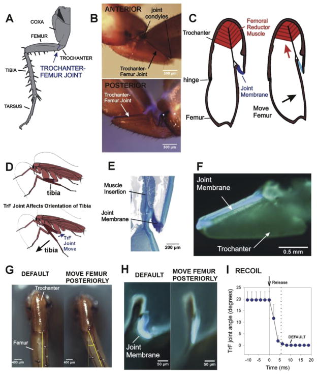
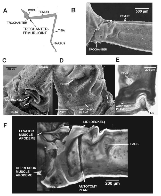
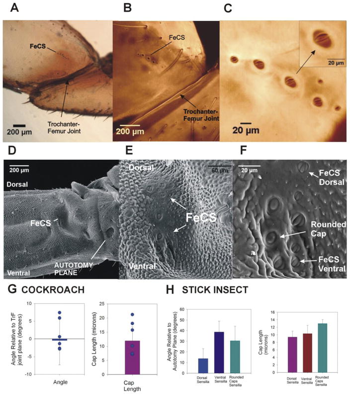
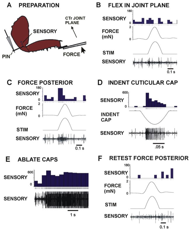
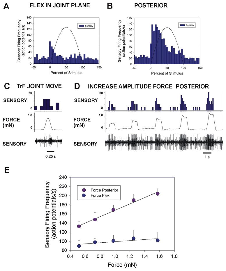
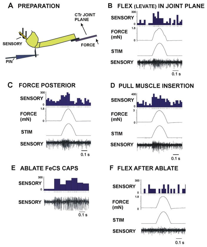
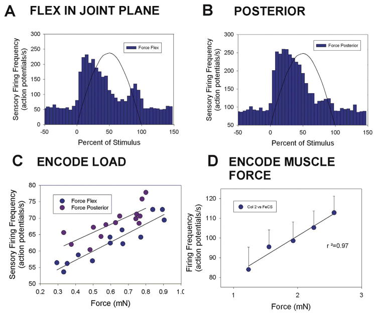
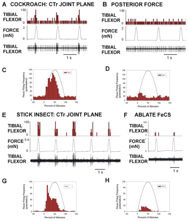
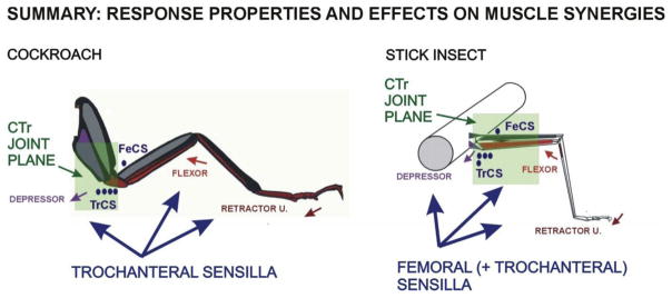
Sense organs that monitor forces in legs can contribute to activation of muscles as synergist groups. Previous studies in cockroaches and stick insects showed that campaniform sensilla, receptors that encode forces via exoskeletal strains, enhance muscle synergies in substrate grip. However synergist activation was mediated by different groups of receptors in cockroaches (trochanteral sensilla) and stick insects (femoral sensilla). The factors underlying the differential effects are unclear as the responses of femoral campaniform sensilla have not previously been characterized. The present study characterized the structure and response properties (via extracellular recording) of the femoral sensilla in both insects. The cockroach trochantero-femoral (TrF) joint is mobile and the joint membrane acts as an elastic antagonist to the reductor muscle. Cockroach femoral campaniform sensilla show weak discharges to forces in the coxo-trochanteral (CTr) joint plane (in which forces are generated by coxal muscles) but instead encode forces directed posteriorly (TrF joint plane). In stick insects, the TrF joint is fused and femoral campaniform sensilla discharge both to forces directed posteriorly and forces in the CTr joint plane. These findings support the idea that receptors that enhance synergies encode forces in the plane of action of leg muscles used in support and propulsion.

Keywords: Campaniform; Force; Insect; Response; Sensitivity; Synergies.


Figures

Fig. 1
Fig. 1. Mobility and specializations of the cockroach trochantero-femoral joint
A. Cockroach middle leg. The trochanter is a small segment upon which large muscles of the coxa and body wall insert. The trochanter articulates with the femur at the trochantero-femoral (TrF) joint. B. Whole mounts of cockroach trochanter – The joint between the trochanteral and femoral segments is a movable hinged articulation, with joint condyles on the anterior side (top). The posterior surface has no condyles and the joint membrane shows blue fluorescence in combined light/UV illumination. C. Left – Diagram of longitudinal section (parallel to the long axis of the trochanter) through trochanter and femur. The reductor femoris muscle is located in the trochanter and inserts onto the femur and the joint membrane. Contraction of the muscle pulls the femur posteriorly and stretches the joint membrane. D. Action of trochanter-femur joint on the distal leg – Posterior movement at the TrF brings the tibia to a more vertical position relative to the substrate. E. Histological section of TrF joint (same plane as 1C) – The joint membrane is thickened and stains intensely with toluidine blue at the insertion of the muscle. F. Fluorescence image of hind leg trochanter split longitudinally and viewed on its inner surface. The membrane shows blue fluorescence in UV illumination. G. Movement at TrF joint – Ventral view of trochanter and femur of cockroach hind leg at rest (default position, left) and after maximum posterior displacement of the TrF joint (right). H. Fluorescence images of TrF joint with the upper half of the trochanter and femur removed in a severed leg (orientation similar to 1E). The joint membrane is curved in the default (rest) position but stretched when the femur is moved posteriorly. I. Elastic recoil – The joint showed rapid recoil to the default position when it was released after posterior displacement.
Fig. 2
Fig. 2. Immobility and specializations of the trochanter-femur joint in stick insects
A. Drawing of stick insect middle leg and trochanter-femur joint.. B. SEM of trochanter and femur in posterior view – The trochanter is functionally fused to the femur with no condyles or joint movement. C. SEM of upper (dorsal) surface of the trochanter – The dorsal surface of the trochanter forms a projection (termed the lid, G., Deckel) which interlocks with a depression on the proximal end of the femur. D. Posterior view (SEM) of proximal femur and lid – The femoral group of campaniform sensilla are located in a depression in the proximal femur, distal to the autotomy plane. E. Dorsal view of whole mount of TrF joint – the cuticle appears darkened and thickened in the region between the autotomy plane and femoral sensilla. F. Confocal projection image of inner (posterior) surface of a bisected coxa, trochanter and femur (the lid remained intact) – The apodeme (tendon) of the Levator Trochanteris muscle inserts upon the ‘lid’’ of the trochanter; the Depressor apodeme inserts upon the reinforced ventral end of the trochanter. The autotomy plane is clearly seen as a thin vertical line between the fused trochanter and femur.
Fig. 3
Fig. 3. Structure of femoral campaniform sensilla in cockroaches and stick insects
A. Posterior view of a whole mount of a cockroach trochanter and femur – The femoral group of campaniform sensilla is located on the posterior surface of the femur, distal to the trochanter-femur joint. B. and C. Confocal projection of images TrF joint – The cuticular caps of the femoral sensilla are arranged in a small, linear row on the proximal femur. At higher magnification (C), the caps of the sensilla are visible inside the cuticular collars. The orientation of the caps is consistent within the group and the cap long axis is approximately parallel to the TrF joint (see inset). D. SEM of posterior view of TrF joint in stick insects – The FeCS are located in a prominent depression of the posterior side of the femur. E and F. Higher magnification views of SEM. E. The caps of the FeCS are relatively dispersed in the distal wall of depression. F. Three subgroups of receptors can be distinguished based upon their position and cap orientation: dorsal sensilla (FeCS Dorsal), ventral sensilla (FeCS Ventral) and sensilla whose caps appear rounded rather than oval-shaped (Rounded Caps). G. Orientations and cap sizes of cockroach femoral campaniform sensilla – These graphs plot the mean orientation (left) and long axis length (right) of the caps of the femoral campaniform sensilla (points show range of values from one representative animal). The sensilla are generally oriented parallel to trochanter-femur joint and have a range of cap sizes (histogram at right). H. Orientations and cap sizes of stick insect femoral campaniform sensilla – Three types of receptors could be distinguished based upon their orientation (left) and cap shape. Rounded caps had the largest lengths (right).
Fig. 4
Fig. 4. Responses of cockroach femoral campaniform sensilla to imposed forces
A. Preparation. The coxa of left hind leg of an intact cockroach was mounted via staples and the distal segments severed at the femoro tibial joint. All trochanter campaniform sensilla were ablated but the femoral group (FeCS) remained intact. Forces were applied to the femur in different directions with movement resisted by a pin (PIN) inserted into the proximal trochanter. Sensory activity was recorded with wires (SENSORY) in the distal coxa after severing the main leg nerve proximally. B. Flexion in the coxo-trochanteral joint plane – Forces applied in the joint plane produced only transient sensory discharges. C. Forces perpendicular (posterior) to the joint plane – Vigorous bursts were obtained to forces perpendicular to joint plane. D. and E. Indentation of the caps of single sensilla (D) and cap ablations (E) produced action potentials of amplitude equivalent to those seen in tests of responses to forces. F. No sensory discharges were seen when forces (posterior) were applied to the leg after cap ablation. (Sensory histograms in B–F: action potentials/second).
Fig. 5
Fig. 5. Plots of responses and encoding of force magnitude in cockroach femoral campaniform sensilla
A. and B. Cumulative plots of sensory discharges to forces applied in the plane of the CTr joint in the direction of joint flexion (A. n = 75 tests, N = 3 animals) and in a posterior direction, perpendicular to the joint plane (B. n = 85 tests, N = same 3 animals). Minimal sensory discharges occurred in the plane while intense firing was elicited in a posterior direction. C. Movement resisted by joint stiffness – Brief sensory discharges could be elicited when the TrF joint was not fully engaged. D. Amplitude series – Forces were applied in a posterior direction at varying amplitudes but the same rate of rise and decline with the TrF joint fully engaged E. Encoding of force magnitude – Plot of the mean sensory discharges (largest units) to forces applied as ramp and hold functions to forces in the joint plane (flexion) and posteriorly, perpendicular to the plane (flexion n = 8 repetitions of 5 levels; posterior n = 11 repetitions of 5 levels, N = 1). The femoral sensilla effectively encode force magnitude in a posterior direction but discharges were minimal in the plane of the coxo-trochanteral joint. (Sensory histograms in C, D: action potentials/second).
Fig. 6
Fig. 6. Responses of stick insect femoral campaniform sensilla to imposed forces
A. Preparation. The coxa of an intact stick insect left middle leg was mounted via staples and distal segments removed at the femoro-tibial joint. Forces (FORCE) were applied to the distal femur in different directions with movement is resisted by a pin (PIN) inserted into the proximal trochanter. The pin was attached to a motor to mimic contractions of the depressor muscle. Sensory activity was recorded from the main leg nerve with oil hook electrodes (SENSORY) proximal to the coxa (nerve crushed proximally). All trochanteral groups were ablated but the FeCS remained intact. B. Flexion (Levation) in the coxo-trochanteral joint plane – Forces applied to distal femur in the plane of movement of the CTr joint produced intense sensory discharges. C. Forces perpendicular (posterior) to the joint plane – Vigorous firing was also obtained to forces perpendicular to joint plane applied in a posterior direction. D. Pull on depressor insertion – Forces applied at the depressor insertion (via the pin and motor) also produced sensory discharges of similar amplitudes. E. Ablations of the caps of the femoral campaniform sensilla produced intense firing. F. No sensory responses were seen when forces were applied to the leg after cap ablation. (Sensory histograms in CD: potentials/second).
Fig. 7
Fig. 7. Plots of responses and encoding of load and muscle forces in stick insect femoral campaniform sensilla
A. and B. Cumulative plots of sensory discharges to forces applied in the plane of the CTr joint in the direction of joint flexion (levation) and in a posterior direction perpendicular to the joint plane. Intense discharges were elicited both in a posterior direction and in the plane of the CTr joint. C. Encoding of load – Plot of the mean sensory firing frequencies to forces applied as ramp and hold functions to forces in the joint plane (flexion) and perpendicular to the plane. The femoral sensilla effectively encode force magnitude in the plane of the coxo-trochanteral joint and in a posterior direction perpendicular to the plane. D. Encoding of muscle force – Responses to depressor muscle forces were obtained by applying forces to the pin in the trochanter that were resisted in the distal femur by the force probe. The mean discharge frequency effectively encoded the muscle forces.
Fig. 8
Fig. 8. Evaluation of contribution of femoral campaniform sensilla to synergist discharges in the tibial flexor muscle in cockroaches and stick insects
Forces were applied to the femur in preparations in which the main leg nerve was not severed and motor innervation remained intact. A. and B. Forces applied in cockroaches in the plane of the CTr joint produced intense bursts in the tibial flexor muscle (A) but did not occur to forces directed posteriorly (B). C. and D. Histograms of mean responses in synergist muscle – Activation of synergist discharge in tibial flexor muscle was elicited by forces in the joint plane (C, n = 323 repetitions from N = 3 preparations) but only small transient firing was present in some preparations to posterior forces (D, n = 279 repetitions from the same animals). E. and F. Effects of ablation of femoral campaniform sensilla in stick insects – Forces applied in the CTr joint plane in stick insects produced bursts of activity in the flexor muscle in intact preparations (E). Only weak discharges occurred if the femoral campaniform sensilla had been ablated prior to the tests (F). G. and H. Histograms of flexor activation – Flexor activation to forces in the plane of the CTr joint (F) was greatly reduced but not entirely eliminated by ablation of the femoral campaniform sensilla (G, n = 174 tests in N = 3 animals; H, n = 190 tests in N = 3 animals). (Flexor muscle histograms in A, B, D, F: muscle potentials/second).
Fig. 9
Fig. 9. Summary diagram of force receptors and muscle synergies in stick insects and cockroaches
Activation of muscle synergists is correlated with response sensitivity to forces in the CTr joint plane. See text for discussion.

Similar articles

Cited by

References

    1. Akay T, Bässler U, Gerharz P, Büschges A. The role of sensory signals from the insect coxa-trochanteral joint in controlling motor activity of the femur-tibia joint. J Neurophysiol. 2001;85:594–604. - PubMed
    1. Akay T. Ph D Thesis. University of Cologne; 2002. The Role of Sensory Signals for Interjoint Coordination in Stick Insect Legs (Carausius morosus and Cuniculina impigra)
    1. Ayali A, Borgmann A, Büschges A, Couzin-Fuchs E, Daun-Gruhn S, Holmes P. The comparative investigation of the stick insect and cockroach models in the study of insect locomotion. Curr Opin Insect Sci. 2015;12:1–10.
    1. Bässler U. Neural Basis of Elementary Behavior in Stick Insects. Springer; Berlin: 1983.
    1. Bässler U, Rohrbacher J, Karg G, Breutel G. Interruption of searching movements of partly restrained front legs of stick insects, a model situation for the start of a stance phase? Biol Cybern. 1991;65:507–514.

LinkOut - more resources