

Review
doi: 10.1038/aps.2017.42.
Epub 2017 May 29.
Conjugated polymer nanomaterials for theranostics
Affiliations
- PMID: 28552910
- PMCID: PMC5520193
- DOI: 10.1038/aps.2017.42
Item in Clipboard
Review
Conjugated polymer nanomaterials for theranostics
Acta Pharmacol Sin.
2017 Jun.
Abstract
Conjugated polymer nanomaterials (CPNs), as optically and electronically active materials, hold promise for biomedical imaging and drug delivery applications. This review highlights the recent advances in the utilization of CPNs in theranostics. Specifically, CPN-based in vivo imaging techniques, including near-infrared (NIR) imaging, two-photon (TP) imaging, photoacoustic (PA) imaging, and multimodal (MM) imaging, are introduced. Then, CPN-based photodynamic therapy (PDT) and photothermal therapy (PTT) are surveyed. A variety of stimuli-responsive CPN systems for drug delivery are also summarized, and the promising trends and translational challenges are discussed.
Figures
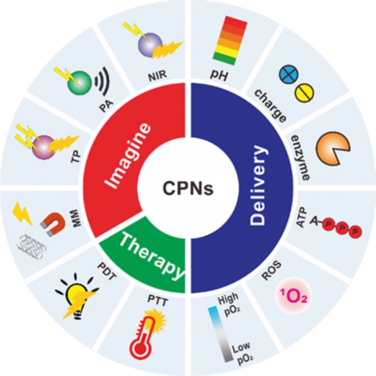
Schematic of utilizing conjugated polymer nanomaterials (CPNs) for theranostics.
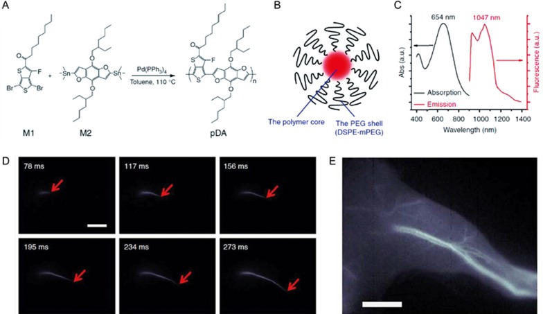
(A) Synthesis of the conjugated polymer pDA. (B) The nanoparticle (pDA-PEG) with a hydrophobic conjugated polymer core and a hydrophilic PEG shell. (C) Absorption and emission spectra of pDA-PEG. (D) Ultrafast second near-infrared (NIR-II) imaging of arterial blood flow. (E) The NIR-II fluorescence image of the same mouse hindlimb after full perfusion of pDA-PEG containing blood into the hindlimb. The scale bars are 5 mm. Reproduced with permission from Ref .
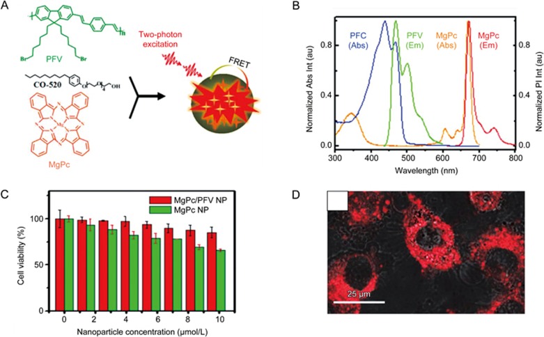
(A) Schematic preparation procedures of MgPc/PFV NPs. (B) Normalized absorption and emission spectra of PFV NPs and MgPc NPs. (C) In vitro cytotoxicity of HepG2 cancer cells treated with MgPc/PFV NPs and MgPc NPs containing the same amount of MgPc for 8 h. (D) TP fluorescent image of HepG2 cancer cells treated with MgPc/PFV NPs. The scale bar is 25 μm. Reproduced with permission from Ref .
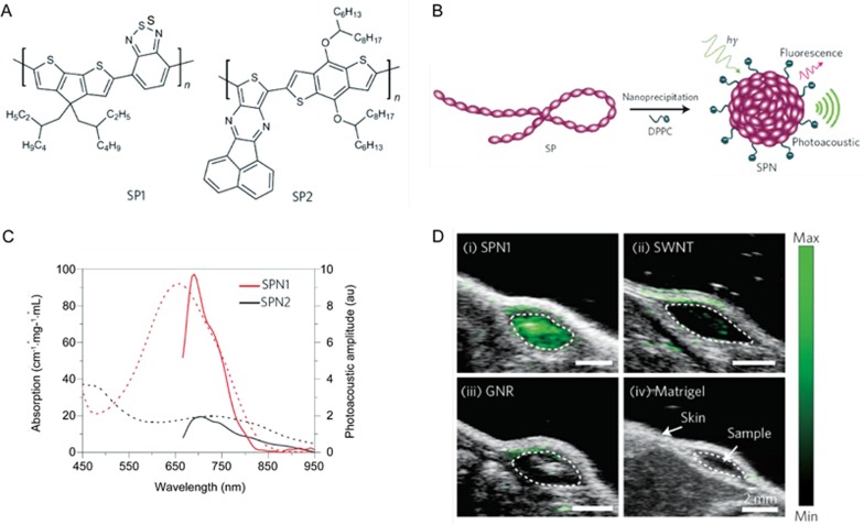
(A) Molecular structures of conjugated polymer SP1 and SP2. (B) Schematic of the preparation of the semiconducting polymer nanoparticles (SPN). (C) Ultraviolet-visible absorption (dashed lines) and photoacoustic (solid lines) spectra of SPNs. (D) Comparison of photoacoustic properties of SPN1 with single-walled carbon nanotubes (SWNTs) and gold nanorods (GNRs). Photoacoustic/ultrasound co-registered images of the nanoparticle-matrigel inclusions in the mice. The images represent transverse slices through the subcutaneous inclusions (dotted circles). The scale bars are 2 mm. Reproduced with permission from Ref .
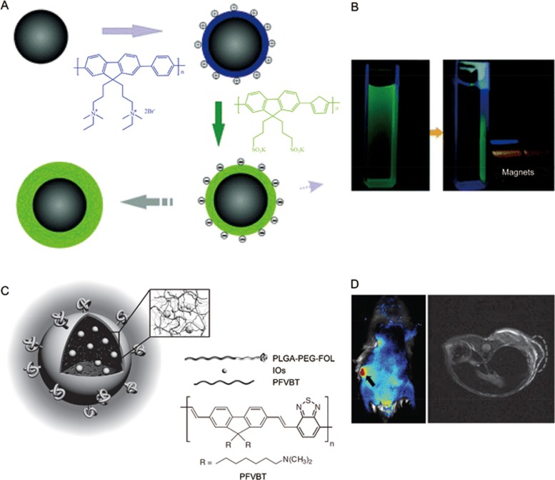
(A) Electrostatic adsorption of the CPs on surfaces of the magnetic nanoparticles to afford magnetic-fluorescent nanoparticles (MF NPs) by layer-by-layer assembly. (B) Images of MF NPs under UV light irradiation without (left) and with magnets (right). (C) Schematic of the conjugated polymer based MF NPs and the chemical structure of conjugated polymer PFVBT. (D) In vivo fluorescence images (left) and magnetic resonance images (right) of the mouse treated with MF NPs. Reproduced with permission from Ref ,.
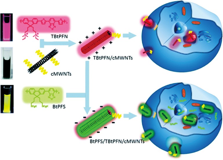
Schematic of the preparation of the charged carbon nanotubes (cMWNTs) and the CPE-cMWNT nanocomposites of the conjugated polyelectrolytes (CPEs) with cMWNTs. Reproduced with permission from Ref .
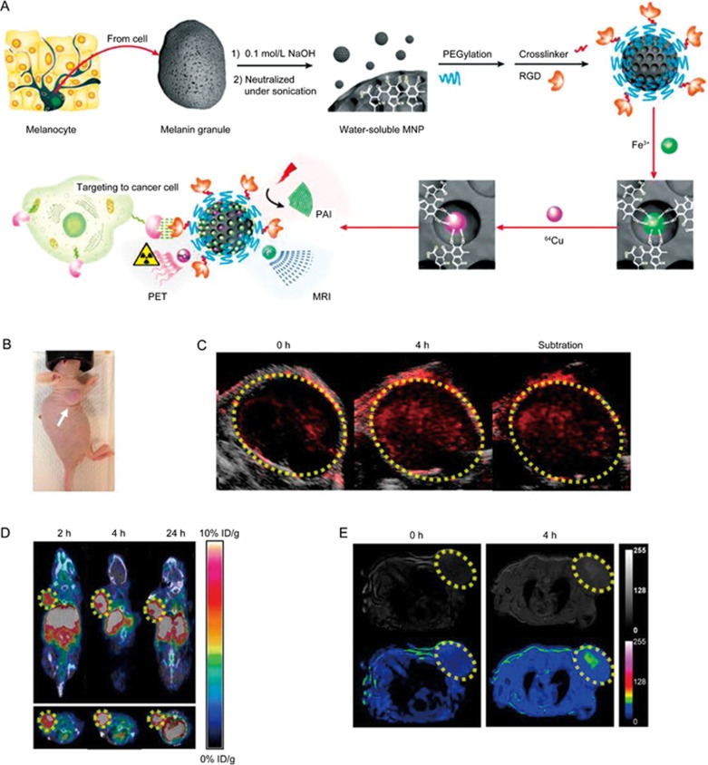
(A) Schematic illustration of the multimodal (MM) imaging melanin nanoplatform (MMPs). (B) Photographic images of U87MG tumor bearing mice. In vivo multimodality imaging of U87MG tumor (region enveloped by yellow dotted line) bearing mice after tail vein injection of 64Cu-Fe-RGD-PEG-MNP, including (C) photoacoustic (PA) imaging, (D) magnetic resonance imaging (MRI), and positron emission tomography (PET), respectively. Reproduced with permission from Ref .
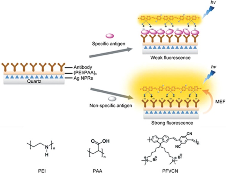
Schematic illustration of the structure and the mechanism of nano-rule detection platform for specific antigen detection assay. Reproduced with permission from Ref .
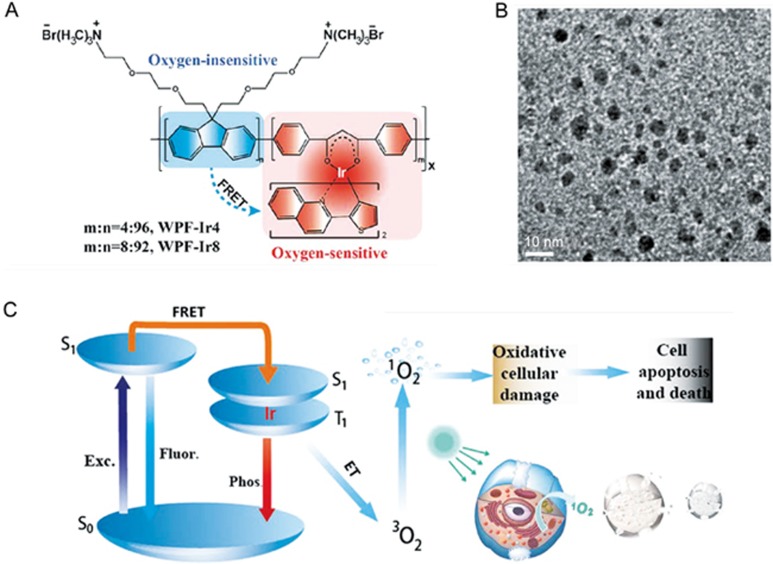
(A) Chemical structures of phosphorescent conjugated polymer with the Ir(III) complexes (Pdots). (B) High resolution transmission electron microscopy (HR-TEM) image of Pdots in aqueous solution. (C) Mechanisms of the Pdots for oxygen sensing and PDT. Reproduced with permission from Ref .
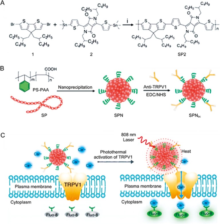
(A) Synthetic route of conjugated polymer SP2. (B) Preparation of the SPN and SPNbc with anti-TRPV1 on the surface. (C) Schematic of SPNbc-mediated photothermal activation of ion channels in neurons. Fluo-8 was used as the in real-time indicator of the intracellular concentration of calcium ions. Reproduced with permission from Ref .
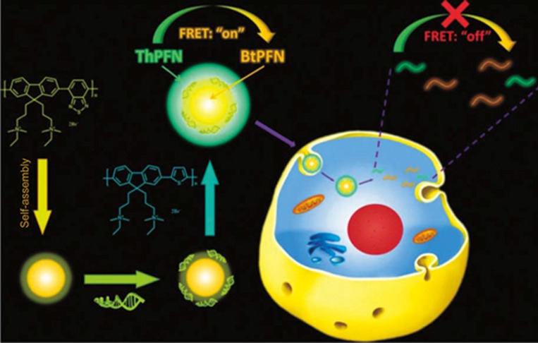
Fluorescent CPNs as siRNA nanocarriers by the sequential electrostatic assembly of siRNA and ThPFN on BtPFN nanoparticles. Reproduced with permission from Ref .
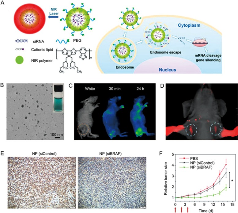
(A) Schematic of the near-infrared (NIR) polymer nanoplatforms (NPs) for siRNA delivery. (B) The TEM image and the digital picture of the NIR NPs. (C) NIR imaging. (D) Sentinel lymph node (SLN) mapping within 10 min after sc injection of NIR NPs into the forepaws. (E) Immunochemical histological observation of BRAF expression in BRAFV600E-mutated 8505C tumor tissue after treatment with different groups: NP (siControl) or NP (siBRAF). BRAF indicates V-Raf murine sarcoma viral oncogene homolog B. (F) Tumor growth curves of PBS-, NP (siControl)-, and NP (siBRAF)-treated the mice. *P<0.05 vs NP (siControl). Reproduced with permission from Ref .
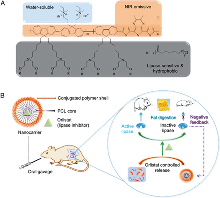
(A) Chemical structure of the conjugated polymer. (B) Schematic of lipase-sensitive conjugated polymer nanocarrier for orlistat delivery via oral gavage: orlistat release triggered by lipase, deactivation of lipase and inhibition of fat digestion, and negative feedback to control the release of the enzyme inhibitor. Reproduced with permission from Ref .
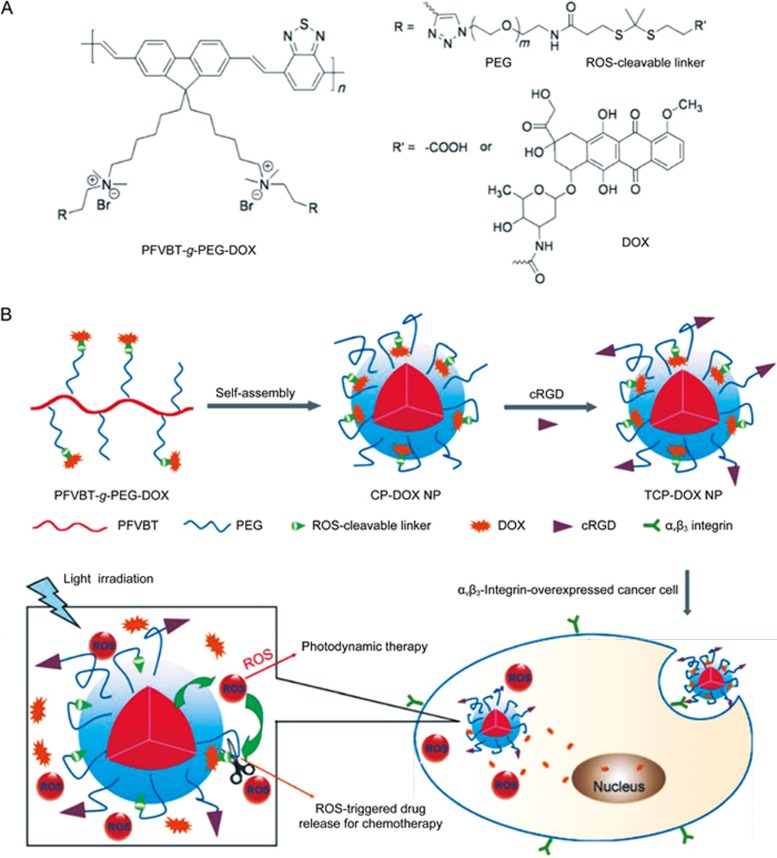
(A) Chemical structure of ROS-activatable conjugated-polyelectrolyte-based polyprodrug. (B) Self-assembled of polyprodrug nanoparticles and the light-activated ROS-responsive drug release for combination chemo-photodynamic therapy. Reproduced with permission from Ref .
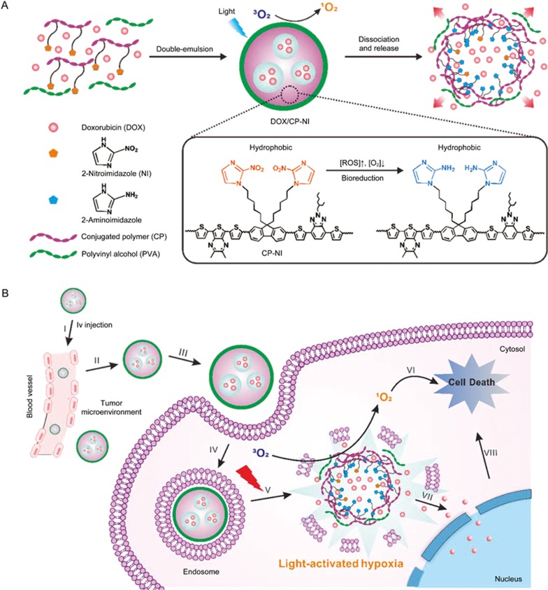
(A) Self-assembled of the light-activated hypoxia-responsive drug-delivery system. (B) Schematic of the nanocarriers for ROS generation and hypoxia-responsive drug release for enhanced synergistic anticancer efficacy. Reproduced with permission from Ref .
Similar articles
-
Near-infrared light-responsive nanomaterials for cancer theranostics.Wiley Interdiscip Rev Nanomed Nanobiotechnol. 2016 Jan-Feb;8(1):23-45. doi: 10.1002/wnan.1347. Epub 2015 Apr 23. Wiley Interdiscip Rev Nanomed Nanobiotechnol. 2016. PMID: 25903643 Review.
-
Gadolinium-Chelated Conjugated Polymer-Based Nanotheranostics for Photoacoustic/Magnetic Resonance/NIR-II Fluorescence Imaging-Guided Cancer Photothermal Therapy.Theranostics. 2019 May 31;9(14):4168-4181. doi: 10.7150/thno.34390. eCollection 2019. Theranostics. 2019. PMID: 31281539 Free PMC article.
-
Peptide-based semiconducting polymer nanoparticles for osteosarcoma-targeted NIR-II fluorescence/NIR-I photoacoustic dual-model imaging and photothermal/photodynamic therapies.J Nanobiotechnology. 2022 Jan 21;20(1):44. doi: 10.1186/s12951-022-01249-4. J Nanobiotechnology. 2022. PMID: 35062957 Free PMC article.
-
Organic Nanotheranostics for Photoacoustic Imaging-Guided Phototherapy.Curr Med Chem. 2019;26(8):1389-1405. doi: 10.2174/0929867324666170921103152. Curr Med Chem. 2019. PMID: 28933283 Review.
-
Conjugated polymer nanoparticles and their nanohybrids as smart photoluminescent and photoresponsive material for biosensing, imaging, and theranostics.Mikrochim Acta. 2022 Feb 3;189(3):83. doi: 10.1007/s00604-021-05153-w. Mikrochim Acta. 2022. PMID: 35118576 Review.
Cited by
-
Supramolecular Polysaccharide Nanotheranostics that Inhibit Cancer Cells Growth and Monitor Targeted Therapy Response.Nanotheranostics. 2020 May 18;4(3):156-172. doi: 10.7150/ntno.44703. eCollection 2020. Nanotheranostics. 2020. PMID: 32483521 Free PMC article.
-
Photoacoustic Drug Delivery.Sensors (Basel). 2017 Jun 15;17(6):1400. doi: 10.3390/s17061400. Sensors (Basel). 2017. PMID: 28617354 Free PMC article. Review.
-
Light-activated nanomaterials for tumor immunotherapy.Front Chem. 2022 Oct 7;10:1031811. doi: 10.3389/fchem.2022.1031811. eCollection 2022. Front Chem. 2022. PMID: 36277335 Free PMC article. Review.
-
Polyfluorene-Based Multicolor Fluorescent Nanoparticles Activated by Temperature for Bioimaging and Drug Delivery.Nanomaterials (Basel). 2019 Oct 18;9(10):1485. doi: 10.3390/nano9101485. Nanomaterials (Basel). 2019. PMID: 31635330 Free PMC article.
-
White light-induced cell apoptosis by a conjugated polyelectrolyte through singlet oxygen generation.RSC Adv. 2018 Mar 1;8(17):9218-9222. doi: 10.1039/c8ra00774h. eCollection 2018 Feb 28. RSC Adv. 2018. PMID: 35541876 Free PMC article.
References
-
- Sariciftci NS, Smilowitz L, Heeger AJ, Wudl F. Photoinduced electron transfer from a conducting polymer to buckminsterfullerene. Science 1992; 258: 1474–6. - PubMed
-
- Swager TM. The molecular wire approach to sensory signal amplification. Acc Chem Res 1998; 31: 201–7.
-
- Petros RA, DeSimone JM. Strategies in the design of nanoparticles for therapeutic applications. Nat Rev Drug Discov 2010; 9: 615–27. - PubMed
Publication types
MeSH terms
Substances
LinkOut - more resources
Full Text Sources
Other Literature Sources
Miscellaneous
