. 2017 May 26;13(5):e1006826.
doi: 10.1371/journal.pgen.1006826. eCollection 2017 May.

Asymmetric regulation of quorum-sensing receptors drives autoinducer-specific gene expression programs in Vibrio cholerae

Affiliations

Asymmetric regulation of quorum-sensing receptors drives autoinducer-specific gene expression programs in Vibrio cholerae

Amanda Hurley et al. PLoS Genet. .

Abstract

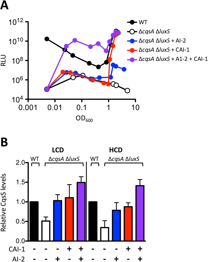
Quorum sensing (QS) is a mechanism of chemical communication that bacteria use to monitor cell-population density and coordinate group behaviors. QS relies on the production, detection, and group-wide response to extracellular signal molecules called autoinducers. Vibrio cholerae employs parallel QS circuits that converge into a shared signaling pathway. At high cell density, the CqsS and LuxPQ QS receptors detect the intra-genus and inter-species autoinducers CAI-1 and AI-2, respectively, to repress virulence factor production and biofilm formation. We show that positive feedback, mediated by the QS pathway, increases CqsS but not LuxQ levels during the transition into QS-mode, which amplifies the CAI-1 input into the pathway relative to the AI-2 input. Asymmetric feedback on CqsS enables responses exclusively to the CAI-1 autoinducer. Because CqsS exhibits the dominant QS signaling role in V. cholerae, agonism of CqsS with synthetic compounds could be used to control pathogenicity and host dispersal. We identify nine compounds that share no structural similarity to CAI-1, yet potently agonize CqsS via inhibition of CqsS autokinase activity.


Conflict of interest statement

The authors have declared that no competing interests exist.

Figures

Fig 1
Fig 1. Simplified V. cholerae QS circuit.
At LCD, the transmembrane receptors CqsS (red) and LuxPQ (blue) act as kinases that autophosphorylate and transfer phosphate, via LuxU, to LuxO. LuxO~P activates transcription of genes encoding the Qrr1-4 sRNAs. Qrr1-4 activate translation of AphA and repress translation of HapR, which are, respectively, the major LCD and HCD transcription factors. At HCD, in the presence of the CAI-1 and AI-2 autoinducers, CqsS and LuxPQ are inhibited for kinase activity so they act as phosphatases that strip phosphate from LuxO via LuxU. Dephosphorylated LuxO is inactive, Qrr1-4 sRNA are not transcribed, and therefore, HapR protein is produced while AphA protein is not. HapR represses virulence factor production and biofilm formation. The figure shows the direction of phosphoryl flow in the LCD state. The CAI-1 and AI-2 structures are shown. VpsS and CqsR (gray) are two newly discovered receptors that act in parallel to CqsS and LuxPQ. The VpsS and CqsR ligands are not known.
Fig 2
Fig 2. CqsS production increases at high cell density.
A) Light production from V. cholerae carrying WT CqsS (C6706str2, circles) and CqsS::3XFLAG (AH330, squares) and the QS-controlled luxCDABE operon. Relative light units (RLU) are defined as counts/min ml-1 per OD600. The panel shows representative data from n = 3 experiments. B) Quantitative western blot showing the indicated amounts of purified CqsS protein (left) and the amounts of CqsS present in V. cholerae cells collected at the specified OD600 (right). The bottom panel shows the RNA Pol α subunit loading control. Lysate from 0.045 OD600 of cells was loaded per well. C) CqsS dimers per V. cholerae cell at the specified OD600 were quantified from panel B by dividing the number of CqsS dimers in each sample by the cell density of the culture, which had been calculated from the colony forming units (CFUs). Error bars represent standard errors of the mean. D) CqsS (closed circles) and LuxQ dimers (open circles) quantified from V. cholerae strain AH420 (carries CqsS::3XFLAG and LuxQ::3XFLAG) during growth and normalized to the CqsS level at OD600 = 0.2, see S2 Fig. Error bars represent standard errors of the mean. E) Relative CqsS dimers per cell in the specified V. cholerae strains at LCD (OD600 = 0.2) normalized to WT levels, see S3A Fig. Error bars represent standard errors of the mean. We note that these errors are smaller than those shown in Fig 2C because they represent relative protein levels rather than absolute protein levels. All experiments were performed three times.
Fig 3
Fig 3. CAI-1 levels dictate the QS response in vivo.
A) Concentration of CAI-1 in WT V. cholerae (AH330: carries CqsS::3XFLAG) cell-free culture fluids at the specified OD600 calculated from a standard curve using known quantities of synthetic CAI-1 (see S5 Fig). The experiment was performed in triplicate and error bars represent standard errors of the mean. B) CAI-1 production was determined using the V. cholerae CAI-1 reporter strain WN1102 following administration of 30% cell-free culture fluids prepared from the specified V. cholerae strains collected at LCD (OD600 = 0.2). Values were normalized to the bioluminescence emission induced by WT cell-free culture fluids. The experiment was performed three times and error bars represent standard errors of the mean. Unpaired student’s t test comparing WT to ΔhapR and WT to LuxO D61A ΔhapR produced p values of 0.0005 and 0.002, respectively. The differences between the WT and the LuxO D61E values were not significant (p = 0.1223). C) Light production from WT V. cholerae (AH330, black) compared to the cqsA mutant (AH370: ΔcqsA, carries CqsS::3XFLAG, white is the no-addition control) in response to the indicated amounts of synthetic CAI-1 (multiple colors). Relative light units (RLU) are defined as counts/min ml-1 per OD600. The assay was repeated three times and the data shown are from a representative experiment.
Fig 4
Fig 4. The CAI-1-CqsS system overrides the AI-2-LuxPQ system.
A) Bioluminescence output from the QS-controlled luxCDABE operon in WT V. cholerae (AH330: carries CqsS::3XFLAG, black) and the double autoinducer synthase mutant (AH371: ΔcqsA ΔluxS, carries CqsS::3XFLAG, white) in response to AI-2 (blue), CAI-1 (red), or both AI-2 and CAI-1 (purple) at saturating 1 μM and 5 μM concentrations, respectively. Relative light units (RLU) are defined as counts/min ml-1 per OD600. The assay was repeated three times and the data shown are from one representative experiment. B) Western blot quantitation of CqsS levels in the ΔcqsA ΔluxS V. cholerae strain AH371 normalized to WT AH330 levels, see S7B Fig. Colors and treatments as in panel A. LCD and HCD conditions are OD600 = 0.2 and 2.0, respectively. All experiments were performed in triplicate. Error bars represent standard errors of the mean.
Fig 5
Fig 5. The CAI-1-CqsS pathway controls specific genes.
qRT-PCR of hapA at HCD (OD600 = 2.0) in: A) WT (C6706str2), ΔhapR (MM239), and ΔcqsA ΔluxS (MM914) V. cholerae strains and B) WT, LuxQ+AI-2+ (WN3627: ΔcqsS ΔvpsS ΔcqsR) and LuxQ+AI-2- (AH464: ΔcqsS ΔvpsS ΔcqsR ΔluxS) strains. AI-2 and CAI-1 were provided at 1 μM and 5 μM, respectively. Means and standard errors of the mean for three independent experiments are shown. Transcript levels were normalized to hfq mRNA.
Fig 6
Fig 6. Structures and activities of synthetic CqsS agonists.
Structures (A and D), activities (B and E), EC50 values and 95% confidence intervals (CI) (C and F) of Class 1A and Class 1B CqsS synthetic agonists, respectively. In panels B and E, light-production from the V. cholerae CAI-1 reporter strain WN1102 is shown in response to increasing concentrations of CAI-1 (red) and the designated synthetic compounds. The reported EC50 for CAI-1 is 35 nM [31], we measure it here to be 38 nM. Relative light units (RLU) are defined as counts/min ml-1 per OD600. All experiments were performed in triplicate. Error bars showing standard errors of the mean are present, albeit small, in panels B and E.
Fig 7
Fig 7. Synthetic agonists inhibit CqsS autophosphorylation.
A) Inhibition of autophosphorylation of CqsS D618N by 1 μM CAI-1 (red), Class 1A compound #1 (gray), and Class 1B compound #10 (white). Reactions were subjected to gel electrophoresis and each gel contained an independent DMSO control. Gels from a single experiment were exposed simultaneously. Band intensities for CqsS D618N~P were normalized to the maximum phosphorylation in presence of DMSO (black), see S11A Fig. Experiments were performed in triplicate and error bars represent standard errors of the mean. B) Concentration-dependent inhibition of WT CqsS autophosphorylation by CAI-1 and representative synthetic agonists, see S11B Fig. Experiments were performed in duplicate and error bars represent standard errors of the mean. C) CqsS-dependent repression of qrr4-gfp expression following addition of DMSO, 1 μM CAI-1, compound #1, or compound #10 when WT CqsS or the designated CqsS variant is present in V. cholerae. Experiments were performed in triplicate and error bars represent standard errors of the mean. We note that slightly different EC50/IC50 values are obtained in the two assays used in this figure and in the bioluminescence assay used in Fig 6B and 6E. Different EC50 values are obtained because the QS components measured in the assays function in different tiers of the cascade and are related in a non-linear fashion.

Similar articles

Cited by

References

    1. Ng W-L, Bassler BL. Bacterial Quorum-Sensing Network Architectures. Annu Rev Genet. 2015;197–222. - PMC - PubMed
    1. Papenfort K, Bassler BL. Quorum sensing signal-response systems in Gram-negative bacteria. Nat Rev Microbiol. 2016;14(9):576–88. doi: 10.1038/nrmicro.2016.89 - DOI - PMC - PubMed
    1. Higgins DA, Pomianek ME, Kraml CM, Taylor RK, Semmelhack MF, Bassler BL. The major Vibrio cholerae autoinducer and its role in virulence factor production. Nature. 2007;450(7171):883–6. doi: 10.1038/nature06284 - DOI - PubMed
    1. Chen X, Schauder S, Potier N, Van Dorsselaer A, Pelczer I, Bassler BL, et al. Structural identification of a bacterial quorum-sensing signal containing boron. Nature. 2002;415(s):545–9. - PubMed
    1. Wei Y, Perez LJ, Ng W-L, Semmelhack MF, Bassler BL. Mechanism of Vibrio cholerae autoinducer-1 biosynthesis. ACS Chem Biol. 2011;6(4):356–65. doi: 10.1021/cb1003652 - DOI - PMC - PubMed