. 2017 May 10:12:3617-3636.
doi: 10.2147/IJN.S124661. eCollection 2017.

The metal nanoparticle-induced inflammatory response is regulated by SIRT1 through NF-κB deacetylation in aseptic loosening

Affiliations

The metal nanoparticle-induced inflammatory response is regulated by SIRT1 through NF-κB deacetylation in aseptic loosening

Zhantao Deng et al. Int J Nanomedicine. .

Abstract

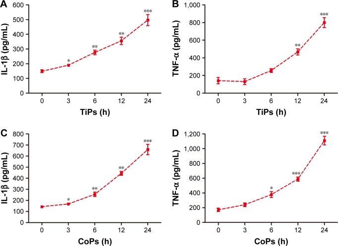
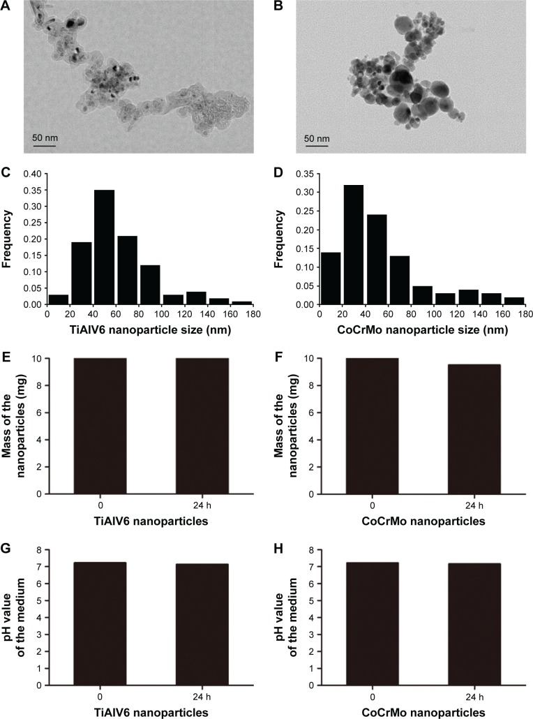
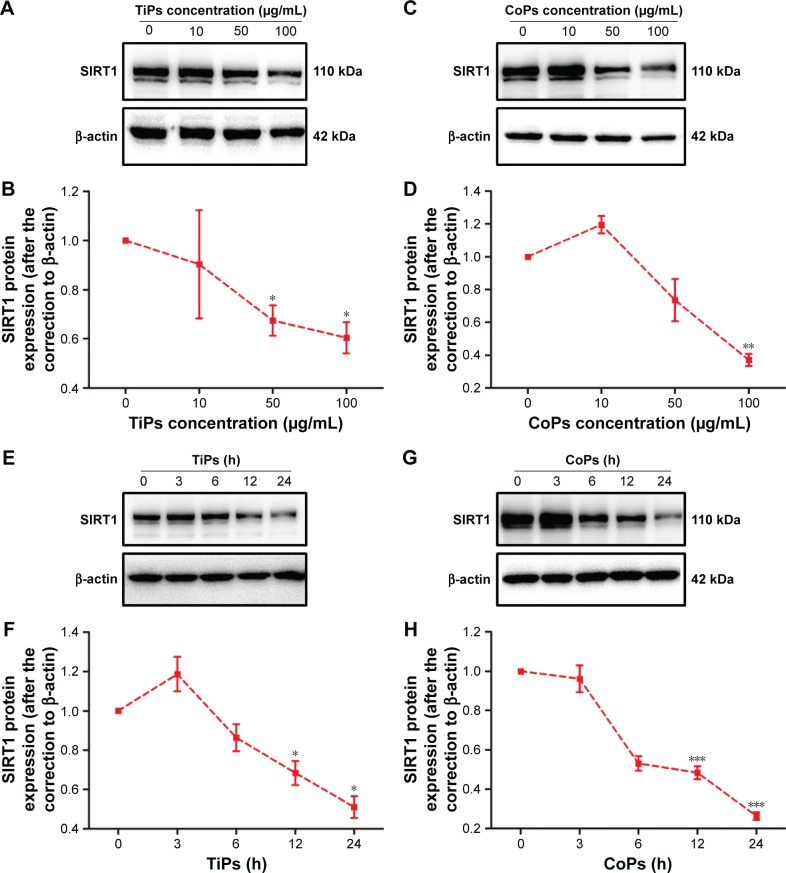
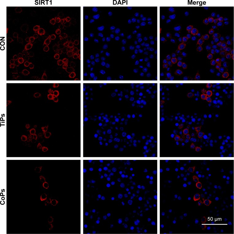
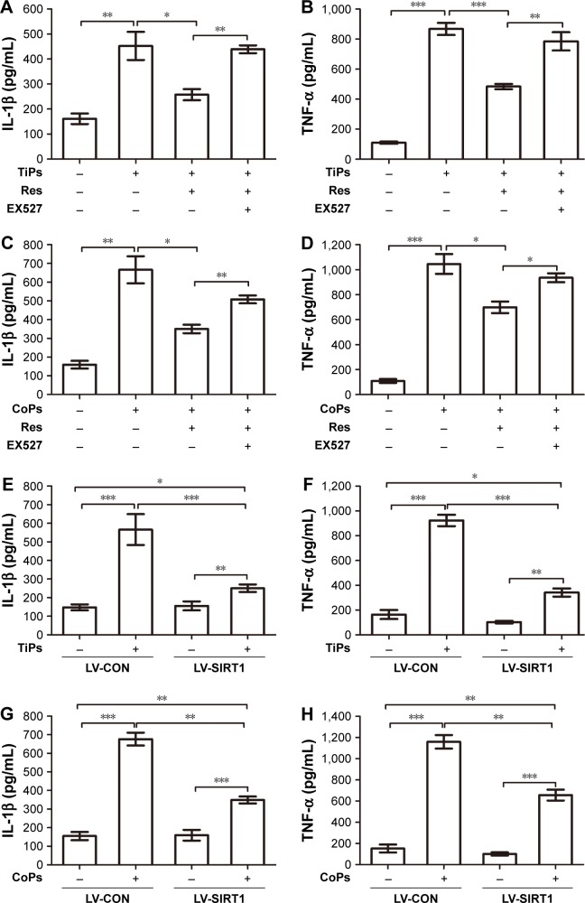
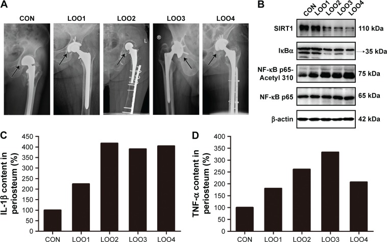
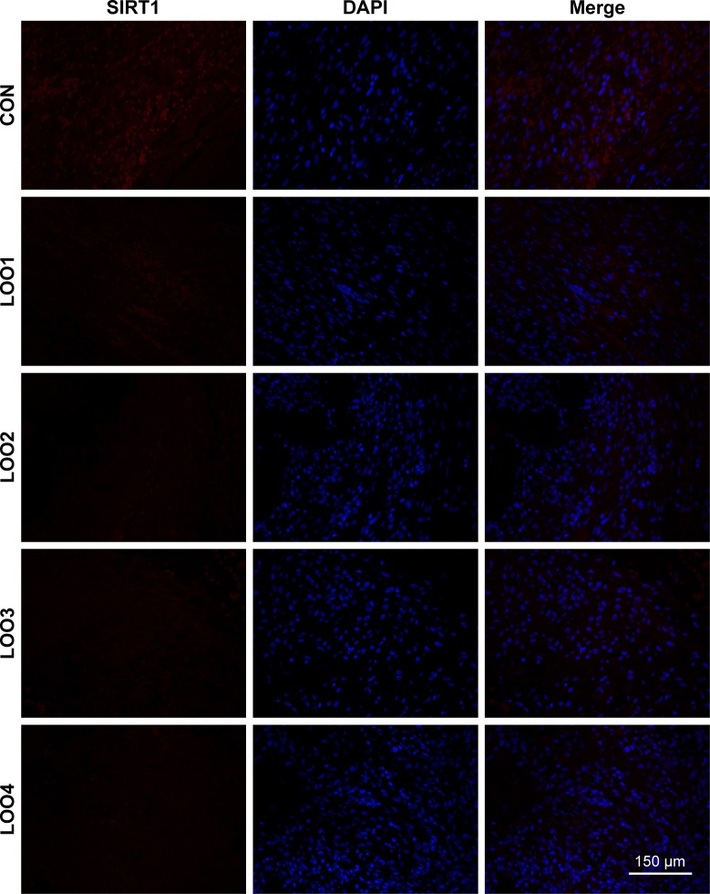
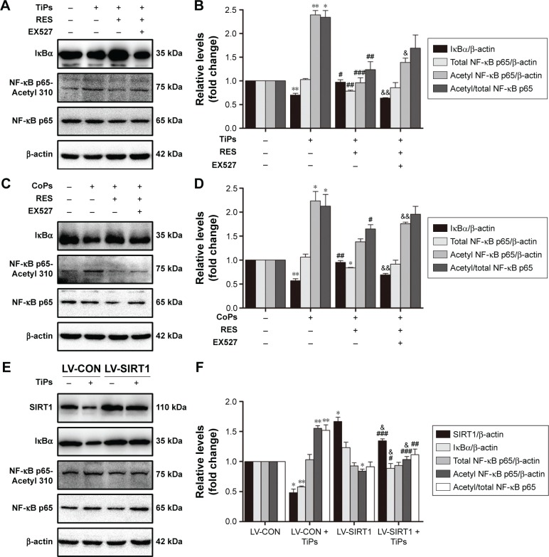
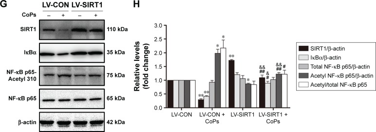
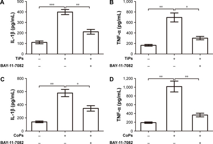
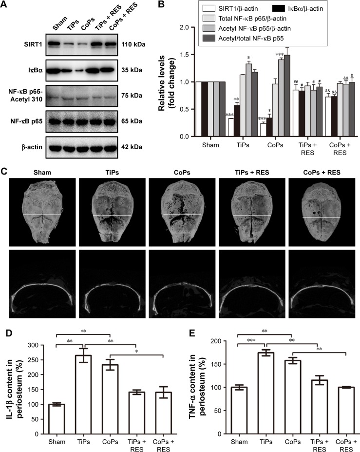
Aseptic loosening is the most common cause of total hip arthroplasty (THA) failure, and osteolysis induced by wear particles plays a major role in aseptic loosening. Various pathways in multiple cell types contribute to the pathogenesis of osteolysis, but the role of Sirtuin 1 (SIRT1), which can regulate inflammatory responses through its deacetylation, has never been investigated. We hypothesized that the downregulation of SIRT1 in macrophages induced by metal nanoparticles was one of the reasons for osteolysis in THA failure. In this study, the expression of SIRT1 was examined in macrophages stimulated with metal nanoparticles from materials used in prosthetics and in specimens from patients suffering from aseptic loosening. To address whether SIRT1 downregulation triggers these inflammatory responses, the effects of the SIRT1 activator resveratrol on the expression of inflammatory cytokines in metal nanoparticle-stimulated macrophages were tested. The results demonstrated that SIRT1 expression was significantly downregulated in metal nanoparticle-stimulated macrophages and clinical specimens of prosthesis loosening. Pharmacological activation of SIRT1 dramatically reduced the particle-induced expression of inflammatory cytokines in vitro and osteolysis in vivo. Furthermore, SIRT1 regulated particle-induced inflammatory responses through nuclear factor kappa B (NF-κB) acetylation. Thus, the results of this study suggest that SIRT1 plays a key role in metal nanoparticle-induced inflammatory responses and that targeting the SIRT1 pathway may lead to novel therapeutic approaches for the treatment of aseptic prosthesis loosening.

Keywords: NF-κB; SIRT1; aseptic loosening; inflammatory response; metal nanoparticle.


Conflict of interest statement

Disclosure The authors report no conflicts of interest in this work.

Figures

Figure 1
Figure 1
Metal nanoparticle-induced inflammatory responses in the supernatant of Raw264.7 cells. Notes: (AD) The expression of IL-1β and TNF-α produced by Raw264.7 cells following incubation with TiPs (100 μg/mL) or CoPs (100 μg/mL) at the indicated time periods (0, 3, 6, 12, or 24 h). *P<0.05, **P<0.05, ***P<0.001 versus time 0. The data of all the experiments are represented as the mean ± SEM from three independent experiments. Abbreviations: IL, interleukin; TNF, tumor necrosis factor; TiPs, TiAl6V4 particles; CoPs, CoCrMo particles; SEM, standard error of the mean.
Figure 2
Figure 2
The characteristics of metal nanoparticles. Notes: (A, B) TEM images of TiPs and CoPs. (C, D) The particle size distribution of TiPs and CoPs was calculated by SimplePCI software (Compix). (E, F) A total of 10 mg of TiPs and CoPs were cultured in 100 mL of DMEM, and the mass of the particles was detected after 24 h. (G, H) A total of 10 mg of TiPs and CoPs were cultured in 100 mL of DMEM, and the pH value of the medium was detected. Abbreviations: TEM, transmission electron microscope; TiPs, TiAl6V4 particles; CoPs, CoCrMo particles; DMEM, Dulbecco’s Modified Eagle’s Medium.
Figure 3
Figure 3
SIRT1 expression was downregulated after exposure to metal nanoparticles. Notes: (A, C) SIRT1 protein levels after Raw264.7 cells were incubated with TiPs or CoPs at various concentrations (0, 10, 50, or 100 μg/mL). (B, D) The densities of the Western blot bands in (A) and (C) were quantified using the Gene Tools software. *P<0.05, **P<0.01 versus concentration 0. (E, G) SIRT1 protein levels after Raw264.7 cells were incubated with 100 μg/mL TiPs or CoPs for the indicated time periods (0, 3, 6, 12, or 24 h). (F, H) The density of the Western blot bands in (E) and (G) were quantified using the Gene Tools software. *P<0.05, ***P<0.001 versus time 0. The data of all the experiments are represented as the mean ± SEM from three independent experiments. Abbreviations: SIRT1, Sirtuin 1; TiPs, TiAl6V4 particles; CoPs, CoCrMo particles; SEM, standard error of the mean.
Figure 4
Figure 4
Immunofluorescence was performed to examine the expression of SIRT1 in the RAW264.7 cells. Notes: The cells were treated with 100 μg/mL TiPs or CoPs for 24 h. Red, SIRT1; blue, DAPI nuclear staining. Abbreviations: SIRT1, Sirtuin 1; TiPs, TiAl6V4 particles; CoPs, CoCrMo particles; DAPI, 4,6-diamidino-2-phenylindole; CON, control.
Figure 5
Figure 5
SIRT1 regulates the induction of inflammatory cytokine expression by metal nanoparticles in the supernatant of Raw264.7 cells. Notes: (AD) Inhibition of the expression of inflammatory cytokines by resveratrol. The expression of inflammatory cytokines in Raw264.7 cells incubated with resveratrol (10 μM) for 12 h prior to stimulation with TiPs (100 μg/mL) or CoPs (100 μg/mL) for another 24 h. EX527 (5 μM) was used to reverse the effects of resveratrol. *P<0.05, **P<0.05, ***P<0.001. (EH) Inhibition of the expression of inflammatory cytokines by SIRT1 overexpression. Control cells and SIRT1-overexpressing cells were stimulated with TiPs (100 μg/mL) or CoPs (100 μg/mL) for 24 h. *P<0.05, **P<0.05, ***P<0.001. The data of all the experiments are represented as the mean ± SEM from three independent experiments. Abbreviations: SIRT1, Sirtuin 1; TiPs, TiAl6V4 particles; CoPs, CoCrMo particles; SEM, standard error of the mean; Res, resveratrol; LV-CON, lentivirus control.
Figure 6
Figure 6
SIRT1 was associated with the induction of inflammatory cytokine expression by metal particles in clinical specimens of aseptic loosening. Notes: (A) Radiographic films of patients with aseptic loosening. The arrow indicates the site where the specimens were obtained. (B) Western blots of clinical specimens of aseptic loosening. (C, D) The expression of inflammatory cytokines of clinical specimens of aseptic loosening. The cytokine concentrations were quantified by ELISA. Abbreviations: SIRT1, Sirtuin 1; ELISA, enzyme-linked immunosorbent assay; LOO, aseptic loosening; IκB, inhibitor of κB; NF-κB, nuclear factor kappa B; IL, interleukin; TNF, tumor necrosis factor.
Figure 7
Figure 7
SIRT1 was downregulated in the clinical specimens of patients with aseptic loosening. Notes: Immunofluorescence was performed to examine the expression of SIRT1 in the clinical specimens of patients with aseptic loosening. Red, SIRT1; blue, DAPI nuclear staining. Abbreviations: SIRT1, Sirtuin 1; DAPI, 4,6-diamidino-2-phenylindole; LOO, aseptic loosening; CON, control.
Figure 8
Figure 8
The SIRT1–NF-κB pathway was involved in the inflammation responses induced by metal nanoparticles. Notes: (A) Western blots performed after Raw264.7 cells were treated with PBS (control), TiPs, TiPs + resveratrol, TiPs + resveratrol + EX527. (B) The density of the Western blot bands in (A) was quantified using Gene Tools software. *P<0.05; **P<0.01 versus control. #P<0.05, ##P<0.01, ###P<0.001 versus TiPs. &P<0.05, &&P<0.01 versus TiPs + resveratrol. (C) Western blots performed after Raw264.7 cells were treated with PBS (control), CoPs, CoPs + resveratrol, CoPs + resveratrol + EX527. (D) The density of the Western blot bands in (C) was quantified using the Gene Tools software. *P<0.05; **P<0.01 versus control. #P<0.05, ##P<0.01 versus CoPs. &&P<0.01 versus CoPs + resveratrol. (E) Western blots performed after control cells and SIRT1 overexpression cells were treated with TiPs. (F) The density of the Western blot bands in (E) was quantified using the Gene Tools software. *P<0.05, **P<0.01 versus LV-CON. #P<0.05, ##P<0.01, ###P<0.001 versus LV-CON + TiPs. &P<0.05 versus LV-SIRT1. (G) Western blots performed after control cells and SIRT1 overexpression cells were treated with CoPs. (H) The density of the Western blot bands in (G) was quantified using Gene Tools software. *P<0.05, **P<0.01 versus LV-CON. #P<0.05, ##P<0.01 versus LV-CON + CoPs. &P<0.05, &&P<0.01 versus LV-SIRT1. The data of all the experiments are represented as the mean ± SEM from three independent experiments. Abbreviations: SIRT1, Sirtuin 1; NF-κB, nuclear factor kappa B; PBS, phosphate-buffered saline; TiPs, TiAl6V4 particles; CoPs, CoCrMo particles; SEM, standard error of the mean; IκB, inhibitor of κB; RES, resveratrol.
Figure 8
Figure 8
The SIRT1–NF-κB pathway was involved in the inflammation responses induced by metal nanoparticles. Notes: (A) Western blots performed after Raw264.7 cells were treated with PBS (control), TiPs, TiPs + resveratrol, TiPs + resveratrol + EX527. (B) The density of the Western blot bands in (A) was quantified using Gene Tools software. *P<0.05; **P<0.01 versus control. #P<0.05, ##P<0.01, ###P<0.001 versus TiPs. &P<0.05, &&P<0.01 versus TiPs + resveratrol. (C) Western blots performed after Raw264.7 cells were treated with PBS (control), CoPs, CoPs + resveratrol, CoPs + resveratrol + EX527. (D) The density of the Western blot bands in (C) was quantified using the Gene Tools software. *P<0.05; **P<0.01 versus control. #P<0.05, ##P<0.01 versus CoPs. &&P<0.01 versus CoPs + resveratrol. (E) Western blots performed after control cells and SIRT1 overexpression cells were treated with TiPs. (F) The density of the Western blot bands in (E) was quantified using the Gene Tools software. *P<0.05, **P<0.01 versus LV-CON. #P<0.05, ##P<0.01, ###P<0.001 versus LV-CON + TiPs. &P<0.05 versus LV-SIRT1. (G) Western blots performed after control cells and SIRT1 overexpression cells were treated with CoPs. (H) The density of the Western blot bands in (G) was quantified using Gene Tools software. *P<0.05, **P<0.01 versus LV-CON. #P<0.05, ##P<0.01 versus LV-CON + CoPs. &P<0.05, &&P<0.01 versus LV-SIRT1. The data of all the experiments are represented as the mean ± SEM from three independent experiments. Abbreviations: SIRT1, Sirtuin 1; NF-κB, nuclear factor kappa B; PBS, phosphate-buffered saline; TiPs, TiAl6V4 particles; CoPs, CoCrMo particles; SEM, standard error of the mean; IκB, inhibitor of κB; RES, resveratrol.
Figure 9
Figure 9
NF-κB inhibitor suppresses metal nanoparticle-induced inflammatory responses in macrophages. Notes: (AD) The expression of inflammatory cytokines in the supernatant of Raw264.7 cells after pretreatment with BAY-11-7082 (10 μM) for 8 h prior to stimulation with TiPs (100 μg/mL) or CoPs (100 μg/mL) for another 24 h. The cytokine concentrations were quantified by ELISA. *P<0.05, **P<0.01, ***P<0.001. The data of all the experiments are represented as the mean ± SEM from three independent experiments. Abbreviations: NF-κB, nuclear factor kappa B; TiPs, TiAl6V4 particles; CoPs, CoCrMo particles; SEM, standard error of the mean; ELISA, enzyme-linked immunosorbent assay; IL, interleukin; TNF, tumor necrosis factor.
Figure 10
Figure 10
SIRT1 mediated PIO and local inflammatory responses in animal models. Notes: (A) Western blots performed in the periosteum of animals treated with PBS (sham), TiPs, CoPs, TiPs + resveratrol, or CoPs + resveratrol. (B) The density of the Western blot bands in (A) was quantified using the Gene Tools software. *P<0.05, **P<0.01, ***P<0.001 versus sham operation group. #P<0.05, ##P<0.01 versus TiPs group. &P<0.05, &&P<0.01 versus CoPs group. (C) Representative images of micro-CT with 3-dimensional reconstructed images (top panel) from animals treated with PBS (sham), TiPs, CoPs, TiPs + resveratrol, or CoPs + resveratrol. In the second panel, the white horizontal line indicates cross-sectional views of the reconstructed images. (D, E) The levels of TNF-α and IL-1β were detected by ELISA in the periosteal tissues of animals treated with PBS (sham), TiPs, CoPs, TiPs + resveratrol, or CoPs + resveratrol. *P<0.05, **P<0.01, ***P<0.001. Data are represented as the mean ± SEM. n=6 mice per group. Abbreviations: SIRT1, Sirtuin 1; PIO, particle-induced osteolysis; PBS, phosphate-buffered saline; TiPs, TiAl6V4 particles; CoPs, CoCrMo particles; CT, computed tomography; TNF, tumor necrosis factor; IL, interleukin; ELISA, enzyme-linked immunosorbent assay; SEM, standard error of the mean; IκB, inhibitor of κB; NF-κB, nuclear factor kappa B; RES, resveratrol.

Similar articles

Cited by

References

    1. Prokopovich P. Interactions between mammalian cells and nano- or micro-sized wear particles: physico-chemical views against biological approaches. Adv Colloid Interface Sci. 2014;213:36–47. - PubMed
    1. Deng Z, Wang Z, Jin J, et al. SIRT1 protects osteoblasts against particle-induced inflammatory responses and apoptosis in aseptic prosthesis loosening. Acta Biomater. 2017;49:541–554. - PubMed
    1. Purdue PE, Koulouvaris P, Potter HG, Nestor BJ, Sculco TP. The cellular and molecular biology of periprosthetic osteolysis. Clin Orthop Relat Res. 2007;454:251–261. - PubMed
    1. Wang R, Wang Z, Ma Y, et al. Particle-induced osteolysis mediated by endoplasmic reticulum stress in prosthesis loosening. Biomaterials. 2013;34(11):2611–2623. - PubMed
    1. Gallo J, Raska M, Mrazek F, Petrek M. Bone remodeling, particle disease and individual susceptibility to periprosthetic osteolysis. Physiol Res. 2008;57(3):339–349. - PubMed

MeSH terms

LinkOut - more resources