. 2017 May 11:11:28.
doi: 10.3389/fnsys.2017.00028. eCollection 2017.

Dm5-HT2B: Pharmacological Characterization of the Fifth Serotonin Receptor Subtype of Drosophila melanogaster

Affiliations

Dm5-HT2B: Pharmacological Characterization of the Fifth Serotonin Receptor Subtype of Drosophila melanogaster

Wolfgang Blenau et al. Front Syst Neurosci. .

Abstract

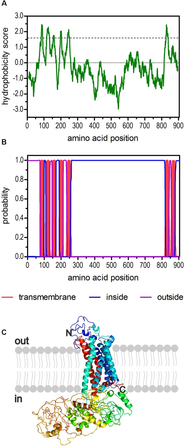
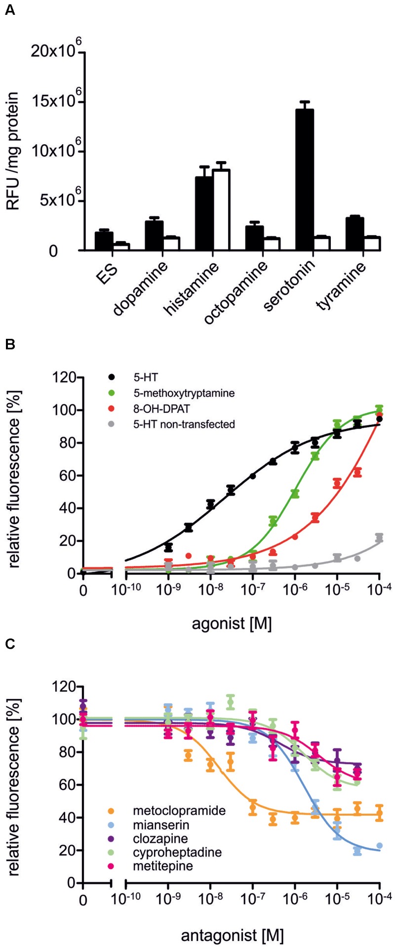
Serotonin (5-hydroxytryptamine, 5-HT) is an important regulator of physiological and behavioral processes in both protostomes (e.g., insects) and deuterostomes (e.g., mammals). In insects, serotonin has been found to modulate the heart rate and to control secretory processes, development, circadian rhythms, aggressive behavior, as well as to contribute to learning and memory. Serotonin exerts its activity by binding to and activating specific membrane receptors. The clear majority of these receptors belong to the superfamily of G-protein-coupled receptors. In Drosophila melanogaster, a total of five genes have been identified coding for 5-HT receptors. From this family of proteins, four have been pharmacologically examined in greater detail, so far. While Dm5-HT1A, Dm5-HT1B, and Dm5-HT7 couple to cAMP signaling cascades, the Dm5-HT2A receptor leads to Ca2+ signaling in an inositol-1,4,5-trisphosphate-dependent manner. Based on sequence similarity to homologous genes in other insects, a fifth D. melanogaster gene was uncovered coding for a Dm5-HT2B receptor. Knowledge about this receptor's pharmacological properties is very limited. This is quite surprising because Dm5-HT2B has been attributed to distinct physiological functions based on genetic interference with its gene expression. Mutations were described reducing the response of the larval heart to 5-HT, and specific knockdown of Dm5-HT2B mRNA in hemocytes resulted in a higher susceptibility of the flies to bacterial infection. To gain deeper understanding of Dm5-HT2B's pharmacology, we evaluated the receptor's response to a series of established 5-HT receptor agonists and antagonists in a functional cell-based assay. Metoclopramide and mianserin were identified as two potent antagonists that may allow pharmacological interference with Dm5-HT2B signaling in vitro and in vivo.

Keywords: 4; 5-trisphosphate; Ca2+; G protein-coupled receptor; biogenic amine; cAMP; cellular signaling; inositol-1; insect; second messenger.


Figures

FIGURE 1
FIGURE 1
Structural characteristics of the deduced amino acid sequence of Dm5-HT2B. (A) Hydrophobicity profile of Dm5-HT2B. The profile was calculated according to the algorithm of Kyte and Doolittle (1982) using a window size of 19 amino acids. Peaks with scores greater than 1.6 (dashed line) indicate possible transmembrane regions. (B) Prediction of transmembrane domains with TMHMM server v. 2.0 (Krogh et al., 2001). Putative transmembrane domains are indicated in red. Extracellular regions are shown as purple line, intracellular regions as blue line. (C) The primary sequence of Dm5-HT2B was submitted to Phyre2 (Kelley et al., 2015). The 3D model of the receptor is color-coded (rainbow). The extracellular N-terminus and the intracellular C-terminus are labeled.
FIGURE 2
FIGURE 2
Amino acid sequence alignment of Dm5-HT2B and orthologous receptors from Apis mellifera (Am5-HT2B, CBX90121), Rhodnius prolixus (Rp5-HT2B, AKQ13312), and Panulirus interruptus (Pi5-HT2B, AAS57919). Identical residues (≥75%) are shown as white letters against black, whereas conservatively substituted residues are shaded. Putative transmembrane domains (TM1–TM7) are indicated by gray bars. Potential N-glycosylation sites (formula image
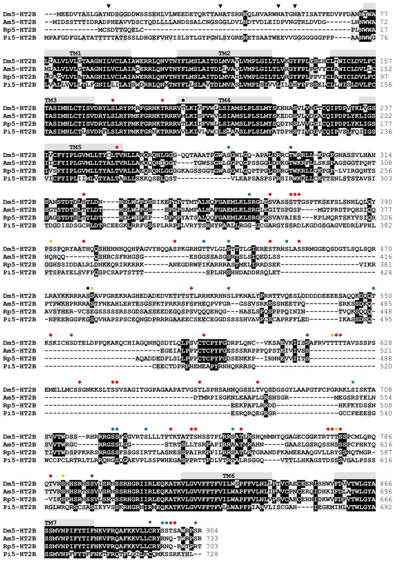
), PKA phosphorylation sites (formula image), PKC phosphorylation sites (formula image), phosphorylation sites for both PKA and PKC (formula image), PKG phosphorylation sites (formula image), phosphorylation sites for both PKA and PKG (formula image), phosphorylation sites for all three kinases (formula image), and putative palmitoylation sites () of Dm5-HT2B are indicated. The amino acid position is given on the right.
FIGURE 3
FIGURE 3
Bayesian phylogeny of 5-HT receptors. Alignments were performed using Clustal W (Thompson et al., 1994) by using the core amino-acid sequences lacking the variable regions of the N- and C-terminus and the third cytoplasmic loop. Human rhodopsin (Hs RHOD) and D. melanogaster FMRFamide receptor (Dm FMRFaR) were used to root the tree. Numbers at branches represent the posterior probabilities. Receptor subclasses are highlighted by distinct colors. Abbreviations of species in alphabetical order: Aa Aedes aegypti, Ac Aplysia californica, Ag Anopheles gambiae, Ak Aplysia kurodai, Am Apis mellifera, Ap Acyrthosiphon pisum, Bm Bombyx mori, Bt Bombus terrestris, Cv Cimex lectularius, Cv Calliphora vicina, Dm Drosophila melanogaster, Dr Danio rerio, Hs Homo sapiens, Ms Manduca sexta, Pa Periplaneta americana, Pc Procambarus clarkii, Pi Panulirus interruptus, Pr Pieris rapae, Rn Rhodnius neglectus, Tc Tribolium castaneum, Ti Triatoma infestans.
FIGURE 4
FIGURE 4
Pharmacological properties of Dm5-HT2B. (A) Effect of different biogenic amines on Ca2+-dependent Fluo-4 fluorescence in Dm5-HT2B-expressing and non-transfected HEK 293 cells. Bars represent changes in relative fluorescence units (RFU/mg protein) in Dm5-HT2B-expressing (black bars) and non-transfected HEK 293 cells (white bars). Biogenic amines were applied in a concentration of 10-6 M. Mean values ± SD were calculated from octuplicate determinations. ES, extracellular solution. (B) Concentration-dependent effects of serotonin on Dm5-HT2B-expressing (black) and non-transfected HEK 293 cells (gray) as well as of 5-methoxytryptamine (green) and 8-OH-DPAT (red) on Dm5-HT2B-expressing cells. Data from representative experiments are shown. (Each data point represents the mean ± SD of an octuplicatedetermination. The relative fluorescence signal (%) for measurementswith serotonin was normalized to the value measured in the presenceof 10-4 M serotonin in Dm5-HT2B-expressing cells (=100%). The relative fluorescence signals (%) for measurements with 5-methoxytryptamine and 8-OH-DPAT were normalized to the value measured in the presence of 10-4 M of the respective ligand (=100%). (C) Concentration-dependenteffects of potential antagonists on serotonin-stimulated Dm5-HT2B-evoked Ca2+ signals. Increasing concentrations (10-9 M to 10-4 M) of receptor antagonists were added to the receptor-expressing cell line. The Ca2+-dependent Fluo-4 signals were registered and normalized to thefluorescence evoked with 10-7 M serotonin (=100%). Data from representative experiments are shown. Each data point represents the mean ± SD of an octuplicate determination.)

Similar articles

Cited by

References

    1. Alekseyenko O. V., Chan Y. B., Fernandez M. P., Bülow T., Pankratz M. J., Kravitz E. A. (2014). Single serotonergic neurons that modulate aggression in Drosophila. Curr. Biol. 24 2700–2707. 10.1016/j.cub.2014.09.051 - DOI - PMC - PubMed
    1. Alekseyenko O. V., Kravitz E. A. (2014). Serotonin and the search for the anatomical substrate of aggression. Fly 8 200–205. 10.1080/19336934.2015.1045171 - DOI - PMC - PubMed
    1. Alekseyenko O. V., Lee C., Kravitz E. A. (2010). Targeted manipulation of serotonergic neurotransmission affects the escalation of aggression in adult male Drosophila melanogaster. PLoS ONE 5:e10806 10.1371/journal.pone.0010806 - DOI - PMC - PubMed
    1. Arakawa S., Gocayne J. D., McCombie W. R., Urquhart D. A., Hall L. M., Fraser C. M., et al. (1990). Cloning, localization, and permanent expression of a Drosophila octopamine receptor. Neuron 4 343–354. - PubMed
    1. Ballesteros J., Weinstein H. (1995). Integrated methods for the construction of three-dimensional models of structure-function relations in G protein-coupled receptors. Methods Neurosci. 25 366–428.

LinkOut - more resources