. 2017 May 12:8:57.
doi: 10.3389/fgene.2017.00057. eCollection 2017.

Non-coding RNAs in the Ovarian Follicle

Affiliations

Non-coding RNAs in the Ovarian Follicle

Rosalia Battaglia et al. Front Genet. .

Abstract

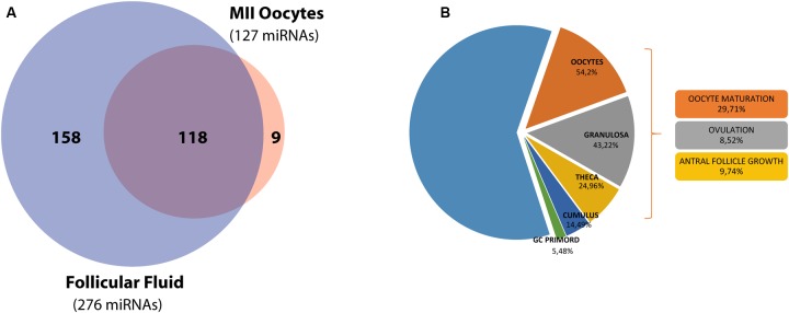
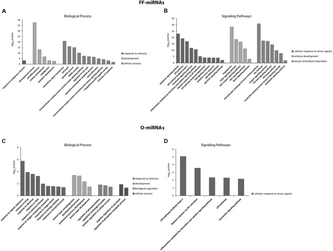
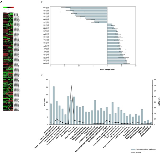
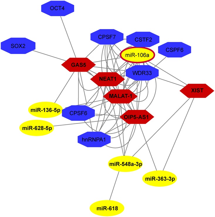
The mammalian ovarian follicle is the complex reproductive unit comprising germ cell, somatic cells (Cumulus and Granulosa cells), and follicular fluid (FF): paracrine communication among the different cell types through FF ensures the development of a mature oocyte ready for fertilization. This paper is focused on non-coding RNAs in ovarian follicles and their predicted role in the pathways involved in oocyte growth and maturation. We determined the expression profiles of microRNAs in human oocytes and FF by high-throughput analysis and identified 267 microRNAs in FF and 176 in oocytes. Most of these were FF microRNAs, while 9 were oocyte specific. By bioinformatic analysis, independently performed on FF and oocyte microRNAs, we identified the most significant Biological Processes and the pathways regulated by their validated targets. We found many pathways shared between the two compartments and some specific for oocyte microRNAs. Moreover, we found 41 long non-coding RNAs able to interact with oocyte microRNAs and potentially involved in the regulation of folliculogenesis. These data are important in basic reproductive research and could also be useful for clinical applications. In fact, the characterization of non-coding RNAs in ovarian follicles could improve reproductive disease diagnosis, provide biomarkers of oocyte quality in Assisted Reproductive Treatment, and allow the development of therapies for infertility disorders.

Keywords: follicular fluid; human oocyte; lncRNAs; microRNAs; ovarian follicle.


Figures

FIGURE 1
FIGURE 1
miRNA distribution in the ovarian follicle and functional analysis of the target genes. (A) Venn diagram shows the overlap between miRNA sets in hFF and the mature MII oocyte. (B) A second diagram shows miRNA target expression and function inside the ovarian follicle.
FIGURE 2
FIGURE 2
Significant overrepresentation of GOs, in terms of Biological Processes, and signaling pathways for miRNAs identified in hFF (FF-miRNAs) and the MII oocytes (O-miRNAs) are shown in (A,B) and (C,D), respectively. The significance values are reported as -log10 (P-value).
FIGURE 3
FIGURE 3
miRNA expression in hFF and human oocytes. (A) Heat map of normalized miRNA expression data (–DCT values) of 118 Common-miRNAs for hFF and oocyte samples. The red and green colors represent up and down regulated miRNA expression levels, respectively. Equally expressed miRNAs are indicated in black. (B) Relative expression levels of 27 and 11 miRNAs that were differentially expressed between FF and oocytes. (C) Signaling pathway enrichment analysis for common miRNAs with KEGG against listed target genes. The probability values are reported as –log10 (P-value).
FIGURE 4
FIGURE 4
ncRNA network in human MII oocytes. The Network, drawn by NPInter v3.0, shows the interaction among lncRNAs and miRNAs involved in stemness, RNA maturation and epigenetics.

Similar articles

Cited by

References

    1. Bartel D. P. (2004). MicroRNAs: genomics, biogenesis, mechanism, and function. Cell 116 281–297. - PubMed
    1. Batagov A. O., Kuznetsov V. A., Kurochkin I. V. (2011). Identification of nucleotide patterns enriched in secreted RNAs as putative cis-acting elements targeting them to exosome nano-vesicles. BMC Genomics 12(Suppl. 3):S18 10.1186/1471-2164-12-S3-S18 - DOI - PMC - PubMed
    1. Battaglia R., Vento M. E., Ragusa M., Barbagallo D., La Ferlita A., Di Emidio G., et al. (2016). MicroRNAs are stored in human MII oocyte and their expression profile changes in reproductive aging. Biol. Reprod. 95 1–13. 10.1095/biolreprod.116.142711 - DOI - PubMed
    1. Bond C. S., Fox A. H. (2009). Paraspeckles: nuclear bodies built on long noncoding RNA. J. Cell Biol. 186 637–644. 10.1083/jcb.200906113 - DOI - PMC - PubMed
    1. Briggs S. F., Dominguez A. A., Chavez S. L., Reijo Pera R. A. (2015). Single-cell XIST expression in human preimplantation embryos and newly reprogrammed female induced pluripotent stem cells. Stem Cells 33 1771–1781. 10.1002/stem.1992 - DOI - PMC - PubMed