Review
. 2017 Apr;12(4):518-524.
doi: 10.4103/1673-5374.205000.

Axon degeneration: make the Schwann cell great again

Affiliations
Review

Axon degeneration: make the Schwann cell great again

Keit Men Wong et al. Neural Regen Res. 2017 Apr.

Abstract

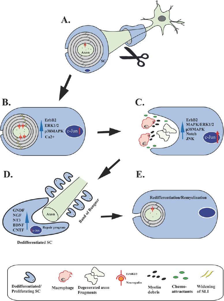
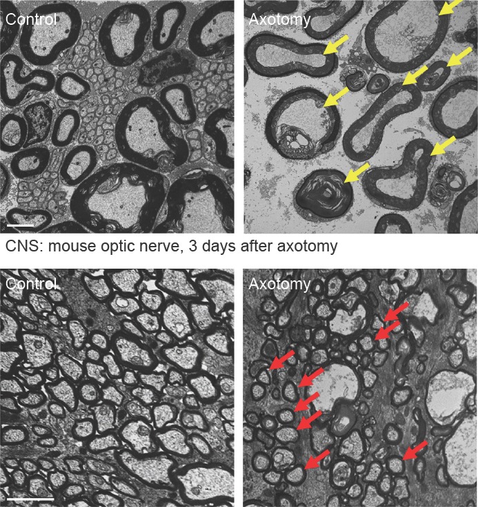
Axonal degeneration is a pivotal feature of many neurodegenerative conditions and substantially accounts for neurological morbidity. A widely used experimental model to study the mechanisms of axonal degeneration is Wallerian degeneration (WD), which occurs after acute axonal injury. In the peripheral nervous system (PNS), WD is characterized by swift dismantling and clearance of injured axons with their myelin sheaths. This is a prerequisite for successful axonal regeneration. In the central nervous system (CNS), WD is much slower, which significantly contributes to failed axonal regeneration. Although it is well-documented that Schwann cells (SCs) have a critical role in the regenerative potential of the PNS, to date we have only scarce knowledge as to how SCs 'sense' axonal injury and immediately respond to it. In this regard, it remains unknown as to whether SCs play the role of a passive bystander or an active director during the execution of the highly orchestrated disintegration program of axons. Older reports, together with more recent studies, suggest that SCs mount dynamic injury responses minutes after axonal injury, long before axonal breakdown occurs. The swift SC response to axonal injury could play either a pro-degenerative role, or alternatively a supportive role, to the integrity of distressed axons that have not yet committed to degenerate. Indeed, supporting the latter concept, recent findings in a chronic PNS neurodegeneration model indicate that deactivation of a key molecule promoting SC injury responses exacerbates axonal loss. If this holds true in a broader spectrum of conditions, it may provide the grounds for the development of new glia-centric therapeutic approaches to counteract axonal loss.

Keywords: Wallerian degeneration; dedifferentiation; glia; myelin; neurodegeneration; oligodendrocytes.


Figures

Figure 1
Figure 1
Schwann cell (SC) responses following axotomy. Schematic illustrating distinct stages of SC responses following axotomy and during axonal regeneration and remyelination. (A) Under basal conditions the axon is encapsulated by a compact myelin sheath, established by Neuregulin-ErbB2/3 signaling during development. (B) Upon injury the first reactions in the SC, already minutes to 1 hour after axotomy, include widening of Schmidt-Lanterman incisures (yellow stripes in compact myelin), activation of the ErbB2 receptor tyrosine kinase (red anchors), activation of p38- and Erk1/2 mitogen-activated protein kinase (MAPK) signaling, and rapid increase of cytoplasmic calcium levels in the SC. Increased expression of c-Jun is also observed few hours after nerve injury. Actin progressively polymerizes in widened Schmidt-Lanterman incisures (SLI) (not depicted). These changes are accompanied by a general increase of the SC body size. (C) Following a latency phase with axons showing no morphological evidence of degeneration, axons then abruptly disintegrate leaving axon fragments behind. In parallel, during Wallerian degeneration (WD) there is formation of myelin debris secondary to rapid disassembly of the myelin sheaths. SCs release cytokines and chemokines that attract macrophages. In addition, SCs robustly upregulate injury response pathways including MAPK kinase pathways, Notch signaling, as well as further expression increases of c-Jun. (D) Axonal regeneration following WD is underway with SCs forming bands of Bungner and releasing surface molecules (not shown) and a multitude of neurotrophic factors including glial cell-derived neurotrophic factor (GDNF), nerve growth factor (NGF), neurotrophin-3 (NT3), brain-derived neurotrophic factor (BDNF), and ciliary neurotrophic factor (CNTF). This release is regulated by c-Jun in the SC. (E) SCs eventually conclude their repair program and redifferentiate to promote completion of nerve repair. Regenerating axons are remyelinated by SCs to restore normal nerve function.
Figure 2
Figure 2
Wallerian degeneration is protracted in the central nervous system. Transmission electron microscopy of control and axotomized transverse mouse sciatic nerves (upper row) and optic nerves (lower row) representing Wallerian degeneration of the peripheral nervous system (PNS) and central nervous system (CNS), respectively. Note complete degeneration of all PNS axons with granular disintegration of the axoplasm in the distal nerve stump three days following axotomy (yellow arrows depict examples). In contrast, many CNS axons are structurally preserved three days following optic nerve axotomy (red arrows depict examples). This indicates that Wallerian degeneration of axons progresses slower in the CNS than in the PNS. Scale bars: 2 μm.

References

    1. Agthong S, Kaewsema A, Tanomsridejchai N, Chentanez V. Activation of MAPK ERK in peripheral nerve after injury. BMC Neurosci. 2006;7:45. - PMC - PubMed
    1. Arthur-Farraj PJ, Latouche M, Wilton DK, Quintes S, Chabrol E, Banerjee A, Woodhoo A, Jenkins B, Rahman M, Turmaine M, Wicher GK, Mitter R, Greensmith L, Behrens A, Raivich G, Mirsky R, Jessen KR. c-Jun reprograms Schwann cells of injured nerves to generate a repair cell essential for regeneration. Neuron. 2012;75:633–647. - PMC - PubMed
    1. Avery MA, Rooney TM, Pandya JD, Wishart TM, Gillingwater TH, Geddes JW, Sullivan PG, Freeman MR. WldS prevents axon degeneration through increased mitochondrial flux and enhanced mitochondrial Ca2+ buffering. Curr Biol. 2012;22:596–600. - PMC - PubMed
    1. Babetto E, Beirowski B, Russler EV, Milbrandt J, DiAntonio A. The Phr1 ubiquitin ligase promotes injury-induced axon self-destruction. Cell Rep. 2013;3:1422–1429. - PMC - PubMed
    1. Babetto E, Beirowski B, Janeckova L, Brown R, Gilley J, Thomson D, Ribchester RR, Coleman MP. Targeting NMNAT1 to axons and synapses transforms its neuroprotective potency in vivo. J Neurosci. 2010;30:13291–13304. - PMC - PubMed