. 2017:2017:2541275.
doi: 10.1155/2017/2541275. Epub 2017 May 3.

Human Adipose-Derived Stem Cells Exhibit Enhanced Proliferative Capacity and Retain Multipotency Longer than Donor-Matched Bone Marrow Mesenchymal Stem Cells during Expansion In Vitro

Affiliations

Human Adipose-Derived Stem Cells Exhibit Enhanced Proliferative Capacity and Retain Multipotency Longer than Donor-Matched Bone Marrow Mesenchymal Stem Cells during Expansion In Vitro

Kimberley L Burrow et al. Stem Cells Int. 2017.

Abstract

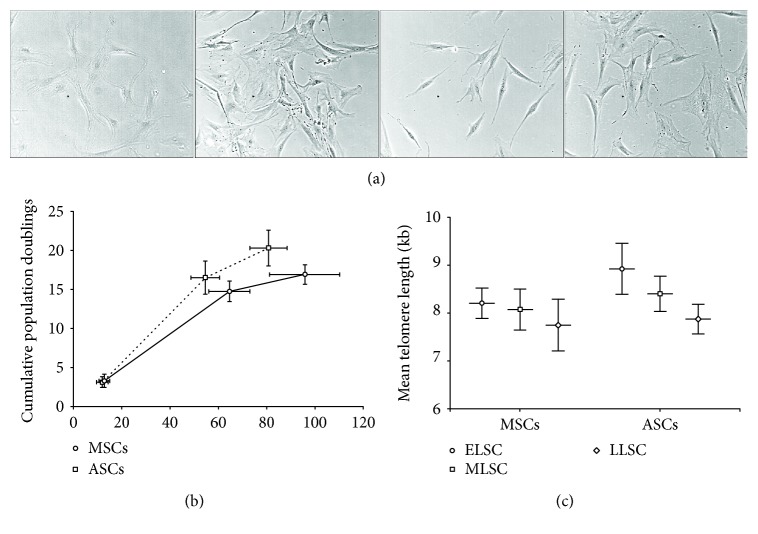
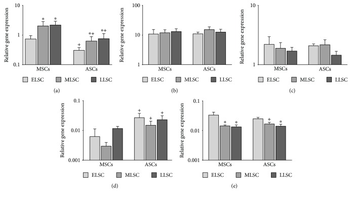
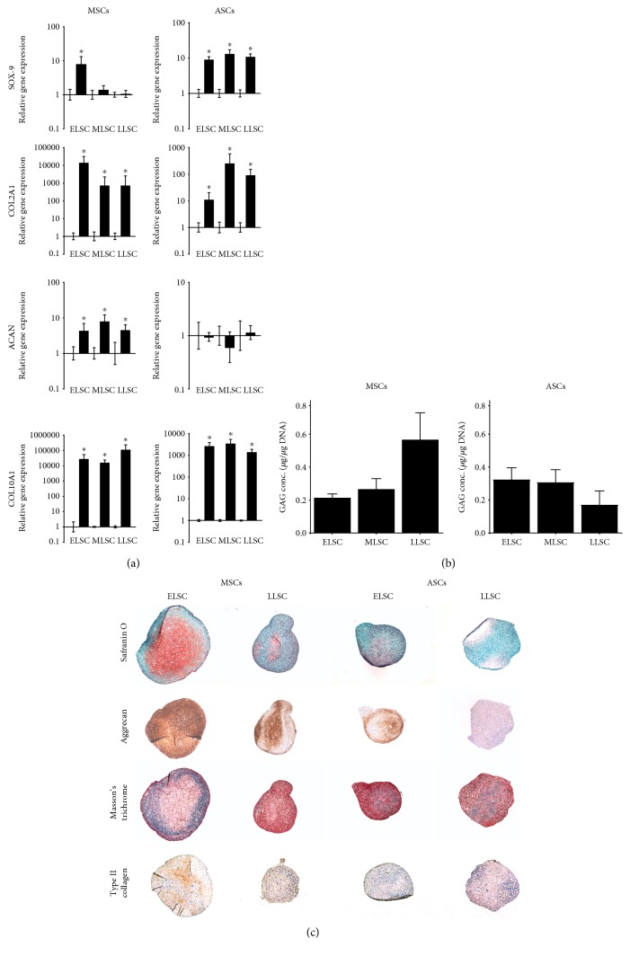
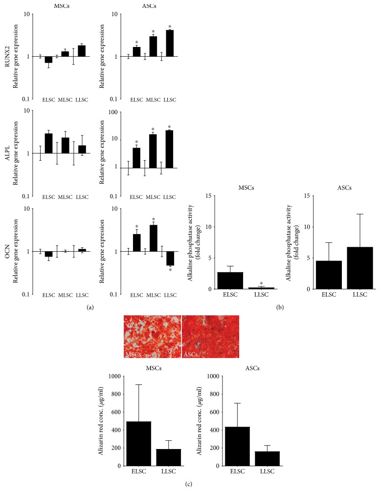
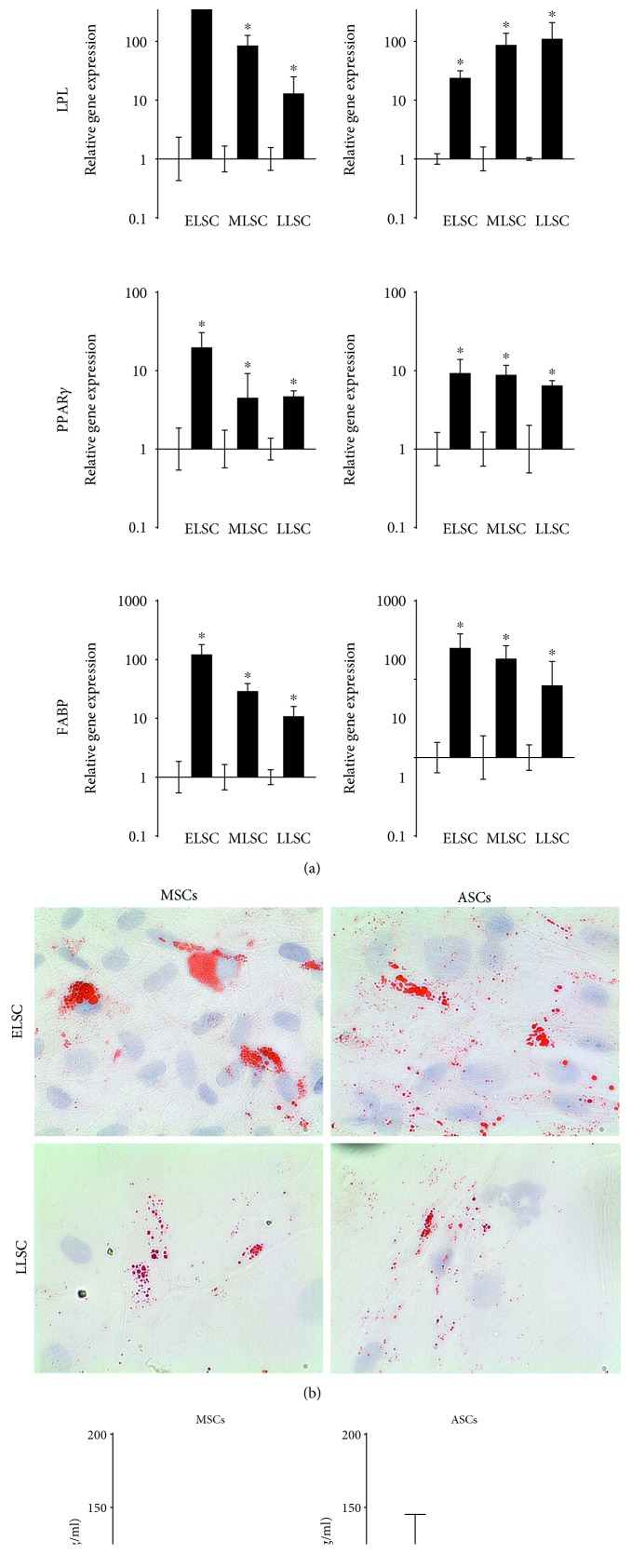
Bone marrow-derived mesenchymal stem cells (MSCs) and adipose-derived multipotent/mesenchymal stem cells (ASCs) have been proposed as the ideal cell types for a range of musculoskeletal tissue engineering and regenerative medicine therapies. However, extensive in vitro expansion is required to generate sufficient cells for clinical application and previous studies have demonstrated differences in the proliferative capacity and the impact of expansion on differentiation capacity of both MSCs and ASCs. Significantly, these studies routinely use cells from different donors, making direct comparisons difficult. Importantly, this study directly compared the proliferative capacity and multipotency of human MSCs and ASCs from the same donors to determine how each cell type was affected by in vitro expansion. The study identified that ASCs were able to proliferate faster and undergo greater population doublings than donor-matched MSCs and that senescence was primarily driven via telomere shortening and upregulation of p16ink4a. Both donor-matched MSCs and ASCs were capable of trilineage differentiation early in cultures; however, while differentiation capacity diminished with time in culture, ASCs retained enhanced capacity compared to MSCs. These findings suggest that ASCs may be the most appropriate cell type for musculoskeletal tissue engineering and regenerative medicine therapies due to their enhanced in vitro expansion capacity and limited loss of differentiation potential.


Figures

Figure 1
Figure 1
Assessment of MSC and ASC CD profile. (a) Expression of positive markers CD73, CD90, and CD105 was assessed using flow cytometry, along with expression of a panel of negative markers (CD14, CD20, CD34, and CD45) in donor-matched MSC and ASC samples (n = 5). Expression was compared to IgG controls. MSC and ASC numbers 1–5 represent donor-matched samples. (b) Expression of CD markers was further compared between ELSC and LLSC with no significant difference in expression identified with time in culture. Plots show representative data from one donor-matched sample and ELSC and LLSC compared to an IgG control.
Figure 2
Figure 2
Population kinetics. (a) Representative phase-contrast microscopy images of donor-matched MSCs and ASCs during monolayer culture at ELSC and LLSC. (b) Cumulative population doublings over time in culture demonstrating enhanced proliferation rate in ASCs compared to donor-matched MSCs (N = 5). Points represent ELSC, MLSC, and LLSC, ±SEM for both CPD and days in culture. (c) Mean telomere length in donor-matched MSCs and ASCs at ELSC, MLSC, and LLSC (±SEM) demonstrating longer telomeres in ASCs compared to MSCs and a decrease in mean telomere length with time in culture in both cell types (N = 5). ∗ represents P < 0.05 at MLSC or LLSC compared to ELSC. + represents P < 0.05 in ASCs compared to MSCs at the same LSC.
Figure 3
Figure 3
qPCR analysis of senescence marker genes p16ink4a (a), p21 (b), and p53 (c) and pluripotency marker genes Nanog (d) and Oct-4 (e) in donor-matched MSCs and ASC at ELSC, MLSC, and LLSC. Data presented and relative gene expression normalised to the mean of the reference genes MRPL19 and EIF2B1 and plotted on a log scale (N = 5). Values are mean ± SEM. ∗ represents P < 0.05 at MLSC or LLSC compared to ELSC. + represents P < 0.05 in ASCs compared to MSCs at the same LSC.
Figure 4
Figure 4
Assessment of the influence of monolayer expansion of the chondrogenic potential of donor-matched MSCs and ASCs. (a) Quantitative real-time PCR analysis of chondrogenic marker genes SOX-9, COL2A1, ACAN, and COL10A1 at ELSC, MLSC, and LLSC in MSCs and ASCs following pellet culture for 21 days in chondrogenic media. Data was normalised to the mean of the reference genes MRPL19 and EIF2B1 and presented as relative fold change in gene expression compared to day 0 controls plotted on a log scale. N = 5. Values are mean ± SEM. ∗ = P < 0.05. (b) DMMB analysis of GAG synthesis in MSCs and ASCs at ELSC and LLSC. Data represents mean GAG concentration from two pellets for each cell sample (μg/μg DNA) ± SEM. N = 5. ∗ = P < 0.05 compared to day 0 control. (c) Representative histological images of ELSC and LLSC in MSC and ASC pellets following 21 days in culture in a chondrogenic media. All images taken at equivalent magnification to allow comparison of size of pellets.
Figure 5
Figure 5
Assessment of the influence of monolayer expansion of the osteogenic potential of donor-matched MSCs and ASCs. (a) qPCR analysis of osteogenic marker at ELSC, MLSC, and LLSC in MSCs and ASCs following culture for 21 days in osteogenic media. Data was normalised to the mean of the reference genes MRPL19 and EIF2B1 and presented as relative fold change in gene expression compared to day 0 controls plotted on a log scale. N = 5. Values are mean ± SEM. ∗ = P < 0.05. (b) Quantification of alkaline phosphatase activity in MSCs and ASCs from triplicate wells at ELSC and LLSC. Data represent mean fold change in activity ± SEM compared to day 0 controls. N = 5. ∗ = P < 0.05. (c) Representative images of alizarin red staining of ELSC MSCs and ASCs following 0 days and 21 days in culture in osteogenic media and quantification of alizarin red staining in MSCs and ASCs at ELSC and LLSC. Data represent mean alizarin red concentration ± SEM. N = 5. ∗ = P < 0.05.
Figure 6
Figure 6
Assessment of the influence of monolayer expansion of the adipogenic potential of donor-matched MSCs and ASCs. (a) qPCR analysis of adipogenic marker genes at ELSC, MLSC, and LLSC in MSCs and ASCs following culture for 21 days in adipogenic media. Data was normalised to the mean of the reference genes MRPL19 and EIF2B1 and presented as relative fold change in gene expression compared to day 0 controls plotted on a log scale. N = 5. Values are mean ± SEM. ∗ = P < 0.05. (b) Representative images of oil red O staining in ELSC and LLSC MSCs and ASCs following 21 days in culture in adipogenic media. (c) Quantification of oil red O staining in MSCs and ASCs from triplicate wells at ELSC, MLSC, and LLSC. Data represents mean oil red O concentration (μg/ml) ± SEM. N = 5.

Similar articles

Cited by

References

    1. Ashton B. A., Allen T. D., Howlett C. R., Eaglesom C. C., Hattori A., Owen M. Formation of bone and cartilage by marrow stromal cells in diffusion chambers in vivo. Clinical Orthopaedics and Related Research. 1980;151(151):294–307. - PubMed
    1. Caplan A. I. Mesenchymal stem cells. Journal of Orthopaedic Research: Official Publication of the Orthopaedic Research Society. 1991;9(5):641–650. doi: 10.1002/jor.1100090504. - DOI - PubMed
    1. Friedenstein A. J., Chailakhyan R. K., Gerasimov U. V. Bone marrow osteogenic stem cells: in vitro cultivation and transplantation in diffusion chambers. Cell and Tissue Kinetics. 1987;20(3):263–272. - PubMed
    1. Friedenstein A. J., Deriglasova U. F., Kulagina N. N., et al. Precursors for fibroblasts in different populations of hematopoietic cells as detected by the in vitro colony assay method. Experimental Hematology. 1974;2(2):83–92. - PubMed
    1. Richardson S. M., Kalamegam G., Pushparaj P. N., et al. Mesenchymal stem cells in regenerative medicine: focus on articular cartilage and intervertebral disc regeneration. Methods. 2016;99:69–80. doi: 10.1016/j.ymeth.2015.09.015. - DOI - PubMed

LinkOut - more resources