. 2015 Jan 1;6(1):181-189.
doi: 10.1039/c4sc02424a. Epub 2014 Sep 16.

Ultrafast delocalization of excitation in synthetic light-harvesting nanorings

Affiliations

Ultrafast delocalization of excitation in synthetic light-harvesting nanorings

Chaw-Keong Yong et al. Chem Sci. .

Abstract

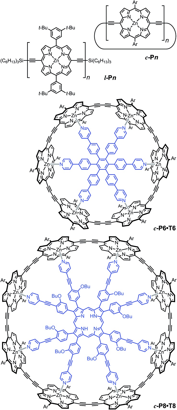
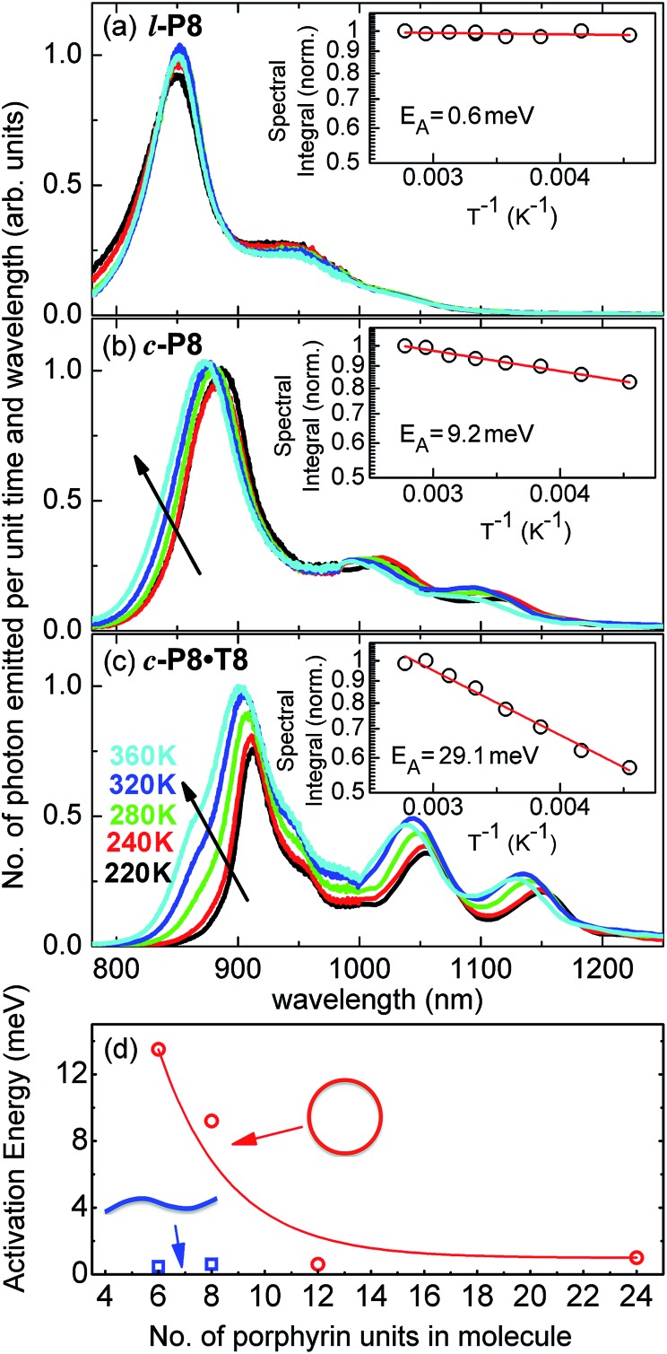
Rings of chlorophyll molecules harvest sunlight remarkably efficiently during photosynthesis in purple bacteria. The key to their efficiency lies in their highly delocalized excited states that allow for ultrafast energy migration. Here we show that a family of synthetic nanorings mimic the ultrafast energy transfer and delocalization observed in nature. π-Conjugated nanorings with diameters of up to 10 nm, consisting of up to 24 porphyrin units, are found to exhibit excitation delocalization within the first 200 fs of light absorption. Transitions from the first singlet excited state of the circular nanorings are dipole-forbidden as a result of symmetry constraints, but these selection rules can be lifted through static and dynamic distortions of the rings. The increase in the radiative emission rate in the larger nanorings correlates with an increase in static disorder expected from Monte Carlo simulations. For highly symmetric rings, the radiative rate is found to increase with increasing temperature. Although this type of thermally activated superradiance has been theoretically predicted in circular chromophore arrays, it has not previously been observed in any natural or synthetic systems. As expected, the activation energy for emission increases when a nanoring is fixed in a circular conformation by coordination to a radial template. These nanorings offer extended chromophores with high excitation delocalization that is remarkably stable against thermally induced disorder. Such findings open new opportunities for exploring coherence effects in nanometer molecular rings and for implementing these biomimetic light-harvesters in man-made devices.


Figures

Fig. 1
Fig. 1. (a and b) Two orthogonal views of a snapshot of c-P24 from a molecular dynamics simulation at 300 K (using the MM+ force field implemented in HyperChem). (c) Molar extinction (black line) and normalized photoluminescence (blue dashed line) spectra at 295 K of c-P24 cyclic porphyrins in toluene/1% pyridine. The emission was recorded after excitation at 770 nm (1.61 eV). The insert shows the chemical structure of c-P24 (Ar = 3,5-bisoctyloxyphenyl).
Fig. 2
Fig. 2. Structures of cyclic oligomers c-Pn , linear oligomers l-Pn , the 6-porphyrin nanoring template complex c-P6·T6 and the 8-porphyrin nanoring template complex c-P8·T8. Template units are shown in blue. “Ar” indicated 3,5-di(tert-butyl)phenyl.
Fig. 3
Fig. 3. Plot of the photon energy of the peak Q x PL band for linear oligomers ( l-Pn , n = 3, 4, 5, 6, 8 and 18; blue curve) and cyclic oligomers ( c-Pn , n = 6, 8, 12 and 24; red curve) recorded in toluene/1% pyridine at 295 K against the number of porphyrin units in the oligomer (n). The solid lines are fits according to eqn (2), while the error bar represents the peak fitting uncertainty for all samples.
Fig. 4
Fig. 4. (a) PL emission transients of c-P24 in toluene/1% pyridine for excitation at a photon wavelength of 770 nm and detection at 880 nm. Solution samples were excited with pulse polarization either parallel (I para, black squares) or perpendicular (I perp, red circles) to the detection polarization, as illustrated in the inset. (b) The derived PL polarization anisotropy γ, defined by eqn (1) as a function of time after excitation at 770 nm and 820 nm, for detection at 880 nm. Measurements with excitation at 770 nm and detection at 870 nm give identical results to those with excitation at 770 nm and detection at 880 nm.
Fig. 5
Fig. 5. (a) Initial PL polarization anisotropy within the first few hundred femtoseconds after excitation at 770 nm, as a function of the number of porphyrins contained in the molecule, for free nanorings (red open circles), template-bound nanorings (black crossed hexagons) and linear oligomers (blue open squares) in solution. (b) Radiative transition rate as a function of the number of porphyrins. The transition rates for free porphyrin nanorings, template-bound porphyrin nanorings and porphyrin linear oligomers are represented by the red open circles, black crossed hexagons and blue open squares, respectively. (c) Ensemble-averaged deviation of nanorings from circularity, where a and b are the long and short axes of each nanoring. was determined from Monte Carlo modeling at 290 K as described in the main text, using parameters defined by correlation with conformations observed in STM images. All lines are guides to the eye.
Fig. 6
Fig. 6. Schematic diagram illustrating the processes leading to coupled electronic-vibrational transitions in porphyrin nanorings. Large rings are more flexible, and hence static distortions from symmetry yield lowest vibronic transitions that are allowed within the standard Franck–Condon approximation. For small rings, Herzberg–Teller intensity borrowing from higher-lying allowed transitions yields emission from the lowest state through involvement of a non-totally symmetric vibration quantum.
Fig. 7
Fig. 7. Steady-state PL spectra for (a) l-P8, (b) c-P8 in toluene/1% pyridine and (c) c-P8·T8 in toluene, at solution temperatures of 220 K (black line), 240 K (red), 260 K (green), 320 K (blue) and 360 K (cyan). The insets show the spectral integral over these spectra (which is proportional to the total number of photons emitted from the sample) as a function of inverse temperature. From these data, the shown activation energy E A for thermally enhanced emission was extracted through exponential fitting. (d) Plot of the activation energy, E A, as a function of the number of porphyrins in the molecule for non-templated, cyclic (open circles) and linear porphyrin oligomers (full squares). Lines are guides to the eye.

References

    1. Cogdell R. J., Gall A., Köhler J. Q. Rev. Biophys. 2006;39:227. - PubMed
    1. McDermott G., Prince S. M., Freer A. A., Hawthornthwaite-Lawless A. M., Papiz M. Z., Cogdell R. J., Isaacs N. W. Nature. 1995;374:517. - PubMed
    1. van Oijen A. M., Ketelaars M., Köhler J., Aartsma T. J., Schmidt J. Science. 1999;285:400. - PubMed
    1. Engel G. S., Calhoun T. R., Read E. L., Ahn T.-K., Mančal T., Cheng Y.-C., Blankenship R. E., Fleming G. R. Nature. 2007;446:782. - PubMed
    1. Herek J. L., Wohlleben W., Cogdell R. J., Zeidler D., Motzkus M. Nature. 2002;417:533. - PubMed