

A Comprehensive Study of Retinal Vessel Classification Methods in Fundus Images
- PMID: 28553578
- PMCID: PMC5437764
A Comprehensive Study of Retinal Vessel Classification Methods in Fundus Images
Abstract
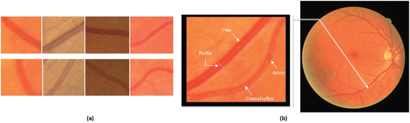
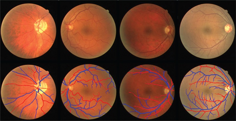
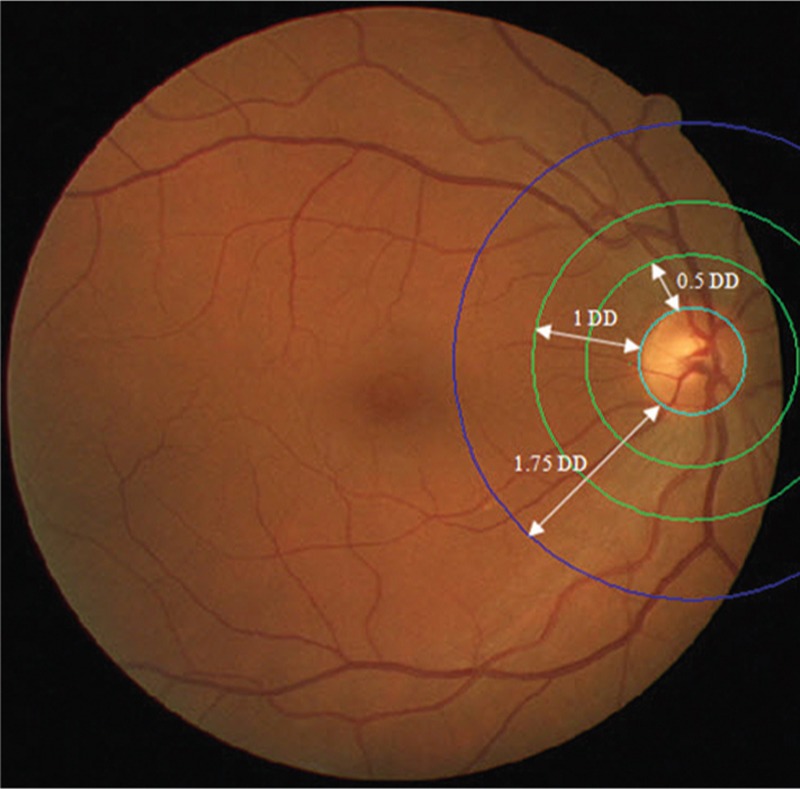
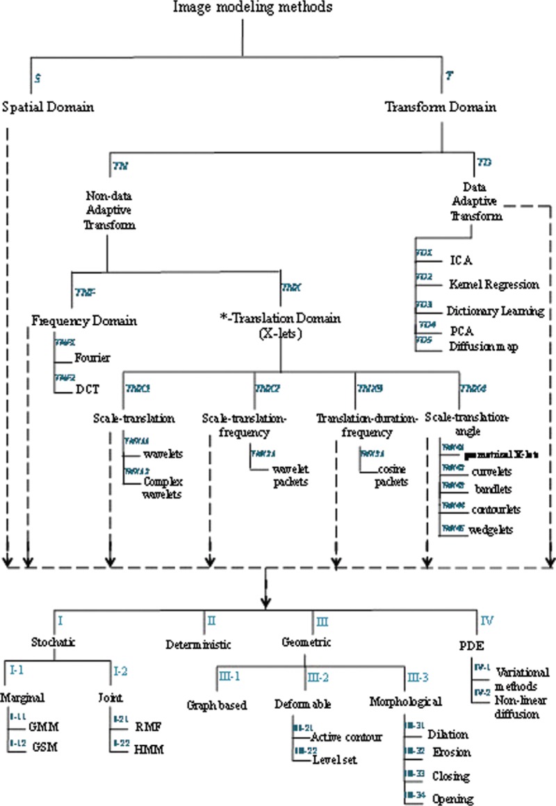
Nowadays, it is obvious that there is a relationship between changes in the retinal vessel structure and diseases such as diabetic, hypertension, stroke, and the other cardiovascular diseases in adults as well as retinopathy of prematurity in infants. Retinal fundus images provide non-invasive visualization of the retinal vessel structure. Applying image processing techniques in the study of digital color fundus photographs and analyzing their vasculature is a reliable approach for early diagnosis of the aforementioned diseases. Reduction in the arteriolar-venular ratio of retina is one of the primary signs of hypertension, diabetic, and cardiovascular diseases which can be calculated by analyzing the fundus images. To achieve a precise measuring of this parameter and meaningful diagnostic results, accurate classification of arteries and veins is necessary. Classification of vessels in fundus images faces with some challenges that make it difficult. In this paper, a comprehensive study of the proposed methods for classification of arteries and veins in fundus images is presented. Considering that these methods are evaluated on different datasets and use different evaluation criteria, it is not possible to conduct a fair comparison of their performance. Therefore, we evaluate the classification methods from modeling perspective. This analysis reveals that most of the proposed approaches have focused on statistics, and geometric models in spatial domain and transform domain models have received less attention. This could suggest the possibility of using transform models, especially data adaptive ones, for modeling of the fundus images in future classification approaches.
Keywords: Arteries and veins; computer-aided diagnosis; medical image processing; retinal fundus images; retinal vessel classification.
Conflict of interest statement
There are no conflicts of interest.
Figures
References
-
- Nguyen TT, Wang JJ, Wong TY. Retinal vascular changes in pre-diabetes and prehypertension: New findings and their research and clinical implications. Diabetes Care. 2007;30:2708–15. - PubMed
-
- Moss SE, Klein R, Klein BE. The 14-year incidence of visual loss in a diabetic population. Ophthalmology. 1998;105:998–1003. - PubMed
-
- Aiello LM. Perspectives on diabetic retinopathy. Am J Ophthalmol. 2003;136:122–35. - PubMed
Publication types
LinkOut - more resources
Full Text Sources
Other Literature Sources