. 2015 Apr:3:23.
doi: 10.3389/fphy.2015.00023. Epub 2015 Apr 10.

Emerging applications of porphyrins in photomedicine

Affiliations

Emerging applications of porphyrins in photomedicine

Haoyuan Huang et al. Front Phys. 2015 Apr.

Abstract

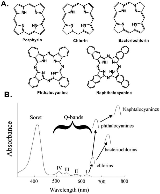
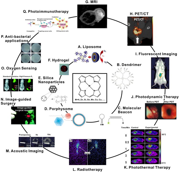
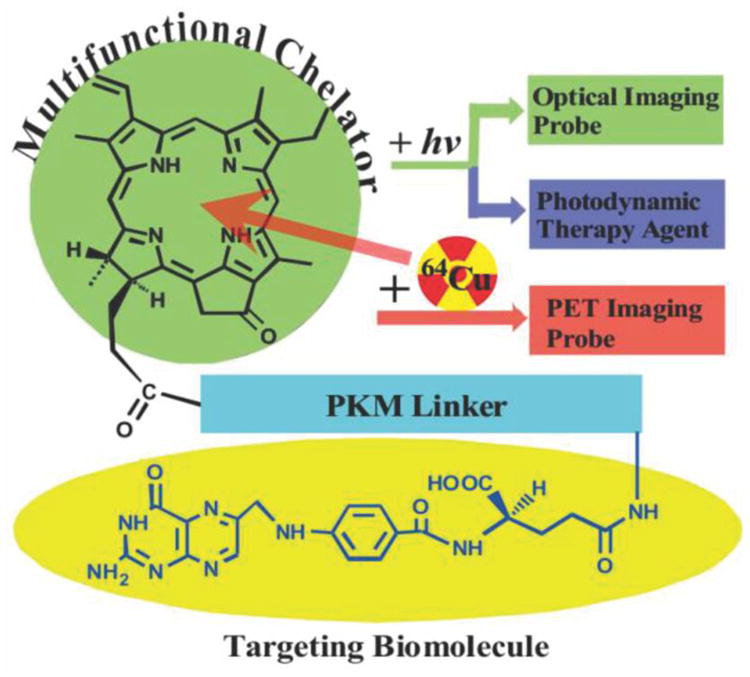
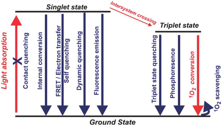
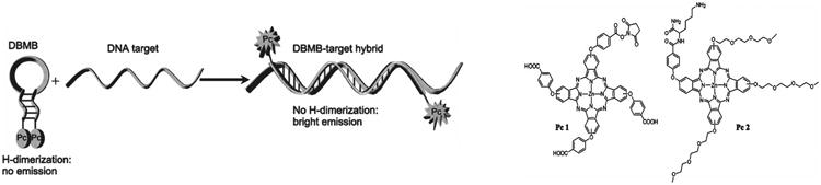
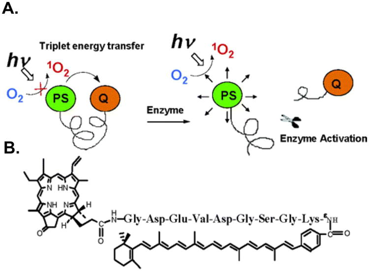
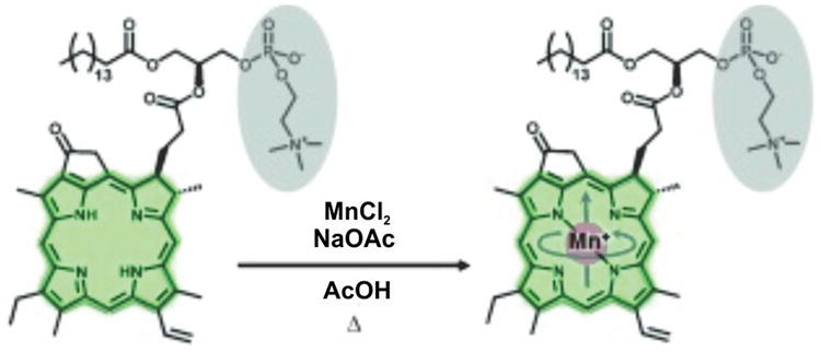
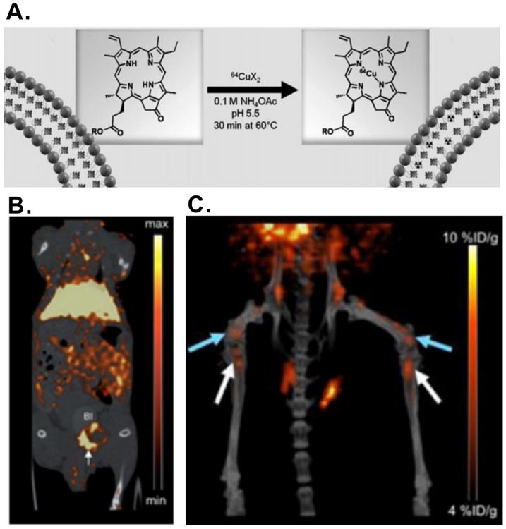
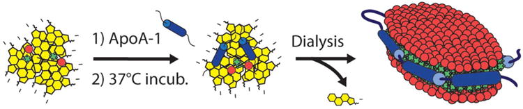
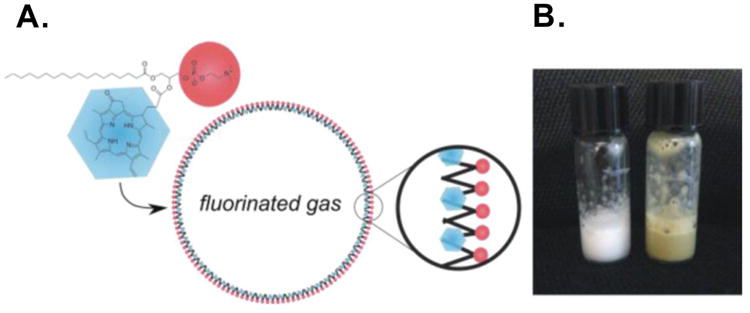
Biomedical applications of porphyrins and related molecules have been extensively pursued in the context of photodynamic therapy. Recent advances in nanoscale engineering have opened the door for new ways that porphyrins stand to potentially benefit human health. Metalloporphyrins are inherently suitable for many types of medical imaging and therapy. Traditional nanocarriers such as liposomes, dendrimers and silica nanoparticles have been explored for photosensitizer delivery. Concurrently, entirely new classes of porphyrin nanostructures are being developed, such as smart materials that are activated by specific biochemicals encountered at disease sites. Techniques have been developed that improve treatments by combining biomaterials with photosensitizers and functional moieties such as peptides, DNA and antibodies. Compared to simpler structures, these more complex and functional designs can potentially decrease side effects and lead to safer and more efficient phototherapies. This review examines recent research on porphyrin-derived materials in multimodal imaging, drug delivery, bio-sensing, phototherapy and probe design, demonstrating their bright future for biomedical applications.


Conflict of interest statement

Conflict of Interest Statement: The authors declare that the research was conducted in the absence of any commercial or financial relationships that could be construed as a potential conflict of interest.

Figures

Figure 1
Figure 1
(A) Structure and (B) Absorption of typical porphyrins, chlorins, bacteriochlorins, phthalocyanine and naphthalocyanines. Arrows show Q-band absorption (Adapted with permission from Berg et al. [6]).
Figure 2
Figure 2. Examples of porphyrin-based biomaterials (inner circle) and applications (outer circle)
(A) Liposomal phthalocyanine delivery [8]; (B) Glycoporphyrin dendrimers [9]; (C) Photodynamic molecular beacons [10]; (D) Porphyrin-phospholipid porphysome. [11]; (E) PpIX-modified mesoporous silica nanoparticle [12]; (F) Pd-porphyrin cross-linked hydrogel [13]; (G)In vivo MRI image of Mn-porphyrin nanoparticles [14]; (H) microPET/CT image of orthotopic PC3 tumor model after injection with 64Cu-porphysomes [15]; (I) Fluorescence tracking of macrophages after injection of porphyrin-modified nanoparticles [16]; (J) Human esophageal cancer treated with PDT [17]; (K) Thermal images of tumor-bearing mice with Pc-loaded nanoparticles exposed to a NIR laser [18]; (L) 8 h (a1) and 24 h (a2) radioimaging of melanoma-bearing mice after injection with 188Re-T3,4CPP [1]; (M) Acoustic images of a tumor-bearing mouse after injection with porphyrin-shell microbubbles [19]; (N) Image-guided surgery with a porphyrin-PEG cross-linked hydrogel [20]; (O) Phosphorescence images of an implanted Pd-porphyrin hydrogel in mice breathing different oxygen levels [13]; (P) Photoirradiation of bacteria under various photosensitizer conditions [21]; (Q) Photoimmunotherapy concept [22]. All figures used with permission from the indicated references.
Figure 3
Figure 3. Pyro-pharmacokinetic modifying linker (peptide sequence)-folate (PPF) structure design (Adapted with permission from Shi et al. [59])
Figure 4
Figure 4. Possible pathways for rational deactivation of an excited state photosensitizer
Simplified singlet oxygen generating diagram with energy levels shown in black and the typical pathway of singlet oxygen generation in red (Adapted with permission from Lovell et al. [64]).
Figure 5
Figure 5. Schematic representation of a dimer based photodynamic molecular beacon is shown on the left and the structure of Pc1 and Pc2 are shown on the right (Adapted with permission from Nesterova et al. [75])
Figure 6
Figure 6
(A) Principle of singlet oxygen scavenging and activation (B) structure of a caspase-3 activatable pyro-peptide-carotenoid photodynamic beacon (Adapted with permission from Chen et al. [79]).
Figure 7
Figure 7. Schematic showing the post chelation process of Mn ion into the pyro-lipid
Adapted with permission from MacDonald et al. [105].
Figure 8
Figure 8
(A) Schematic figure showing the process of chelating 64Cu ion into porphysomes; (B) MicroPET/CT images of coronal single slices through orthotopic PC3 tumor at 24 h after intravenous injection 64Cu-porphysomes; (C) 3D MicroPET/CT images (blue arrow, distal femur metastases, white arrow, proximal tibia metastases) (Adapted with permission from Liu et al. [15, 106]).
Figure 9
Figure 9. Schematic of the self-assembling process resulting in porphysome nanodics (Adapted with permission from Ng et al. [117])
Figure 10
Figure 10. (A) Schematic of the structure of microbubble (B) Photo of microbubbles solutions in room temperature (Adapted with permission from Huynh [19])

References

    1. Zhang Y, Lovell JF. Porphyrins as theranostic agents from prehistoric to modern times. Theranostics. 2012;2:905–15. doi: 10.7150/thno.4908. - DOI - PMC - PubMed
    1. Blamire AM, Ogawa S, Ugurbil K, Rothman D, McCarthy G, Ellermann JM, et al. Dynamic mapping of the human visual cortex by high-speed magnetic resonance imaging. Proc Natl Acad Sci USA. 1992;89:11069–73. doi: 10.1073/pnas.89.22.11069. - DOI - PMC - PubMed
    1. Belliveau JW, Kennedy DN, McKinstry RC, Buchbinder BR, Weisskoff RM, Cohen MS, et al. Functional mapping of the human visual cortex by magnetic resonance imaging. Science. 1991;254:716–9. doi: 10.1126/science.1948051. - DOI - PubMed
    1. Kelkar SS, Reineke TM. Theranostics: combining imaging and therapy. Bioconjug Chem. 2011;22:1879–903. doi: 10.1021/bc200151q. - DOI - PubMed
    1. Josefsen LB, Boyle RW. Unique diagnostic and therapeutic roles of porphyrins and phthalocyanines in photodynamic therapy, imaging and theranostics. Theranostics. 2012;2:916–66. doi: 10.7150/thno.4571. - DOI - PMC - PubMed