. 2017 Jul;13(7):807-813.
doi: 10.1038/nchembio.2392. Epub 2017 May 29.

Structural basis for high-affinity fluorophore binding and activation by RNA Mango

Affiliations

Structural basis for high-affinity fluorophore binding and activation by RNA Mango

Robert J Trachman 3rd et al. Nat Chem Biol. 2017 Jul.

Abstract

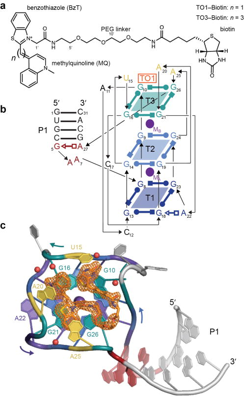
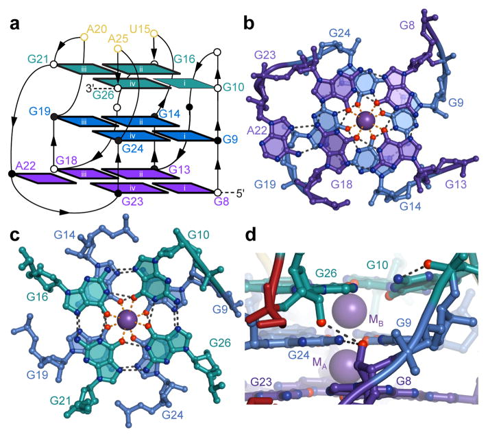
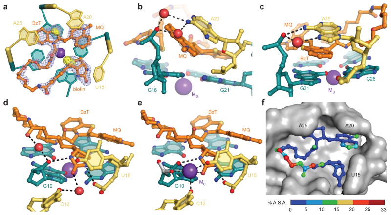
Genetically encoded fluorescent protein tags have revolutionized proteome studies, whereas the lack of intrinsically fluorescent RNAs has hindered transcriptome exploration. Among several RNA-fluorophore complexes that potentially address this problem, RNA Mango has an exceptionally high affinity for its thiazole orange (TO)-derived fluorophore, TO1-Biotin (Kd ∼3 nM), and, in complex with related ligands, it is one of the most redshifted fluorescent macromolecular tags known. To elucidate how this small aptamer exhibits such properties, which make it well suited for studying low-copy cellular RNAs, we determined its 1.7-Å-resolution co-crystal structure. Unexpectedly, the entire ligand, including TO, biotin and the linker connecting them, abuts one of the near-planar faces of the three-tiered G-quadruplex. The two heterocycles of TO are held in place by two loop adenines and form a 45° angle with respect to each other. Minimizing this angle would increase quantum yield and further improve this tool for in vivo RNA visualization.


Conflict of interest statement

COMPETING FINANCIAL INTERSTS

The authors declare no competing financial interests.

Figures

Figure 1
Figure 1
Overall structure of RNA Mango in complex with TO1-Biotin. (a) Chemical structures of TO1-Biotin and TO3-Biotin (ref. 1). The two compounds differ in having one or three benzylidene (methine) carbons, respectively, connecting the two heterocycles of their TO moieties. PEG, polyethyleneglycol linker. (b) Secondary structure of the RNA Mango-TO1-Biotin complex. Thin lines with arrowheads denote connectivity. Base pairs are represented using Leontis-Westhof symbols. The location of the fluorophore and two K+ ions (TO1, MA and MB, respectively) are indicated. Except where noted, this color scheme is used throughout. (c) Cartoon representation of the RNA Mango-TO1-complex. Curved arrows indicate direction of chain, 5′ to 3′. Orange mesh depicts a simulated-annealing omit |Fo|-|Fc| map (TO1-biotin was omitted from the calculation using the final refined atomic coordinates) contoured at 1.5 σ. Purple and red spheres represent K+ ions and water molecules, respectively.
Figure 2
Figure 2
Structure of the G-quadruplex core of RNA Mango. (a) Connectivity and stereochemistry of the RNA Mango G-quadruplex. Except for G10, which adopts the syn- conformation, all nucleotides are anti- (dark nucleobase outlines). Circles denote the pucker of successive backbone riboses (open and black circles, 3′-endo and 2′-endo, respectively). The four guanine stacks are denoted by white lower-case Roman numerals. (b) Ball-and-stick representation of the T1 and T2 tiers and the K+ ion MA. Black and orange dashed lines represent hydrogen bonding and inner-sphere cation coordination, respectively. (c) Tiers T2 and T3 and the K+ ion MB. (d) Detail of a side-view of the quadruplex showing a ribose-zipper-like interaction between G8, G24 and G26 (in tiers T1, T2, and T3, respectively). Buckling of G10 allows its nucleobase to hydrogen bond to its backbone phosphate.
Figure 3
Figure 3
The duplex-quadruplex junction of RNA Mango resembles a GAAA tetraloop. (a) Cartoon representation of the junction with one flanking Watson-Crick base pair from the duplex (gray), and adjacent residues from the G-quadruplex (G8 and G26). b) Hydrogen bonding pattern within the junction. (c) Junction of RNA Mango superimposed on a canonical GAAA tetraloop (gray; PDB 4FNJ; ref. 20).
Figure 4
Figure 4
Structural basis of TO1-Biotin recognition by RNA Mango. (a) Cartoon representation of the ligand binding site, superimposed on the |Fo| – |Fc| electron density map calculated prior to addition of the fluorophore to the crystallographic model (cyan mesh, 2 σ contour). The native anomalous difference Fourier synthesis is shown as a solid yellow surface (3 σ). (b) Detail of a ball-and-stick representation of the complex around the methylquinoline (MQ) of TO1-Biotin. (c) View around the benzothiazole (BzT) of TO1-Biotin. (d) View around the biotin of TO1-Biotin in chain A (Supplementary Fig. 3) of the ASU. (e) View around the biotin of TO1-Biotin in chain B (Supplementary Fig. 3) of the ASU. For clarity, the K+ ion MB is omitted. The K+ ion MC is only present in chain B. (f) Molecular surface of the ligand binding pocket of RNA Mango (grey) with a ball-and-stick representation of TO1-Biotin. The latter is colored according to the percentage of the surface area of each non-hydrogen atom that remains solvent-accessible upon complex formation.
Figure 5
Figure 5
Structure-based analysis of the RNA Mango-TO1-Biotin complex. (a) Effect on fluorophore-binding affinity of mutations in the ligand binding pocket (* Kds in panel (b) were measured using 31-nt RNA constructs with a 4 base pair duplex). (b) Effect on fluorophore-binding affinity of mutations in the tetraloop-like junction motif ( measurements in this panel were with 39–40 nt RNA constructs each containing an 8 base pair duplex). (c) through (f), effect on Kd and relative fluorescence enhancement (FE) of circular permutation (CP) of the connectivity of RNA Mango by either moving the attachment point of the tetraloop-like junction in a full-length (FL) 39–40 nt construct, or by deleting the duplex and junction (Δ). Guanines of the three tiers are represented as squares, colored as in Fig. 1. The four guanine stacks are numbered as in Fig. 2a. The two adenosines that cover the two heterocycles of the TO moiety of the fluorophore are represented as yellow rectangles. All data are the mean of three independent trials ± s.d.

References

    1. Dolgosheina EV, et al. RNA mango aptamer-fluorophore: a bright, high-affinity complex for RNA labeling and tracking. ACS Chem Biol. 2014;9:2412–2420. - PubMed
    1. Ellington AD, Szostak JW. In vitro selection of RNA molecules that bind specific ligands. Nature. 1990;346:818–822. - PubMed
    1. Grate D, Wilson C. Laser-mediated, site-specific inactivation of RNA transcripts. Proc Natl Acad Sci USA. 1999;96:6131–6136. - PMC - PubMed
    1. Babendure JR, Adams SR, Tsien RY. Aptamers switch on fluorescence of triphenylmethane dyes. J Am Chem Soc. 2003;125:14716–14717. - PubMed
    1. Paige JS, Wu KY, Jaffrey SR. RNA mimics of green fluorescent protein. Science. 2011;333:642–646. - PMC - PubMed