

Peridental bone changes after orthodontic tooth movement with fixed appliances: A cone-beam computed tomographic study
- PMID: 28553985
- PMCID: PMC8357220
- DOI: 10.2319/102716-774.1
Peridental bone changes after orthodontic tooth movement with fixed appliances: A cone-beam computed tomographic study
Abstract
Objective: To quantify treatment-related changes in peridental bone height and thickness in orthodontic patients.
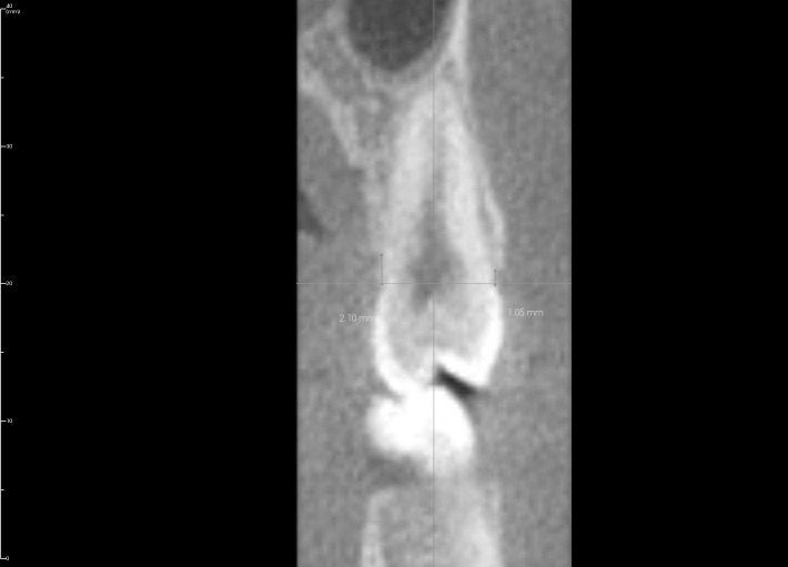
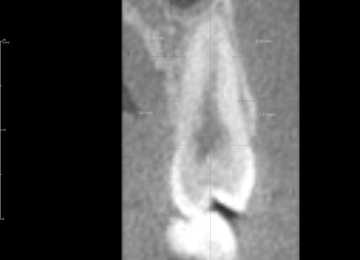
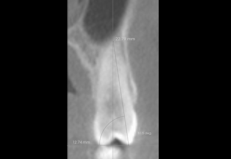
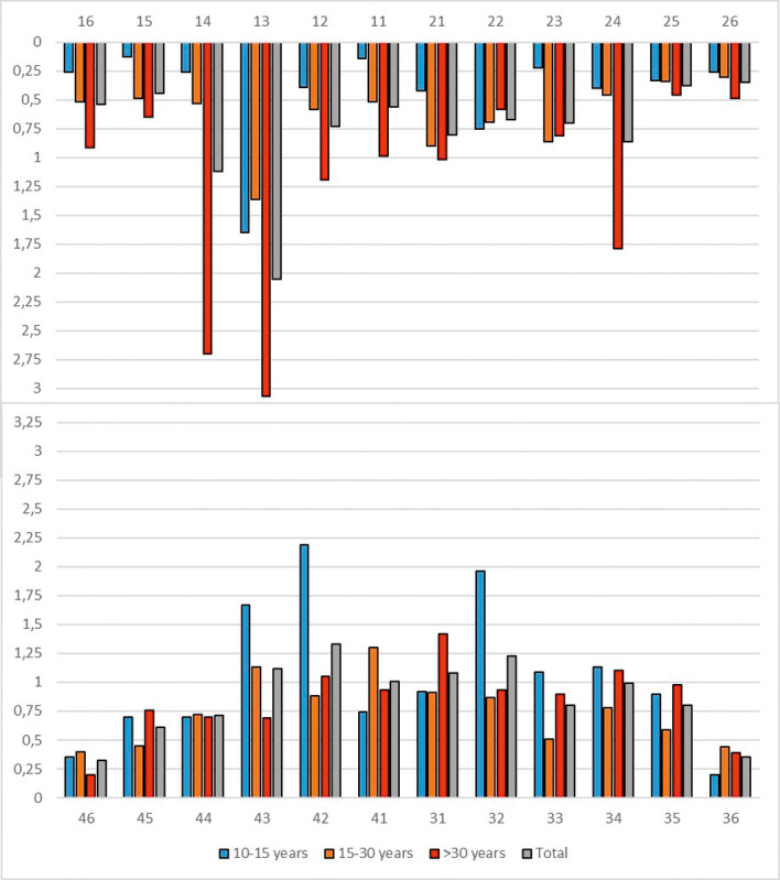
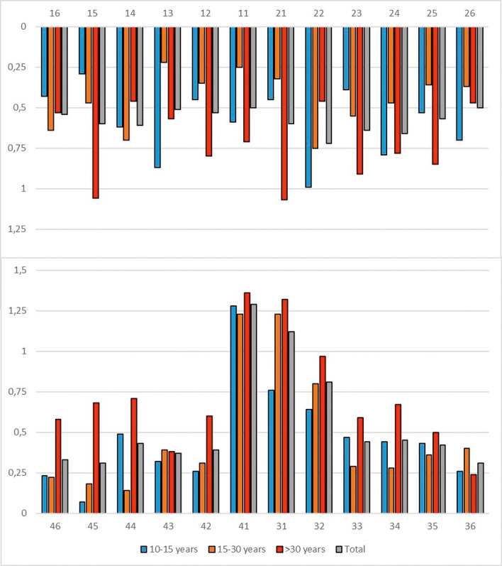
Materials and methods: Cone-beam computed tomographs (CBCTs) of 43 patients (24 female, 19 male; mean age: 25 years, 5 months) who underwent orthodontic treatment with multibracket appliances for at least 1 year were chosen for retrospective evaluation. Dehiscence depth and changes in bone width and tooth inclination were determined for 954 teeth.
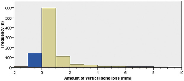
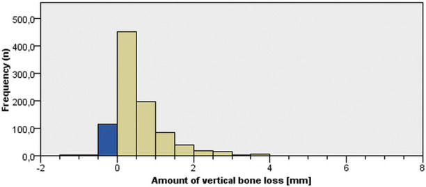
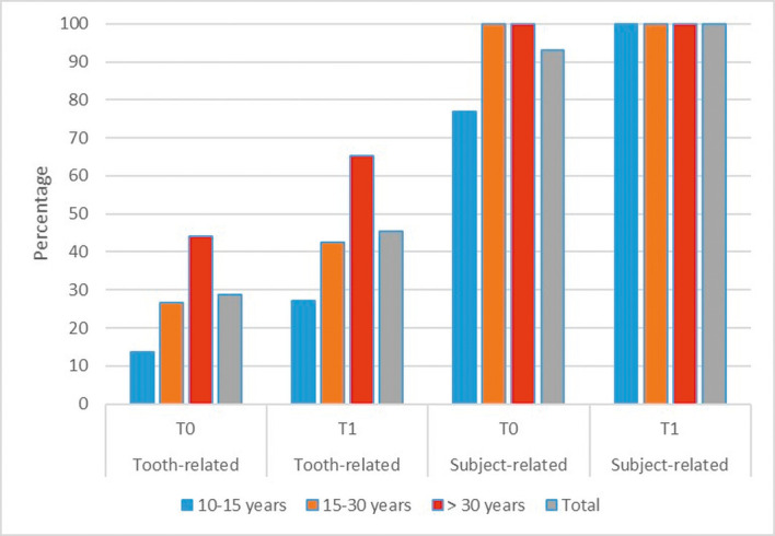
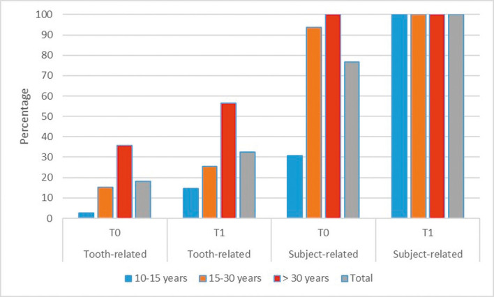
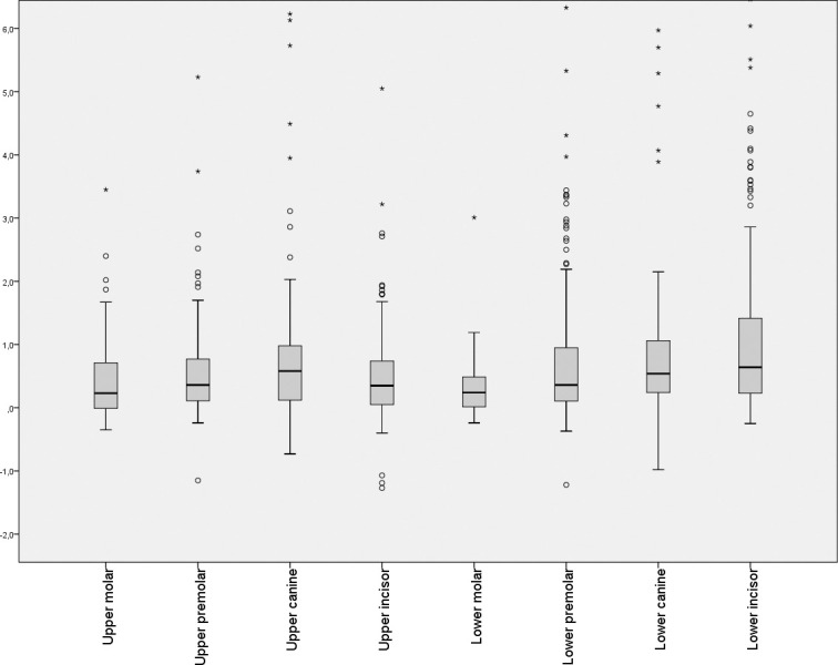
Results: There was a significant decrease in peridental bone height (dehiscence; -0.82 ± 1.47 mm) and bone thickness (-0.56 ± 0.7 and -0.69 ± 0.9 mm at 5 mm and 10 mm apical to the CEJ, respectively) during treatment (P < .001). A significantly greater dehiscence depth with increased vertical bone loss occurred in patients older than 30 years. In patients <30 years old, approximately 20% of the teeth showed defect depths >2 mm before treatment. In 90% of these patients, at least one tooth was affected. The maxillary canines and all mandibular teeth showed a higher risk for vestibular bone loss. Treatment changes in tooth inclination were correlated with horizontal bone loss.
Conclusions: Based on these results, it seems reasonable to recommend that peridental bone in orthodontic patients older than 30 be evaluated on a routine basis due to the risk of increased vertical bone loss. Ninety percent of patients younger than 30 showed reduced bone height (dehiscence) of the periodontium of at least one tooth.
Keywords: Cone-beam computed tomography; Multibracket; Peridental bone.
Figures
References
-
- Garib DG, Yatabe MS, Ozawa TO, Silva Filho OGD. Alveolar bone morphology under the perspective of the computed tomography: defining the biological limits of tooth movement. Dental Press J Orthod. 2010;15(5):192–205.
-
- Mulie RM, Hoeve AT. The limitations of tooth movement within the symphysis, studied with laminagraphy and standardized occlusal films. J Clin Orthod. 1976;10:882–893. - PubMed
-
- Lindhe J, Karring T, Araújo M. Clinical periodontology and implant dentistry. In: Lindhe J, Karring T, Lang N, editors. Anatomy 4th ed. Copenhagen: Blackwell Munksgaard; 2003. pp. 3–48.
-
- Evangelista K, Vasconcelos KDF, Bumann A, Hirsch E, Nitka M, Silva MAG. Dehiscence and fenestration in patients with Class I and Class II Division 1 malocclusion assessed with cone-beam computed tomography. Am J Orthod Dentofacial Orthop. 2010;138:133–135. - PubMed
-
- Baljoon M, Natto S, Bergstrom J. Occurrence of vertical bone defects in dentally aware individuals. Acta Odontol. 2003;61(1):47–51. - PubMed
MeSH terms
LinkOut - more resources
Full Text Sources
Other Literature Sources
