. 2017 Jul 1;25(4):354-361.
doi: 10.4062/biomolther.2016.263.

Role of TAZ in Lysophosphatidic Acid-Induced Migration and Proliferation of Human Adipose-Derived Mesenchymal Stem Cells

Affiliations

Role of TAZ in Lysophosphatidic Acid-Induced Migration and Proliferation of Human Adipose-Derived Mesenchymal Stem Cells

Won Min Mo et al. Biomol Ther (Seoul). .

Abstract

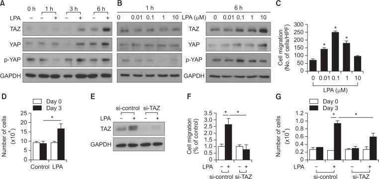
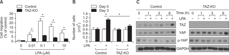
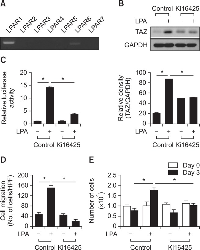
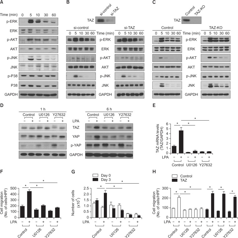
Transcriptional co-activator with a PDZ-binding motif (TAZ) is an important factor in lysophosphatidic acid (LPA)-induced promotion of migration and proliferation of human mesenchymal stem cells (MSCs). The expression of TAZ significantly increased at 6 h after LPA treatment, and TAZ knockdown inhibited the LPA-induced migration and proliferation of MSCs. In addition, embryonic fibroblasts from TAZ knockout mice exhibited the reduction in LPA-induced migration and proliferation. The LPA1 receptor inhibitor Ki16425 blocked LPA responses in MSCs. Although TAZ knockdown or knockout did not reduce LPA-induced phosphorylation of ERK and AKT, the MEK inhibitor U0126 or the ROCK inhibitor Y27632 blocked LPA-induced TAZ expression along with the reduction in the proliferation and migration of MSCs. Our data suggest that TAZ is an important mediator of LPA signaling in MSCs in the downstream of MEK and ROCK signaling.

Keywords: LPA; MEK; Mesenchymal stem cells; ROCK; TAZ.


Conflict of interest statement

CONFLICT OF INTEREST

Authors declare no conflict of interest.

Figures

Fig. 1.
Fig. 1.
Effects of LPA on hippo signaling and the requirement of TAZ for LPA-induced cellular responses in MSCs. (A) The time course of LPA-induced TAZ, YAP, and p-YAP expression is shown. MSCs were treated with serum-free medium containing 10 mM LPA or vehicles for the indicated amount of time. The expression levels of TAZ, YAP, p-YAP, and GAPDH were determined using Western blotting. (B) MSCs were treated with various doses of LPA for 1 h and 6 h. The expression levels of TAZ, YAP, p-YAP, and GAPDH were determined using Western blotting. (C) Migration of MSCs in response to various concentrations of LPA after 12 h incubation was measured using the chemotaxis chamber. Data indicate mean ± SD. *p<0.05 (n=8). (D) The effect of LPA on MSC proliferation is shown. The number of cells were counted and compared with those of the control cells. MSCs exhibited increased LPA-induced proliferation for 3 days. MSCs exhibited increased LPA-induced proliferation for 3 days. The cellular proliferation rate was determined by the cell count using a trypan blue inclusion assay. Data indicate mean ± SD. *p<0.05 (n=6). (E) siRNA-mediated silencing of LPA-induced TAZ expression is shown. MSCs were transfected with control siRNA and a pool of siRNA against TAZ. The expression levels of TAZ and GAPDH were determined using Western blotting. (F) Knockdown of TAZ abrogates LPA (1 uM)-stimulated migration in MSCs. Data indicate mean ± SD. *p<0.05 (n=8). (G) TAZ is required for the LPA-induced cellular proliferation of MSCs. Control and TAZ knockdown-MSCs were treated with LPA (10 μM) for 3 days, followed by cell counting using a trypan blue inclusion assay. Data indicate mean ± SD. *p<0.05 (n=6).
Fig. 2.
Fig. 2.
Role of TAZ in LPA-induced migration and proliferation of MEFs. (A) Wild-type MEFs (control) and TAZ-knockout MEFs (TAZ-KO) were treated with the various concentrations of LPA for 12 h, followed by a chemotactic migration assay. Data indicate mean ± SD. *p<0.05 (n=8). (B) Inhibition of LPA-induced proliferation in the TAZ-knockout MEFs is shown. Wild-type MEFs (control) and TAZ-knockout MEFs (TAZ-KO) were treated with vehicles or LPA (10 μM) for 3 days. The number of cells were counted and compared with those of the control cells. TAZ-knockout MEFs exhibited decreased LPA-induced proliferation for 3 days. The cellular proliferation rate was determined via cell counting using trypan blue inclusion assay. Data indicate mean ± SD. *p<0.05 (n=6). (C) Time-course of LPA-induced TAZ expression is shown. Wild-type MEFs (control) and TAZ-knockout MEFs (TAZ-KO) were treated with vehicle or LPA (10 μM) for the indicated periods. The expression levels of TAZ and GAPDH were determined using Western blotting.
Fig. 3.
Fig. 3.
Involvement of LPAR1 in the LPA-induced migration and proliferation of MSCs. (A) The expression level of the LPA receptors was determined using RT-PCR. (B) MSCs were treated with vehicle control or Ki16425 (10 μM), LPAR1 and 3 inhibitor, followed by Western blotting analysis with indicated antibodies (left panel). Quantification of TAZ expression normalized by GAPDH expression is shown (right panel). Data indicate mean ± SD. *p<0.05 (n=4). (C) YAP/TAZ-responsive reporter (8×GTIIC-luciferase) activities in MSCs after treatment of LPA (10 μM) in the presence or absence of 10 μM Ki16425 are shown. Data indicate mean ± SD. *p<0.05 (n=4). (D) The inhibitory effect of Ki16425 (10 μM) on LPA-induced (1 μM) MSC migration is shown. Data indicate mean ± SD. *p<0.05 (n=8). (E) The inhibitory effect of Ki16425 (10 μM) on LPA-induced (10 μM) MSC proliferation is shown. The cell proliferation rate was determined by cell count using a trypan blue inclusion assay. Data indicate mean ± SD. *p<0.05 (n=6).
Fig. 4.
Fig. 4.
Effects of the MEK inhibitor and the ROCK inhibitor on LPA-induced regulation of hippo signaling in MSCs. (A) The LPA stimulated phosphorylation of MAP kinases in MSCs is shown. MSCs were treated with LPA (10 μM) for the indicated times and subjected to Western blotting analysis with indicated antibodies. (B) siRNA-mediated silencing of TAZ expression is shown. LPA-stimulated phosphorylation of MAP kinases in both the control and TAZ knockdown-MSCs. (C) The wild-type MEFs (control) and TAZ-knockout MEFs (TAZ-KO) were treated with LPA (10 μM) for the indicated times and subjected to Western blotting analysis. The expression level of the indicated antibodies and GAPDH were determined using Western blotting. (D) The effect of U0126 and Y-27632 on LPA-induced TAZ, YAP, and p-YAP expressions are shown. MSCs were treated with vehicle control, U0126 (10 μM) or Y-27632 (10 μM), followed by stimulation with LPA (10 μM) for the indicated times. The expression levels of TAZ, YAP, p-YAP, and GAPDH were determined by Western blotting analysis. (E) Real-time PCR analysis revealed the mRNA expression levels of TAZ in MSCs. MSCs were pretreated with U0126 (10 μM) and Y-27632 (10 μM), followed by treatment with LPA (10 μM) for 6 h. (F) The effect of U0126 (10 μM) and Y-27632 (10 μM) on LPA-induced (1 μM) migration of MSCs. Data indicate mean ± SD. *p<0.05 (n=8). (G) The effect of U0126 (10 μM) and Y-27632 (10 μM) on the LPA-induced (10 μM) cellular proliferation of MSCs. The cellular proliferation rate was determined by cell counting using a trypan blue inclusion assay. Data indicate mean ± SD. *p<0.05 (n=6). (H) TAZ overexpression reversed the inhibitory effect of U0126 (10 μM) or Y27632 (10 μM) on LPA-induced (1 μM) MSCs migration. Data indicate mean ± SD. *p<0.05 (n=8).

Similar articles

Cited by

References

    1. Aggarwal S, Pittenger MF. Human mesenchymal stem cells modulate allogeneic immune cell responses. Blood. 2005;105:1815–1822. doi: 10.1182/blood-2004-04-1559. - DOI - PubMed
    1. Anliker B, Chun J. Cell surface receptors in lysophospholipid signaling. Semin Cell Dev Biol. 2004;15:457–465. doi: 10.1016/j.semcdb.2004.05.005. - DOI - PubMed
    1. Badri L, Lama VN. Lysophosphatidic acid induces migration of human lung-resident mesenchymal stem cells through the β-catenin pathway. Stem Cells. 2012;30:2010–2019. doi: 10.1002/stem.1171. - DOI - PMC - PubMed
    1. Binder BY, Genetos DC, Leach JK. Lysophosphatidic acid protects human mesenchymal stromal cells from differentiation-dependent vulnerability to apoptosis. Tissue Eng. Part A. 2014;20:1156–1164. doi: 10.1089/ten.tea.2013.0487. - DOI - PMC - PubMed
    1. Chan SW, Lim CJ, Guo K, Ng CP, Lee I, Hunziker W, Zeng Q, Hong W. A role for TAZ in migration, invasion, and tumorigenesis of breast cancer cells. Cancer Res. 2008;68:2592–2598. doi: 10.1158/0008-5472.CAN-07-2696. - DOI - PubMed